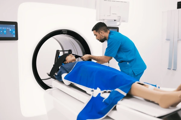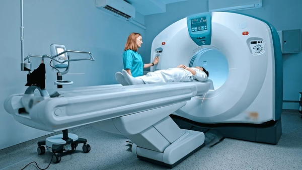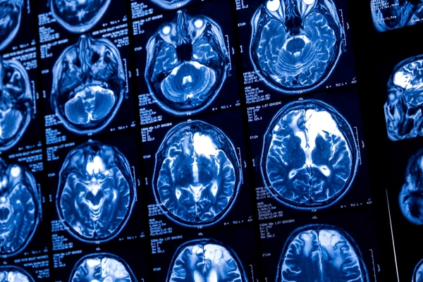
Introduction
When choosing between CT scan vs. MRI, understanding the distinct features and benefits of each tool is crucial. Both play vital roles in diagnosing a variety of medical conditions, but their applications and strengths differ significantly. Knowing these differences can help in selecting the most suitable option for an accurate and effective diagnosis. This article will explore the key differences, advantages, and applications of CT scans and MRIs to guide you in making an informed decision.
What is a CT Scan?
A CT scan, or Computed Tomography scan, is a powerful imaging technique that creates detailed cross-sectional images of the body. It combines X-ray technology and advanced computer algorithms to provide precise visualizations of internal structures, often referred to as “slices” or tomograms.
How a CT Scan Works?
X-ray Transmission: The scanner directs controlled X-ray beams through the body as the patient lies inside the machine.
Data Acquisition: Detectors measure how the X-ray beams are attenuated as they pass through various tissues, capturing essential data.
Rotation and Detection: The gantry rotates around the patient, collecting measurements from multiple angles to ensure comprehensive imaging.
Data Processing: Sophisticated computer algorithms analyze the captured data, transforming it into detailed images for review.
Image Reconstruction: The images can be examined as individual slices or compiled into a three-dimensional model for a complete view of the body’s internal structures.

What is an MRI?
An MRI, or Magnetic Resonance Imaging, is a non-invasive medical technique that creates detailed images of internal body structures. Unlike X-rays or CT scans, MRI uses a strong magnetic field, radio waves, and advanced computing to capture high-resolution images of soft tissues like organs, muscles, and cartilage. This method avoids ionizing radiation, making it a safer option for many patients.
How an MRI Works?
Magnetic Field Alignment: The process begins with the patient placed inside a cylindrical magnet that generates a powerful magnetic field. This field aligns hydrogen protons in the body, particularly those in water molecules within tissues.
Radio Frequency Pulses: Radio frequency (RF) coils emit energy pulses at the Larmor frequency, determined by the magnetic field strength. These pulses disrupt the aligned protons, causing them to resonate.
Signal Detection: As the protons realign after the RF pulse, they emit signals unique to their environment. RF coils detect these signals for further processing.
Image Reconstruction: Complex algorithms process the detected signals to create cross-sectional images, or slices, of the body. These slices can be combined to form detailed 3D representations.
Image Interpretation: Radiologists review the high-resolution images to identify injuries, diseases, or abnormalities in tissues and organs. This helps in diagnosing and monitoring various medical conditions effectively.

CT Scan vs. MRI: Key Differences
Technology and Principle
CT scans use X-rays to create detailed cross-sectional images of the body. The machine takes a series of X-ray images from various angles. A computer then combines these images to produce accurate 3D visuals. On the other hand, MRI technology relies on strong magnetic fields and radio waves to generate body images. It works by aligning hydrogen atoms with the magnetic field and detecting signals emitted as they return to normal.
Image Quality and Contrast
CT scans excel in showing bones, blood vessels, and calcified tissues. They offer high-resolution images of dense structures like bones. In contrast, MRIs provide superior soft tissue contrast, making them ideal for imaging the brain, liver, and muscles. They are especially useful for organs and tissues with little calcium content.
Use of Radiation
CT scans use ionizing radiation, which carries a small risk of long-term health effects. Repeated exposure may slightly increase the risk of cancer. In comparison, MRIs do not use radiation, making them safer for frequent imaging. They are a preferred choice for imaging sensitive areas like the brain and joints.
Scan Time and Availability
CT scans are faster, often taking just a few seconds to minutes per slice. Because of their speed and availability, they are widely used in emergencies. On the other hand, MRI scans take more time, lasting anywhere from 15 to 90 minutes or longer. Fewer facilities have MRI machines, which can limit their accessibility.
Contrast Agents
CT scans frequently use iodine-based contrast agents to improve image clarity and detect specific conditions. Meanwhile, MRIs rely on gadolinium-based agents to enhance soft tissue detail and contrast. These agents work well for highlighting abnormalities in tissues and organs.
Applications of CT Scans vs. MRI
Common Uses
CT scans excel at imaging bones, detecting cancers, and diagnosing blood vessel diseases. Doctors often use them to guide surgical procedures with precision. They are highly effective for quickly assessing injuries or abnormalities in the lungs and abdominal area. This speed makes them invaluable in emergency situations.
MRIs, on the other hand, specialize in soft tissue imaging and are crucial for evaluating the brain and spinal cord. They help diagnose tumors, strokes, and joint disorders with incredible detail. Because they don’t use radiation, MRIs are safer for children, pregnant women, and patients needing frequent imaging. This makes them a preferred choice in these cases.

When to Choose a CT Scan Over a MRI
Speed and Urgency
CT scans deliver fast results, making them a top choice for emergencies. Doctors rely on them for head trauma, chest pain, and abdominal emergencies. Their speed allows quick diagnoses, enabling immediate treatment when time is critical.
MRI scans, however, take longer and are less suitable for urgent situations. They are better for detailed soft tissue imaging when precision matters more than speed.
Imaging Details and Contrast
CT scans offer excellent visualization of bones and blood vessels, especially with iodinated contrast agents. They are highly effective for lung imaging and are widely used in lung cancer screenings due to their precision.
In comparison, MRIs excel at showing soft tissue details without exposing patients to radiation. They are ideal for diagnosing neurological conditions like brain cancer, strokes, and neurodegenerative disorders.
Specific Medical Conditions
CT scans are perfect for quickly assessing the entire body or specific areas like the chest and abdomen. Doctors frequently use them to evaluate fractures, internal bleeding, or conditions like appendicitis and kidney stones.
MRIs work best for conditions that require detailed soft tissue imaging. They are particularly useful for musculoskeletal problems, spinal cord disorders, and cancers like prostate, nasal, and uterine cancers.
Patient Factors
CT scans suit patients who need shorter procedures or those with implants that aren’t MRI-compatible. This makes them ideal for individuals who cannot tolerate long scanning sessions.
MRIs, on the other hand, may not work well for patients with metal implants or severe claustrophobia. Patients who struggle to remain still for extended periods may also face challenges. However, MRIs remain the best option for those needing detailed soft tissue imaging.
FAQ About CT Scans and MRI
Which imaging method is faster?
CT scans take a few minutes, while MRIs usually take 15–90 minutes.
Do CT scans and MRIs expose patients to radiation?
CT scans use radiation; MRIs do not.
Which is better for imaging soft tissues?
MRIs are better for soft tissues; CT scans excel at bones and lungs.
Are there any contraindications for MRI?
Yes, MRIs are unsuitable for patients with metal implants or severe claustrophobia.
How do doctors decide which imaging method to use?
Doctors choose based on the condition, body area, urgency, and patient history.
To get detailed scientific explanations of CT Scan vs. MRI, try Patsnap Eureka.

