Method for promoting maturation of myocardial cells differentiated from multipotential stem cells

A technology of pluripotent stem cells and cardiomyocytes, applied in artificially induced pluripotent cells, biochemical equipment and methods, embryonic cells, etc., to achieve the effects of orderly arrangement, promotion of maturation, and enhanced contractility

Active Publication Date: 2018-05-22
SUZHOU UNIV
4 Cites 10 Cited by

AI-Extracted Technical Summary

Problems solved by technology

In-depth study of the role of RA in the process of induction of cardiomyocytes from pluripotent stem c...

Abstract

The invention relates to a method for promoting maturation of myocardial cells differentiated from multipotential stem cells. In the 0-1 day, the multipotential stem cells which are subcultured to bethe 4-5 generation are subjected to induced differentiation, and a culture medium used in the method contains a GSK-3 inhibitor with the concentration being 2-15 mu M/L; in the 2-3 day, the culture medium is used for conducting induced differentiation continuously; in the 4-5 day, a culture medium containing a Wnt inhibitor with the concentration being 2-10 mu M/L is used for conducting induced differentiation; in the 14-20 days, a culture medium containing retinoic acid with the concentration being 0.2-5 mu M/L is used for conducting induced differentiation continuously, later on, the culturemedium is used for conducting induced differentiation culture on the multipotential stem cells, wherein in the 1-6 days, a first induced differentiation culture medium is used and comprises an RPMI-1640 basic culture medium and B27-insulin, after the 7 day, a second induced differentiation culture medium is used and comprises the RPMI-1640 basic culture medium and B27, or in the whole induced differentiation culture process, a CDM3 induced differentiation culture medium or a serum-free induced differentiation culture medium is used, and the CDM3 induced differentiation culture medium comprises the RPMI-1640 basic culture medium, serum albumin, ascorbic acid and double-antibody.

Application Domain

Culture processSkeletal/connective tissue cells +3

Technology Topic

Double antibodyGSK-3 +12

Image

  • Method for promoting maturation of myocardial cells differentiated from multipotential stem cells
  • Method for promoting maturation of myocardial cells differentiated from multipotential stem cells
  • Method for promoting maturation of myocardial cells differentiated from multipotential stem cells

Examples

  • Experimental program(3)

Example Embodiment

[0036] Example 1 Cultivation and passage of embryonic stem cells
[0037] In this example, the HES3 cell line in hPSCs was used as the experimental object. The HES3 cells were grown on Matrigel-coated culture dishes and diluted with 1:200 DMEM/F12 basal medium for use. Then HES3 cells were planted on Matrigel, cell culture was carried out with mTeSR1 or E8 medium, and the medium was changed every day. When the density of HES3 cells reaches about 80% and the cell clone is large enough, the cells are passaged.
[0038] When passaging, wash twice with DPBS to remove the dead cell debris on the surface, add 0.1mol/L EDTA and place at 37℃, 5% CO 2 Digest in the constant temperature incubator for 7 minutes. After the digestion time is up, observe under an inverted microscope that there are gaps between the adherent cells but have not separated from each other. The cell colonies have become opaque and whitish by naked eyes. Aspirate the digestion solution, add the stem cell culture medium, and use a pipette fan-shaped pipetting to culture At the bottom of the dish, make the stem cells attached to the bottom of the dish fall off, gently and slowly pipette evenly to make a cell suspension. Generally, the ratio of 1:10 or 1:12 is inoculated into Petri dishes coated with Matrigel, and then passaged every 4 days.

Example Embodiment

[0039] Example 2 Embryonic stem cells are induced to differentiate into mature cardiomyocytes
[0040] After the HES3 cells cultured to passage 4 to 5 were washed with DPBS, digested with 0.1mol/L EDTA for 7 minutes, 4 /cm 2 Inoculate in a petri dish at 37℃, 5% CO 2 After 4 days of culture in the incubator, the cell density reached 85% or more, and replaced with RPMI1640+B27-insulin induction medium.
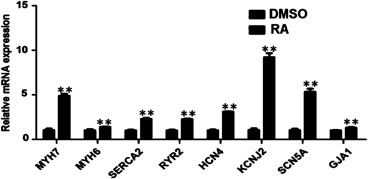
[0041] Retinoic acid treatment group (RA group): On the 0th to 1st day, the induced differentiation was induced by RPMI1640+B27-insulin containing 5μM CHIR99021, and the RPMI1640+B27-insulin was used for incubation on the 2nd to 3rd day; Days were cultured with RPMI1640+B27-insulin containing 5μM Wnt inhibitor. Only RPMI1640+B27-insulin was used for culture on the 6th day. After the 7th day, culture with RPMI1640+B27 every day. Obvious myocardial beating can be observed on the 9th to 10th day of differentiation (D9~D10). On the 14th to 20th day, change to RPMI1640+ containing 1μM retinoic acid (RA) The cells were cultured in B27 induction differentiation medium, and some maturation indicators of cardiomyocytes were detected on the 30th day.
[0042] Set up a control group (DMSO group), the culture process is the same as the above steps, the difference is that the same volume as the treatment group solution is added to the induction medium containing DMSO for culture on the 14th to 20th day.
[0043] In the process of cardiomyocyte induction, RA is processed on the 14th to 20th day, and then the cells are collected on the 30th day, RNA is extracted, and the main structural and functional mature gene expression is detected by real-time fluorescent quantitative PCR (QPCR) instrument. By detecting the expression of these structural and ion channel genes at the mRNA level, compared with the DMSO group, these genes in the RA group showed a significant up-regulation trend, and the difference was significant. The results are as follows figure 1 Shown. This shows that in the process of inducing the whole myocardial differentiation, by adding RA at a specific time and a specific concentration, it is beneficial to promote the maturation of cardiomyocytes, thereby providing effective mature cardiomyocytes for clinical disease treatment and drug screening.
[0044] The expression of cardiomyocyte-specific cardiac troponin cTnT and actininα-actinin was identified by immunofluorescence staining. The induced differentiated cardiomyocytes were digested with 0.25% trypsin and passaged to confocol dishes. The cells adhere to the wall for two days, and then start the experiment. First wash the cells twice with PBS; fix with 4% PFA (paraformaldehyde) for 15 min; wash 3 times with PBST for 5 min each; permeate the membrane with 0.5% TritonX-100 at room temperature for 10 minutes; block with 5% BSA at room temperature for 1 hour ;Incubate the primary antibody overnight at 4°C, the antibody ratio is cTnT 1:200, α-actinin 1:200; wash 3 times with PBST, 5 min each time; incubate the secondary antibody, protect from light at room temperature for 1 hour, the antibody ratio is 1:600; PBST Wash 3 times, 5 min each time; add Hoechst staining, 20 min at room temperature; wash 3 times with PBST, 5 min each time; mount the slides and check with microscope. The result is figure 2 Shown, where, figure 2 A and B are the cTnT expression results of the DMSO group and RA group (Scale bar=20μm), figure 2 C and D are the expression results of α-actinin in DMSO group and RA group respectively (Scalebar=20μm). The results show that after retinoic acid treatment, the myofilaments of cardiomyocytes are significantly longer and orderly arranged, and cardiomyocytes have obvious The muscle segments are neatly arranged.
[0045] The pH value change test of myocardial cells: Take 1 mL of the culture medium of the RA group and DMSO group each in a centrifuge tube, and then take pictures. Before treatment, the color of DMSO and RA groups was pink, after treatment, the color of DMSO group was orange, and the color of RA group was yellow. The results showed that the color of the culture medium of cardiomyocytes treated with retinoic acid had higher acidity value , The metabolism is more vigorous, the cell energy metabolism meter may be used to detect its specific metabolic process in the future, and the results are as follows image 3 Shown.

Example Embodiment

[0046] Example 3 Cultivation, passage and directed induction of induced pluripotent stem cells
[0047] In this example, the HES3 cells in Examples 1 and 2 were replaced with the SCCTM-iPSC-1 cell line in hPSCs as the experimental object. For other specific embodiments, refer to Examples 1 and 2.
[0048] Use patch clamp to record the action potential changes of ventricular myocytes. The cardiomyocytes on the 30th day were digested with 0.25% trypsin for 5 minutes, single cells were plated, and adhered to the wall for 3 days for testing, including the beating frequency of the cardiomyocytes, the amplitude of the action potential, the ascending speed, and the hyperpolarized diastolic potential. According to the experimental results, the results of cardiomyocytes induced by SCCTM-iPSC-1 cells are as follows Figure 4 As shown, Figure 4 A, B, and C are the test results of cardiomyocytes in the DMSO group, the test results of the cardiomyocytes in the RA group, and the comparison chart of the test results under single frequency. Compared with the control, the cardiomyocytes treated with retinoic acid have a slower beating frequency, an increased action potential amplitude (APA), a higher ascending speed (dv/dt), and a super-maximum diastolic blood pressure (MDP) .

PUM

no PUM

Description & Claims & Application Information

We can also present the details of the Description, Claims and Application information to help users get a comprehensive understanding of the technical details of the patent, such as background art, summary of invention, brief description of drawings, description of embodiments, and other original content. On the other hand, users can also determine the specific scope of protection of the technology through the list of claims; as well as understand the changes in the life cycle of the technology with the presentation of the patent timeline. Login to view more.

Similar technology patents

File storage box and compact shelf

PendingCN112890454AAccurate placementOrderly rows
Owner:北京融安特智能科技股份有限公司

Foil winding coil mold for multiple copper bar outgoing lines

InactiveCN102360940AOrderly rowsSimple structure
Owner:天津市特变电工变压器有限公司

Double-ridge welsh onion transplanter

ActiveCN105850319AOrderly rowsevenly distributed
Owner:QINGDAO AGRI UNIV

Controlled circulating water shrimp culture system and working method

PendingCN111700024Apromote maturityShorten the film setting time
Owner:FISHERY MACHINERY & INSTR RES INST CHINESE ACADEMY OF FISHERY SCI

Filter line grid with tungsten sputtered on surface and hafnium oxide deposited in micropores and manufacturing method

PendingCN114699095AThe grid structure is stableOrderly rows
Owner:中建材光芯科技有限公司

Method for preparing microelectrolysis reductive water

ActiveCN104016536AOrderly rowsStrong cohesion
Owner:山东中鲁水务有限公司

Organic formula milk powder for children

PendingCN111713558Apromote maturityimprove visual sensitivity
Owner:HULUN BUIR SHUANGWA DAIRY CO LTD

Classification and recommendation of technical efficacy words

  • promote maturity
  • Orderly rows

Organic formula milk powder for children

PendingCN111713558Apromote maturityimprove visual sensitivity
Owner:HULUN BUIR SHUANGWA DAIRY CO LTD

Controlled circulating water shrimp culture system and working method

PendingCN111700024Apromote maturityShorten the film setting time
Owner:FISHERY MACHINERY & INSTR RES INST CHINESE ACADEMY OF FISHERY SCI

Card label pasting device

PendingCN108190167Astable separationOrderly rows
Owner:宁波聚思智能科技有限公司

Method for preparing microelectrolysis reductive water

ActiveCN104016536AOrderly rowsStrong cohesion
Owner:山东中鲁水务有限公司

Foil winding coil mold for multiple copper bar outgoing lines

InactiveCN102360940AOrderly rowsSimple structure
Owner:天津市特变电工变压器有限公司

Double-ridge welsh onion transplanter

ActiveCN105850319AOrderly rowsevenly distributed
Owner:QINGDAO AGRI UNIV
Why Eureka
Try Eureka