Supercharge Your Innovation With Domain-Expert AI Agents!

Lesion detection method and terminal equipment of an X-ray image

A detection method, X-ray technology, applied in the medical field, can solve the problems of inability to achieve precise positioning, low efficiency of weak supervision target detection methods, and reduce the comprehensiveness of target detection, so as to achieve the effect of improving accuracy

Active Publication Date: 2022-04-01
SHENZHEN UNIV
View PDF5 Cites 0 Cited by
  • Summary
  • Abstract
  • Description
  • Claims
  • Application Information

AI Technical Summary

Problems solved by technology

For the weakly supervised level, the existing weakly supervised target detection technology cannot achieve accurate positioning, and the efficiency of weakly supervised target detection methods is generally low. Among them, the fastest weakly supervised target detection method can only achieve a speed of Detect around two frames
In addition, the current weakly supervised detection of medical data is a single target, which reduces the comprehensiveness of target detection

Method used


Image

  • Lesion detection method and terminal equipment of an X-ray image
  • Lesion detection method and terminal equipment of an X-ray image
  • Lesion detection method and terminal equipment of an X-ray image

Examples


Embodiment Construction

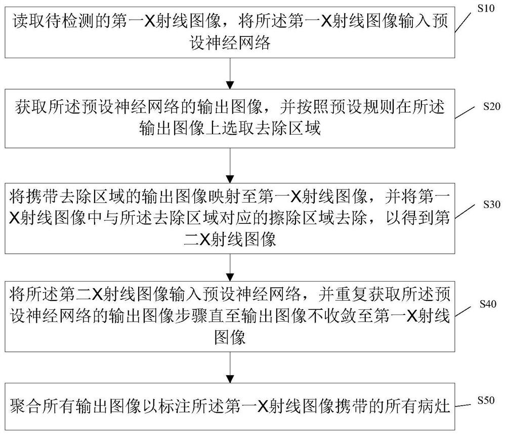
[0038] The present invention provides an X-ray image lesion detection method and terminal equipment. In order to make the purpose, technical solution and effect of the present invention clearer and clearer, the present invention will be further described in detail below with reference to the accompanying drawings and examples. It should be understood that the specific embodiments described here are only used to explain the present invention, not to limit the present invention.

[0039] Those skilled in the art will understand that unless otherwise stated, the singular forms "a", "an", "said" and "the" used herein may also include plural forms. It should be further understood that the word "comprising" used in the description of the present invention refers to the presence of said features, integers, steps, operations, elements and / or components, but does not exclude the presence or addition of one or more other features, Integers, steps, operations, elements, components, and / o...


PUM


Abstract

The invention discloses an X-ray image lesion detection method and a terminal device. The method comprises: inputting a first X-ray image to be detected into a preset neural network; acquiring an output image of the preset neural network, and The rule selects the removal area on the output image; maps the output image carrying the removal area to the first X-ray image to obtain a second X-ray image; inputs the second X-ray image into the preset neural network, and repeatedly obtains the preset Set the output image step of the neural network until the output image does not converge to the first X-ray image; aggregate all output images to label all lesions carried by the first X-ray image. The present invention reacquires a new target area after acquiring the target area by using directions to erase the target area, and aggregates all the acquired target areas into a recognition image, so that multiple targets can be detected and the lesion detection efficiency is improved. accuracy.

Description

technical field [0001] The present invention relates to the field of medical technology, in particular to a lesion detection method and terminal equipment of X-ray images. Background technique [0002] Chest X-ray is one of the most commonly used and cheapest imaging tests for many lung diseases and is used worldwide to screen for a variety of diseases including lung cancer, tuberculosis and pneumonia. Currently, numerous x-ray imaging studies and radiology reports of many thoracic diseases are accumulated and archived in imaging in many modern hospitals. Reading and diagnosing chest X-ray images may be a rudimentary task for a radiologist, but in fact it is a complex problem of reasoning that often requires careful observation and a good knowledge of anatomical principles, physiology, and pathology. In clinical practice, the analysis and diagnosis of chest X-rays heavily rely on the expertise of radiologists with at least many years of professional experience, so it is urg...

Claims


Application Information

Patent Timeline
Patent Type & Authority Patents(China)
IPC IPC(8): G06T7/00G06V10/82
CPCG06T7/0012G06T2207/10116G06T2207/20081G06T2207/20084G06T2207/30096
Inventor 王毅曹逸琴
Owner SHENZHEN UNIV
Features
  • R&D
  • Intellectual Property
  • Life Sciences
  • Materials
  • Tech Scout
  • Unparalleled Data Quality
  • Higher Quality Content
  • 60% Fewer Hallucinations
Learn More