Supercharge Your Innovation With Domain-Expert AI Agents!

A retinal angiography optical contrast imaging system and method

A technology of retinal blood vessels and contrast imaging, which is applied in the field of optics and can solve problems such as complex operations and allergies

Active Publication Date: 2022-06-21
宁波蓝明信息科技有限公司
View PDF7 Cites 0 Cited by
  • Summary
  • Abstract
  • Description
  • Claims
  • Application Information

AI Technical Summary

Problems solved by technology

However, the disadvantage of fluorescein angiography is that its imaging requires the injection of contrast agents, so this method is an invasive, complicated detection method and has risks such as allergies

Method used


Image

  • A retinal angiography optical contrast imaging system and method
  • A retinal angiography optical contrast imaging system and method
  • A retinal angiography optical contrast imaging system and method

Examples


Embodiment Construction

[0010] The specific embodiments of the present invention will be described in detail below with reference to the technical solutions and the accompanying drawings.

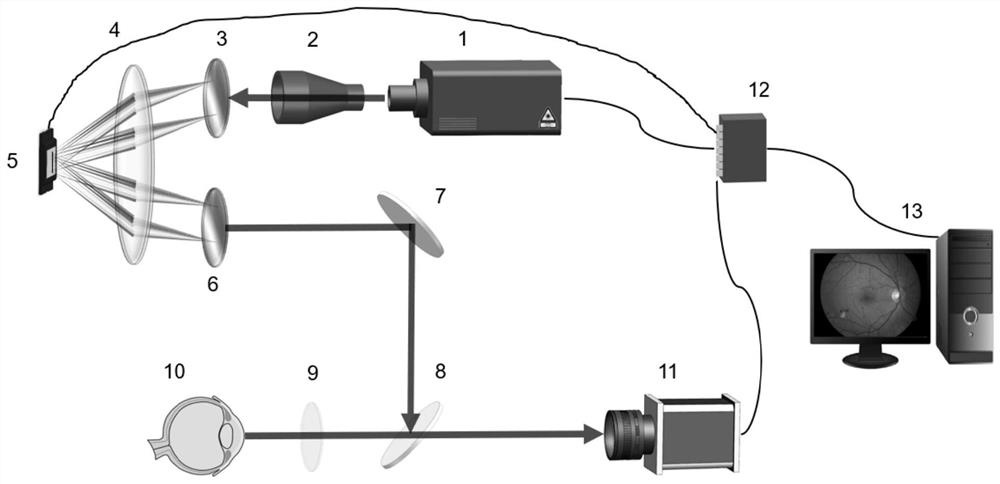
[0011] As shown in the figure, a retinal blood vessel optical angiography imaging system includes a white light source 1, a beam expander 2, a first grating 3, a first lens 4, a digital micromirror device 5, a second grating 6, a mirror 7, a beam splitter Mirror 8, second lens 9, CCD camera 11, data acquisition card 12 and computer 13;

[0012] It is composed of a white light source 1, a beam expander 2, a first grating 3, a first lens 4, a digital micromirror device 5, a second grating 6, a reflector 7, a beam splitter 8 and a second lens 9 for illuminating the eye to be inspected 10 lighting units;

[0013] The imaging unit for imaging the eye 10 to be inspected is composed of the second lens 9, the beam splitter 8 and the CCD camera 11;

[0014] The control unit for controlling the lighting unit and the imagi...


PUM


Abstract

The invention discloses a retinal blood vessel optical contrast imaging system and method, belonging to the field of optical technology. The system includes an illumination unit for illuminating the subject's eye, an imaging unit for collecting retinal image information, and a control unit for encoding and regulating the light emitted by the light source. This method codes and regulates the wavelength of the light incident on the fundus through the coding matrix to form a special illumination light sensitive to retinal blood vessels. By collecting retinal images illuminated by the special light, the contrast of retinal blood vessels can be enhanced. Optical means replace the specific selection process of contrast agent for blood vessels in traditional fundus fluorescein angiography imaging, and realize a simple, non-invasive and high-contrast optical angiography imaging method of retinal vessels.

Description

technical field [0001] The invention belongs to the field of optics, and in particular relates to a retinal blood vessel optical angiography imaging system and method. Background technique [0002] Retinal blood vessels are blood vessels that can be observed non-invasively. Doctors can diagnose fundus-related diseases (such as age-related macular degeneration, diabetic retinopathy, glaucoma, etc.) through structural changes such as the diameter, curvature, and branch shape of retinal blood vessels, and then guide The clinical treatment process can effectively prevent vision loss or blindness. [0003] In clinical practice, retinal blood vessels are usually imaged and observed by methods such as color fundus camera and fundus fluorescein angiography. Among them, the color fundus camera mainly images the fundus surface, and stores the fundus condition in the form of images. Color fundus camera imaging is fast and easy to operate, which greatly facilitates the routine examina...

Claims


Application Information

Patent Timeline
Patent Type & Authority Patents(China)
IPC IPC(8): A61B3/14A61B3/12
CPCA61B3/14A61B3/0016A61B3/12A61B3/0008
Inventor 陈硕孔令敏佟萌萌
Owner 宁波蓝明信息科技有限公司
Features
  • R&D
  • Intellectual Property
  • Life Sciences
  • Materials
  • Tech Scout
  • Unparalleled Data Quality
  • Higher Quality Content
  • 60% Fewer Hallucinations
Learn More