
Vein Ai endoscope analysis method based on VRDS 4D medical image and product

A medical imaging and medical imaging technology, applied in image analysis, medical imaging, medical informatics, etc., can solve the problems that doctors cannot obtain the four-dimensional structure and affect doctors' diagnosis of diseases

Pending Publication Date: 2020-09-01
曹生
View PDF11 Cites 0 Cited by
  • Summary
  • Abstract
  • Description
  • Claims
  • Application Information

AI Technical Summary

Problems solved by technology

However, only by directly viewing the two-dimensional slice data will seriously affect the doctor's diagnosis of the disease, and the doctor cannot obtain an intuitive and real four-dimensional volume structure

Method used

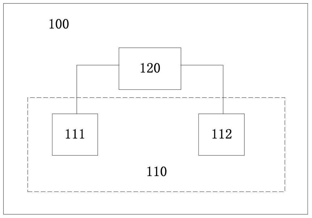

Image

  • Vein Ai endoscope analysis method based on VRDS 4D medical image and product
  • Vein Ai endoscope analysis method based on VRDS 4D medical image and product
  • Vein Ai endoscope analysis method based on VRDS 4D medical image and product

Examples


Embodiment Construction

[0022] In order to enable those skilled in the art to better understand the solution of the present application, the technical solution in the embodiment of the application will be clearly and completely described below in conjunction with the accompanying drawings in the embodiment of the application. Obviously, the described embodiment is only It is a part of the embodiments of this application, not all of them. Based on the embodiments in this application, all other embodiments obtained by persons of ordinary skill in the art without creative efforts fall within the protection scope of this application.

[0023] The terms "first", "second" and the like in the specification and claims of the present application and the above drawings are used to distinguish different objects, rather than to describe a specific order. Furthermore, the terms "include" and "have", as well as any variations thereof, are intended to cover a non-exclusive inclusion. For example, a process, method...


PUM


Abstract

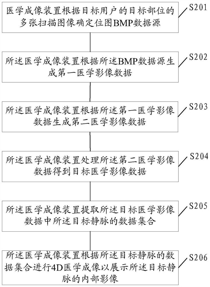
The embodiment of the invention discloses a vein Ai endoscope analysis method based on a VRDS 4D medical image and a product. The vein Ai endoscope analysis method and the product are applied to a medical imaging device. The method comprises the following steps: determining a bitmap BMP data source according to a plurality of scanning images of a target part of a target user, wherein the target part comprises a target vein to be observed and arteries, kidneys and portals associated with the target vein; generating first medical image data according to the BMP data source; generating second medical image data according to the first medical image data; processing the second medical image data to obtain target medical image data; extracting a data set of target veins in the target medical image data; and performing 4D medical imaging according to the data set of the target vein to display an internal image of the target vein. According to the embodiment of the invention, the accuracy andconvenience of inferior vena cava imaging by the medical imaging device can be improved.

Description

technical field [0001] This application relates to the technical field of medical imaging devices, in particular to an Ai endoscopic analysis method and product based on VRDS 4D medical images of veins. Background technique [0002] At present, doctors use computerized tomography (Computed Tomography, CT), magnetic resonance imaging (Magnetic Resonance Imaging, MRI), diffusion tensor imaging (Diffusion Tensor Imaging, DTI), positron emission computed tomography (Positron Emission Computed Tomography, PET) and other technologies to obtain information such as the shape, location, and topological structure of lesion tissue. Doctors still use watching and reading continuous two-dimensional slice data to judge and analyze patients' diseased tissues such as tumors. However, just viewing the two-dimensional slice data will seriously affect the doctor's diagnosis of the disease, and the doctor cannot obtain an intuitive and real four-dimensional volume structure. With the rapid de...

Claims


Application Information

Patent Timeline
IPC IPC(8): G06T7/11G16H30/20
CPCG06T7/11G16H30/20G06T2207/30101G06T2207/10076G16H30/40G16H50/20G16H40/67G06T7/0012G06T2207/10068G06T2207/20084G06T2207/10081G06T2207/10088G06T2207/10104G06T7/12G06T2207/30056G06T2207/30244G06T2207/30084
Inventor 斯图尔特平李戴维伟李
Owner 曹生
Learn More