Detection of lymph node metastasis from gastric carcinoma
a gastric carcinoma and lymph node technology, applied in the field of gastric carcinoma lymph node metastasis detection, can solve the problem of not being consistently expressed in all tumor tissues
- Summary
- Abstract
- Description
- Claims
- Application Information
AI Technical Summary
Benefits of technology
Problems solved by technology
Method used
Image

Examples
example 1
Marker Identification
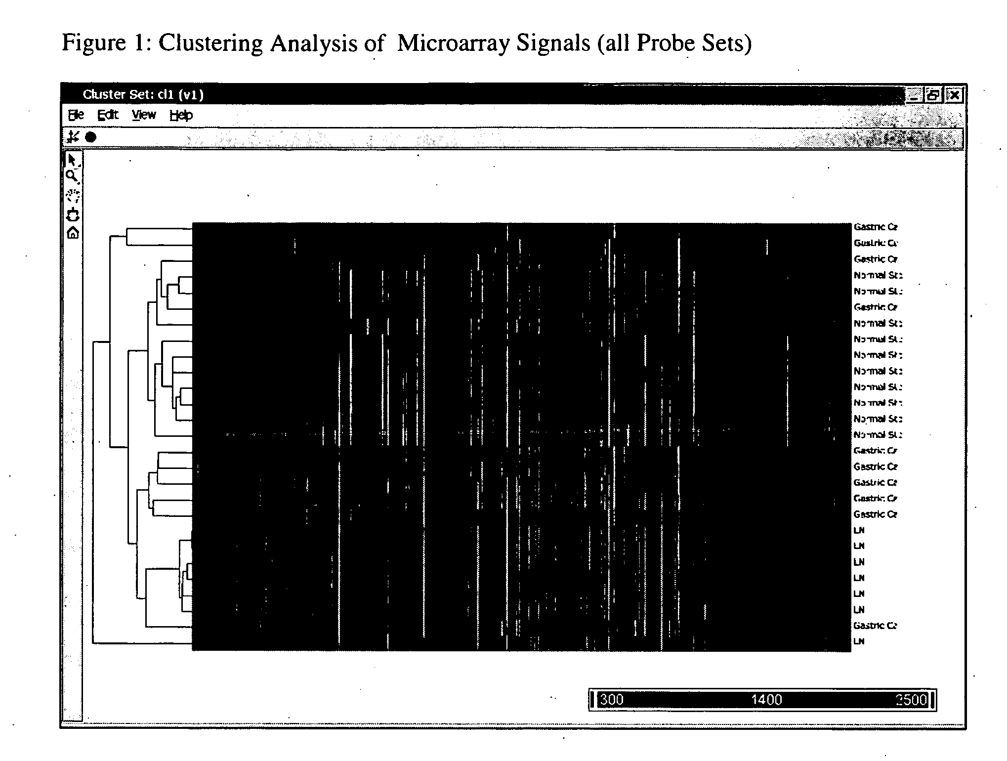
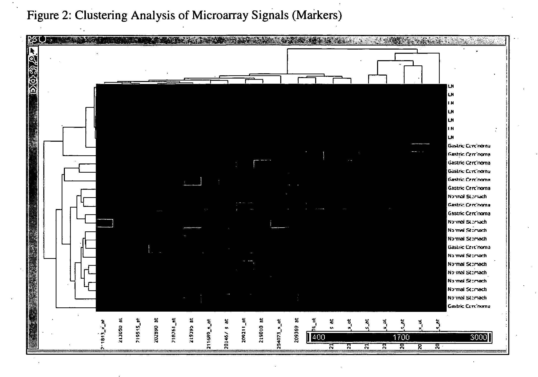
[0070] Freshly frozen samples of 10 gastric carcinomas, 10 normal stomach tissues and 6 histologically confirmed normal lymph nodes were used to identify gene Markers for detection of lymph node metastasis from gastric carcinoma. RNAs were prepared from these tissues with RNeasy Mini Kit (Qiagen, Catalog# 74106) following standard vendor suggested protocols. Biotin labeled aRNA targets were prepared by RNA linear amplification (Ambion, MessageAmp II aRNA Amplification Kit, Catalog# 1751). Prepared targets were applied to Affymetrix HG U133A chips which were processed following standard Affymetrix suggested protocols. Chips results were collected and globally scaled to Target Signal of 600. Hierarchical clustering of microarray results (HG U I 33A chip set) was conducted and the chip results were statistically analyzed using Partek Pro 6.0. ANOVA and fold change analysis were applied to identify genes of interest. Genes with the following characteristics were se...
example 2
Confirmation Study (1)—Microarray Test
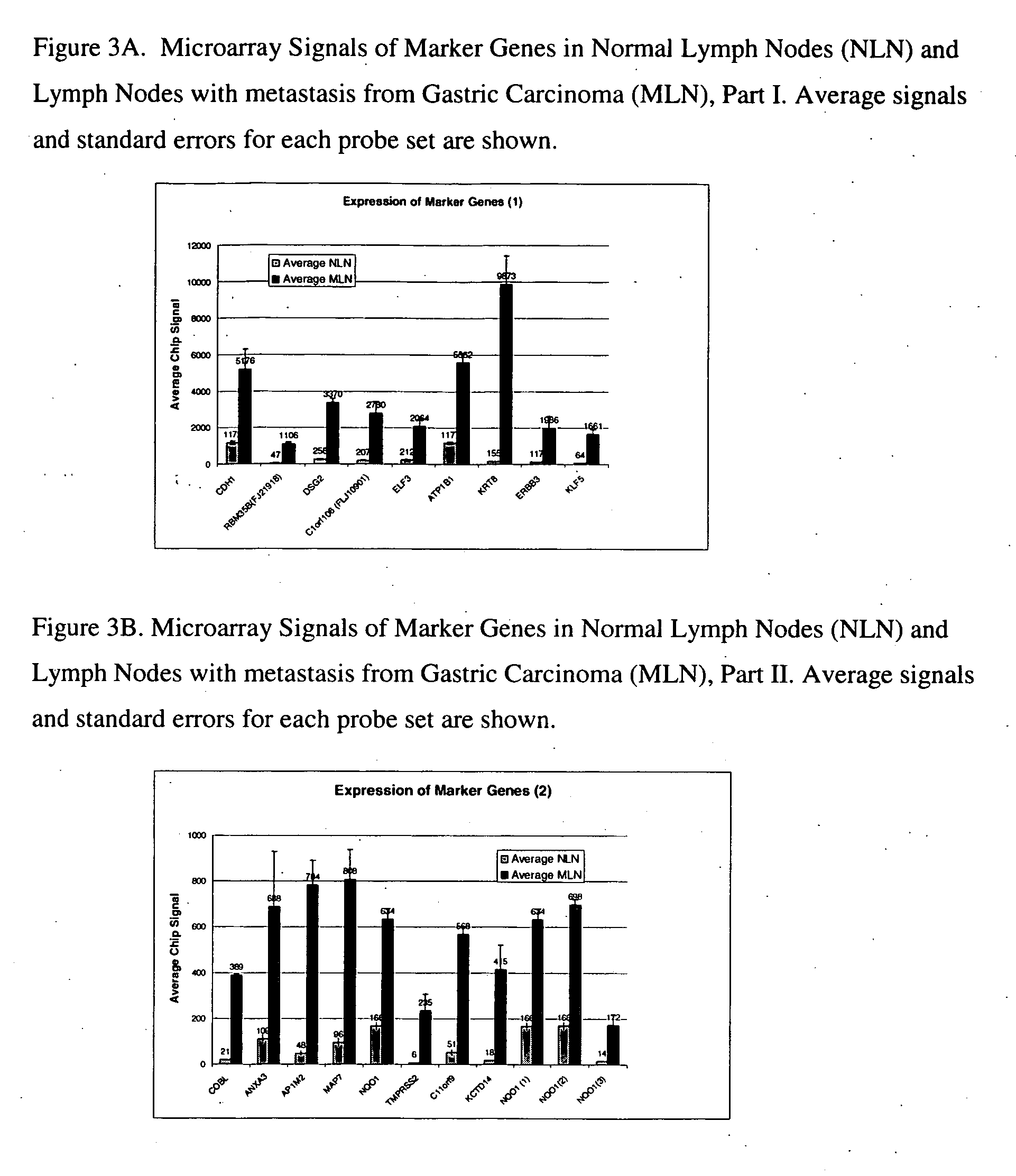
[0074] Confirmation studies were performed to validate the Markers selected in Example 1. These markers were examined to determine whether their expression is maintained in lymph nodes with metastasis from gastric carcinomas of various differentiation statuses. Affymetrix DNA microarrays (HG U 133A) were used in this study. Tissue of 6 normal lymph nodes and 3 lymph nodes with confirmed metastasis from gastric carcinoma were tested. RNAs were prepared from these samples as described above. Biotin-labeled aRNA targets were prepared as in Example 1. Targets were applied to Affymetrix HG U133A chips. Results from the chips were scaled to Target Signal of 600 and analyzed.
[0075] The average signal of each probe set and standard error are plotted in FIG. 3A, FIG. 3B.
example 3
Confirmation Study (2)—Realtime RT-PCR Test
[0076] Studies were conducted to validate the performance of selected gene markers using real time RT-PCR. 10 normal lymph nodes and 16 lymph nodes with histologically confirmed metastasis from gastric carcinoma were tested (including all the samples tested with DNA microarray in Confirmation Study 1). The 10 histologically confirmed normal lymph nodes were from 7 different patients. The 16 lymph nodes with confirmed metastasis from gastric carcinoma were from 13 different patients. The malignancy statuses of the original carcinoma that metastasized to lymph nodes are: 3 well differentiated (contributed to 6 lymph nodes), 7 poorly differentiated (contributed to 7 nodes), 3 moderately differentiated (contributed to 3 lymph nodes).
[0077] All RNAs were prepared as described above. cDNAs were first prepared from 5 μg RNA in 100 μL reaction volume with a High Capacity cDNA Archive Kit (ABI) following the vendor suggested protocol. 2 μL of the ...
PUM
| Property | Measurement | Unit |
|---|---|---|
| diameter | aaaaa | aaaaa |
| volume | aaaaa | aaaaa |
| volume | aaaaa | aaaaa |
Abstract
Description
Claims
Application Information
 - R&D
- Intellectual Property
- Life Sciences
- Materials
- Tech Scout
- Unparalleled Data Quality
- Higher Quality Content
- 60% Fewer Hallucinations
Browse by: Latest US Patents, China's latest patents, Technical Efficacy Thesaurus, Application Domain, Technology Topic, Popular Technical Reports.
© 2025 PatSnap. All rights reserved.Legal|Privacy policy|Modern Slavery Act Transparency Statement|Sitemap|About US| Contact US: help@patsnap.com