Supercharge Your Innovation With Domain-Expert AI Agents!

Endoscope system, processor device of endoscope system, and image processing method

a technology which is applied in the field of endoscope system and processor device of endoscope system, and image processing method, can solve the problems of difficult to reliably distinguish between superficial blood vessels and medium-deep blood vessels

Active Publication Date: 2017-04-25
FUJIFILM CORP
View PDF29 Cites 0 Cited by
  • Summary
  • Abstract
  • Description
  • Claims
  • Application Information

AI Technical Summary

Benefits of technology

Enables reliable extraction and visualization of blood vessels at different depths, improving diagnostic accuracy by allowing for mode-specific processing and enhancement / suppression of blood vessel images, even when observing different body parts.

Problems solved by technology

That is, the appearance or color of the blood vessel may change depending on the part If the color of the blood vessel changes in this way, it is difficult to reliably distinguish superficial blood vessels and medium-deep blood vessels with a blood vessel discrimination method based on the hue disclosed in JP2011-135983A.

Method used


Image

  • Endoscope system, processor device of endoscope system, and image processing method
  • Endoscope system, processor device of endoscope system, and image processing method
  • Endoscope system, processor device of endoscope system, and image processing method

Examples


Embodiment Construction

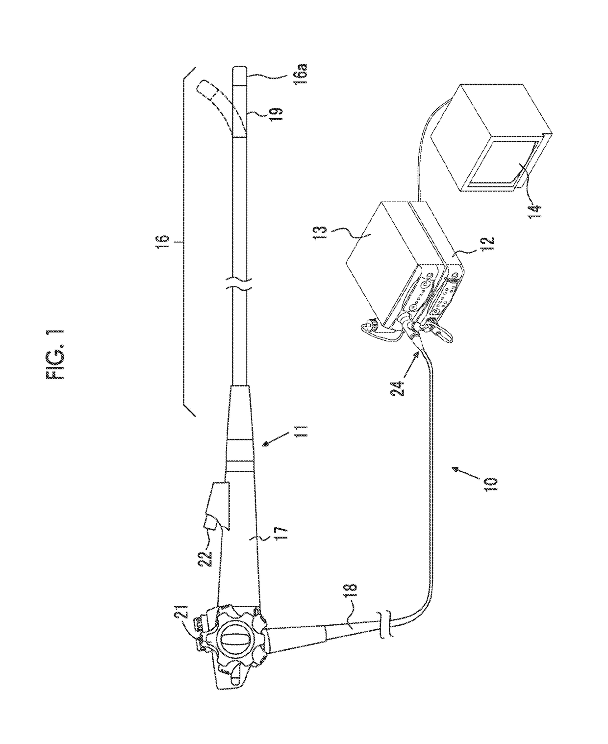
[0037]As shown in FIG. 1, an electronic endoscope system 10 of a first embodiment includes an electronic endoscope 11 that images the inside of a subject, a processor device 12 that generates an endoscope image based on a signal obtained by imaging, a light source device 13 (a form of an illumination unit) that generates light for illuminating the subject, and a monitor 14 that displays an endoscope image. The electronic endoscope 11 includes a flexible insertion unit 16 that is inserted into the body cavity, an operating unit 17 provided at the proximal end of the insertion unit 16, and a universal code 18 that makes a connection between the operating unit 17 and the processor device 12 and the light source device 13.

[0038]The electronic endoscope system 10 has a function of generating a superficial blood vessel enhancement image or suppression image, in which a superficial blood vessel of a subject is enhanced / suppressed, and a medium-deep blood vessel enhancement image or suppres...


PUM


Abstract

Broadband light BB and narrowband light NB are simultaneously irradiated to a subject. A blue signal B, a green signal G, and a red signal R are obtained by imaging the subject using a color CCD 33. A base image is generated from the signals B, G, and R of three colors. A B / G image having a B / G ratio is generated. A superficial blood vessel extraction image is obtained by extracting a pixel, in which the B / G ratio is equal to or less than a boundary value Ls between the mucous membrane and the superficial blood vessel, from the B / G image. A medium-deep blood vessel extraction image is obtained by extracting a pixel, in which the B / G ratio is equal to or greater than a boundary value Ld between the mucous membrane and the medium-deep blood vessel. The boundary values Ls and Ld differ depending on each observation mode.

Description

CROSS-REFERENCE TO RELATED APPLICATIONS[0001]This application is a Continuation of PCT International Application No. PCT / JP2013 / 050359 filed on Jan. 11, 2013, which claims priority under 35 U.S.C §119(a) to Japanese Patent Application No. 2012-013316 filed Jan. 25, 2012. Each of the above applications is hereby expressly incorporated by reference, in its entirety, into the present application.BACKGROUND OF THE INVENTION[0002]1. Field of the Invention[0003]The present invention relates to an endoscope system capable of extracting blood vessels, such as superficial blood vessels and medium-deep blood vessels, in a subject, a processor device of an endoscope system, and an image processing method.[0004]2. Description of the Related Art[0005]In recent medical treatment, diagnosis or the like using an endoscope apparatus has been widely performed. As observation of the inside of a subject using an endoscope apparatus, not only normal observation using white light of broadband light as il...

Claims


Application Information

Patent Timeline
Patent Type & Authority Patents(United States)
IPC IPC(8): A61B1/04A61B1/00G06T5/50A61B1/06
CPCA61B1/0638A61B1/00009A61B1/063G06T5/50A61B1/0646A61B1/0653G06T2207/10024G06T2207/10068G06T2207/20212G06T2207/30004G06T2207/30101A61B1/000094G06T2207/20221
Inventor KAKU, TOSHIHIKOIIDA, TAKAYUKI
Owner FUJIFILM CORP
Features
  • R&D
  • Intellectual Property
  • Life Sciences
  • Materials
  • Tech Scout
  • Unparalleled Data Quality
  • Higher Quality Content
  • 60% Fewer Hallucinations
Learn More