

How to Quantify Radiopaque Density for Calibration
FEB 13, 20269 MIN READ
Generate Your Research Report Instantly with AI Agent
PatSnap Eureka helps you evaluate technical feasibility & market potential.
Radiopaque Density Quantification Background and Objectives
Radiopaque materials have been fundamental to medical imaging since the discovery of X-rays in 1895, enabling visualization of anatomical structures and medical devices that would otherwise be invisible in radiographic procedures. The quantification of radiopaque density represents a critical technical challenge in ensuring accurate calibration of imaging systems and standardization of contrast agents used in diagnostic and interventional procedures. Historically, the field has evolved from subjective visual assessment to sophisticated digital quantification methods, driven by the need for reproducible measurements and quality assurance in clinical settings.
The primary objective of radiopaque density quantification is to establish standardized metrics that correlate material composition and concentration with measurable radiographic opacity. This enables precise calibration of imaging equipment, validation of contrast agent formulations, and quality control of radiopaque medical devices such as catheters, stents, and markers. Accurate quantification is essential for ensuring consistent image quality across different imaging systems and clinical environments, facilitating reliable diagnosis and treatment monitoring.
Current technological goals focus on developing robust methodologies that can account for variables including X-ray energy spectra, detector sensitivity variations, and material composition differences. The challenge extends beyond simple density measurement to encompass spectral characteristics, as radiopaque materials exhibit energy-dependent attenuation properties. Advanced objectives include establishing universal calibration standards that remain valid across multi-vendor platforms and diverse imaging modalities, from conventional radiography to computed tomography and fluoroscopy systems.
The evolution toward quantitative approaches reflects broader trends in medical imaging toward precision medicine and evidence-based practice. Modern requirements demand not only accurate measurement but also traceability to international standards, reproducibility across institutions, and integration with digital imaging workflows. These objectives drive ongoing research into phantom design, computational modeling, and automated analysis algorithms that can transform subjective opacity assessment into objective, quantifiable metrics suitable for regulatory compliance and clinical decision-making.
Market Demand for Calibrated Radiopaque Materials
The medical imaging industry has witnessed sustained growth in demand for calibrated radiopaque materials, driven primarily by the increasing complexity of diagnostic and interventional procedures. Hospitals and imaging centers require standardized phantoms and calibration tools to ensure consistent image quality across different equipment and facilities. This demand stems from regulatory requirements mandating periodic quality assurance testing of radiographic systems, as well as the need for accurate dose measurements in patient safety protocols.
The interventional cardiology and vascular surgery sectors represent particularly robust market segments for calibrated radiopaque materials. As minimally invasive procedures become more prevalent, clinicians depend on precise visualization of catheters, guidewires, and implantable devices. Manufacturers of these medical devices increasingly seek validated calibration standards to optimize the radiopacity of their products during development phases, ensuring adequate visibility without excessive material loading that could compromise mechanical properties.
Dental imaging constitutes another significant application area, where calibrated step wedges and reference phantoms are essential for standardizing exposure parameters and evaluating digital sensor performance. The transition from film-based to digital radiography has intensified the need for objective density quantification methods, as different detector technologies exhibit varying responses to X-ray attenuation.
Research institutions and academic medical centers drive demand for advanced calibration materials that support quantitative imaging studies. The emergence of dual-energy CT and spectral imaging techniques requires sophisticated phantoms with precisely characterized attenuation properties across multiple energy ranges. These applications demand materials with certified density values and minimal batch-to-batch variation to ensure reproducibility in multi-center clinical trials.
The veterinary imaging market, though smaller in scale, shows growing adoption of calibrated radiopaque standards as animal healthcare facilities upgrade to digital systems and implement quality control programs similar to human medicine. Industrial non-destructive testing applications also contribute to market demand, particularly in aerospace and manufacturing sectors where weld inspection and material characterization require traceable calibration references.
Geographic market dynamics reveal concentrated demand in regions with mature healthcare infrastructure and stringent regulatory oversight. However, emerging markets demonstrate accelerating adoption rates as medical imaging capabilities expand and quality standards evolve toward international harmonization.
Current Status and Challenges in Density Quantification
Quantifying radiopaque density for calibration purposes remains a critical yet complex challenge in medical imaging and industrial radiography. Current methodologies predominantly rely on standardized phantoms and reference materials with known densities, which serve as benchmarks for calibration processes. However, significant variability exists across different imaging modalities, including conventional radiography, computed tomography, and fluoroscopy, each requiring distinct calibration approaches and density measurement protocols.
The primary technical challenge lies in establishing consistent and reproducible density measurements across diverse imaging systems and manufacturers. Variations in X-ray beam energy, detector sensitivity, and image reconstruction algorithms introduce substantial inconsistencies in density quantification. Current calibration standards, such as aluminum step wedges and hydroxyapatite phantoms, provide limited dynamic range and may not adequately represent the full spectrum of tissue densities encountered in clinical or industrial applications.
Another significant obstacle involves the nonlinear relationship between material density and radiographic opacity. The Beer-Lambert law provides theoretical foundation, yet practical implementation faces complications from beam hardening effects, scatter radiation, and detector response characteristics. These factors collectively compromise measurement accuracy and limit the precision of density quantification, particularly in regions with high-density gradients or composite materials.
Geographical distribution of advanced calibration capabilities reveals concentration in developed regions with established regulatory frameworks and quality assurance programs. North America and Europe maintain robust standardization protocols through organizations like the American College of Radiology and European Society of Radiology. However, developing regions often lack access to sophisticated calibration equipment and standardized reference materials, creating disparities in measurement reliability and quality control.
Contemporary solutions attempt to address these challenges through software-based corrections and advanced calibration algorithms. Dual-energy imaging techniques and spectral CT methods offer improved material differentiation and density quantification capabilities. Nevertheless, these approaches require substantial computational resources and specialized hardware, limiting widespread adoption. The absence of universally accepted calibration standards and the complexity of cross-platform validation continue to impede progress toward standardized density quantification methodologies across the global imaging community.
Existing Density Quantification Methods
01 Radiopaque materials with controlled density for medical imaging
Radiopaque materials are formulated with specific density characteristics to enhance visibility under X-ray or other imaging modalities. These materials incorporate radiopaque agents such as barium sulfate, bismuth compounds, or iodinated substances at controlled concentrations to achieve optimal contrast. The density of these materials is carefully adjusted to ensure clear visualization of medical devices or anatomical structures during diagnostic or interventional procedures.- Radiopaque materials with controlled density for medical imaging: Radiopaque materials are formulated with specific density characteristics to enhance visibility under X-ray or other imaging modalities. These materials incorporate radiopaque agents such as barium sulfate, bismuth compounds, or iodinated substances at controlled concentrations to achieve optimal contrast. The density of these materials is carefully adjusted to provide clear differentiation from surrounding tissues while maintaining biocompatibility and structural integrity for medical device applications.
- High-density radiopaque compositions for catheter and guidewire applications: Specialized high-density radiopaque compositions are developed for use in catheters, guidewires, and other interventional medical devices. These compositions utilize heavy metal compounds or alloys that provide superior radiopacity while maintaining flexibility and mechanical properties required for minimally invasive procedures. The density optimization ensures that even thin-walled devices remain visible during fluoroscopic guidance.
- Polymer-based radiopaque materials with adjustable density: Polymer matrices are combined with radiopaque fillers to create materials with adjustable density profiles. These formulations allow for precise control of radiopacity by varying the concentration and particle size of radiopaque additives within the polymer system. The resulting materials can be tailored for specific applications requiring different levels of X-ray visibility while maintaining processability and mechanical performance.
- Radiopaque markers and bands with enhanced density contrast: Discrete radiopaque markers and bands are designed with enhanced density to provide precise positional information during medical procedures. These components utilize materials with significantly higher density than surrounding device structures, creating distinct contrast points visible under imaging. The formulations balance high radiopacity with small physical dimensions to minimize device profile while maximizing visibility.
- Gradient density radiopaque structures for improved visualization: Advanced radiopaque structures incorporate gradient density designs where radiopacity varies along the length or across different regions of a medical device. This approach provides enhanced spatial information and orientation feedback during procedures. The gradient is achieved through controlled distribution of radiopaque agents or by using multiple materials with different inherent densities, enabling clinicians to better assess device position and deployment.
02 High-density radiopaque compositions for catheter and guidewire applications
Medical devices such as catheters and guidewires require radiopaque markers or coatings with high density to be clearly visible during fluoroscopic procedures. These compositions typically utilize heavy metal compounds or alloys that provide superior radiopacity while maintaining biocompatibility. The density is optimized to create sufficient contrast without compromising the mechanical properties or flexibility of the device.Expand Specific Solutions03 Adjustable density radiopaque polymeric materials
Polymeric materials can be modified to achieve desired radiopaque density by incorporating radiopaque fillers or additives. The concentration and particle size of these additives can be adjusted to control the overall density and radiopacity of the final product. These materials are particularly useful in applications requiring both flexibility and visibility, such as embolic agents or tissue markers.Expand Specific Solutions04 Multi-layer structures with varying radiopaque density
Medical devices and implants can be designed with multiple layers having different radiopaque densities to provide enhanced visualization and functional properties. These structures may include a core with one density level and outer layers with different density characteristics, allowing for improved imaging while maintaining structural integrity. The gradient or distinct density differences enable better delineation of device boundaries and positioning.Expand Specific Solutions05 Density measurement and standardization of radiopaque materials
Methods and standards for measuring and controlling the density of radiopaque materials ensure consistent imaging performance across different products and applications. These approaches involve specific testing protocols to quantify radiopacity relative to density, establishing benchmarks for material selection and quality control. Standardization helps manufacturers optimize formulations to meet regulatory requirements and clinical needs for adequate visualization.Expand Specific Solutions
Key Players in Radiopaque Material and Imaging Industry
The quantification of radiopaque density for calibration represents a mature technical domain within medical imaging and industrial inspection, currently experiencing steady growth driven by increasing demands for precision diagnostics and security screening applications. The market spans healthcare, customs inspection, and industrial quality control sectors, with established players like Shimadzu Corp., Siemens Medical Solutions USA, and Agilent Technologies demonstrating advanced technological capabilities in medical imaging systems. Industrial applications are led by NUCTECH Co. and Nuctech Beijing Co. in security inspection, while research institutions including Tsinghua University, University of Washington, and Institut de RadioProtection et de Sûreté Nucléaire drive innovation in calibration methodologies. The competitive landscape shows consolidation among major equipment manufacturers alongside specialized providers like Micro Motion and ImaTx, indicating a transitioning market from pure hardware solutions toward integrated software-driven calibration systems with enhanced automation and standardization capabilities.
Shimadzu Corp.
Technical Solution: Shimadzu offers X-ray inspection and analytical systems with sophisticated radiopaque density calibration capabilities. Their methodology combines step-wedge calibration phantoms made from aluminum or acrylic with varying thicknesses to establish density-attenuation relationships. The system utilizes advanced image processing algorithms that perform flat-field correction, scatter reduction, and geometric calibration to ensure measurement linearity. Their calibration software employs least-squares fitting to generate conversion functions between pixel intensity values and material density, with built-in quality assurance protocols for monitoring system stability. The technology is particularly effective for non-destructive testing in industrial quality control and materials characterization applications.
Strengths: Excellent image quality and measurement repeatability, user-friendly calibration interface, strong industrial application support. Weaknesses: Calibration requires specialized phantoms, limited penetration for very dense materials, periodic recalibration needed for optimal performance.
Siemens Medical Solutions USA, Inc.
Technical Solution: Siemens has developed advanced calibration systems for radiopaque density quantification using dual-energy CT imaging technology. Their approach employs phantom-based calibration with known density reference materials, typically containing calcium hydroxyapatite or iodine solutions at varying concentrations. The system utilizes automated segmentation algorithms to identify regions of interest and applies polynomial regression models to establish the relationship between CT Hounsfield Units (HU) and actual material density. Their calibration workflow integrates beam hardening correction algorithms and spectral decomposition techniques to improve measurement accuracy across different X-ray energy levels, enabling precise quantification of radiopaque materials in both clinical and industrial applications.
Strengths: High accuracy with dual-energy imaging, automated calibration workflow, excellent integration with clinical systems. Weaknesses: Expensive equipment requirements, limited to specific CT scanner models, requires regular phantom-based recalibration.
Core Innovations in Radiopaque Calibration Standards
Method for calibrating a radiometric density measuring apparatus
PatentInactiveUS20200393391A1
Innovation
- A one-point calibration method is developed by calculating the mass attenuation coefficient using the half-value thickness of gamma radiation in an empty container and a known calibration medium, allowing for precise determination of the calibration curve without the need for multiple density measurements.


Calibration of the transformation of spectral X-ray attenuation values in density and atomic number information
PatentInactiveUS6997610B2
Innovation
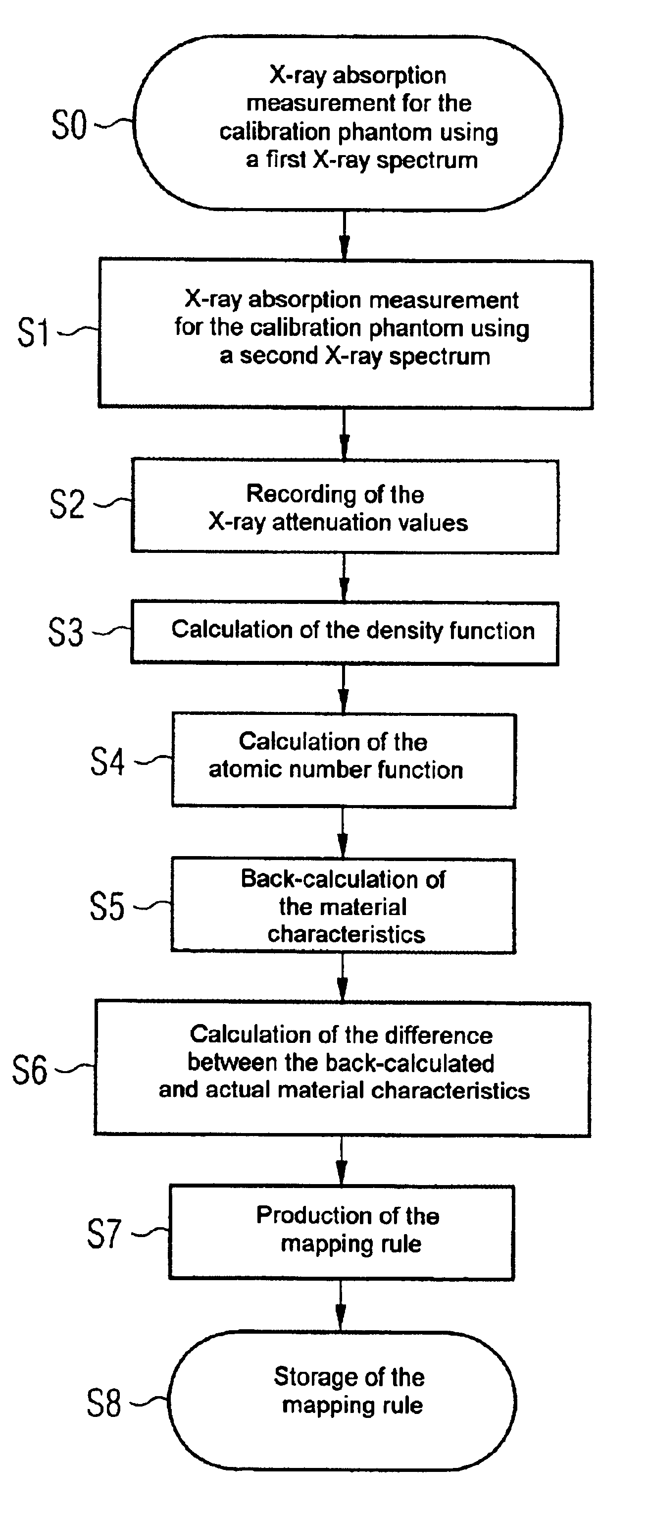
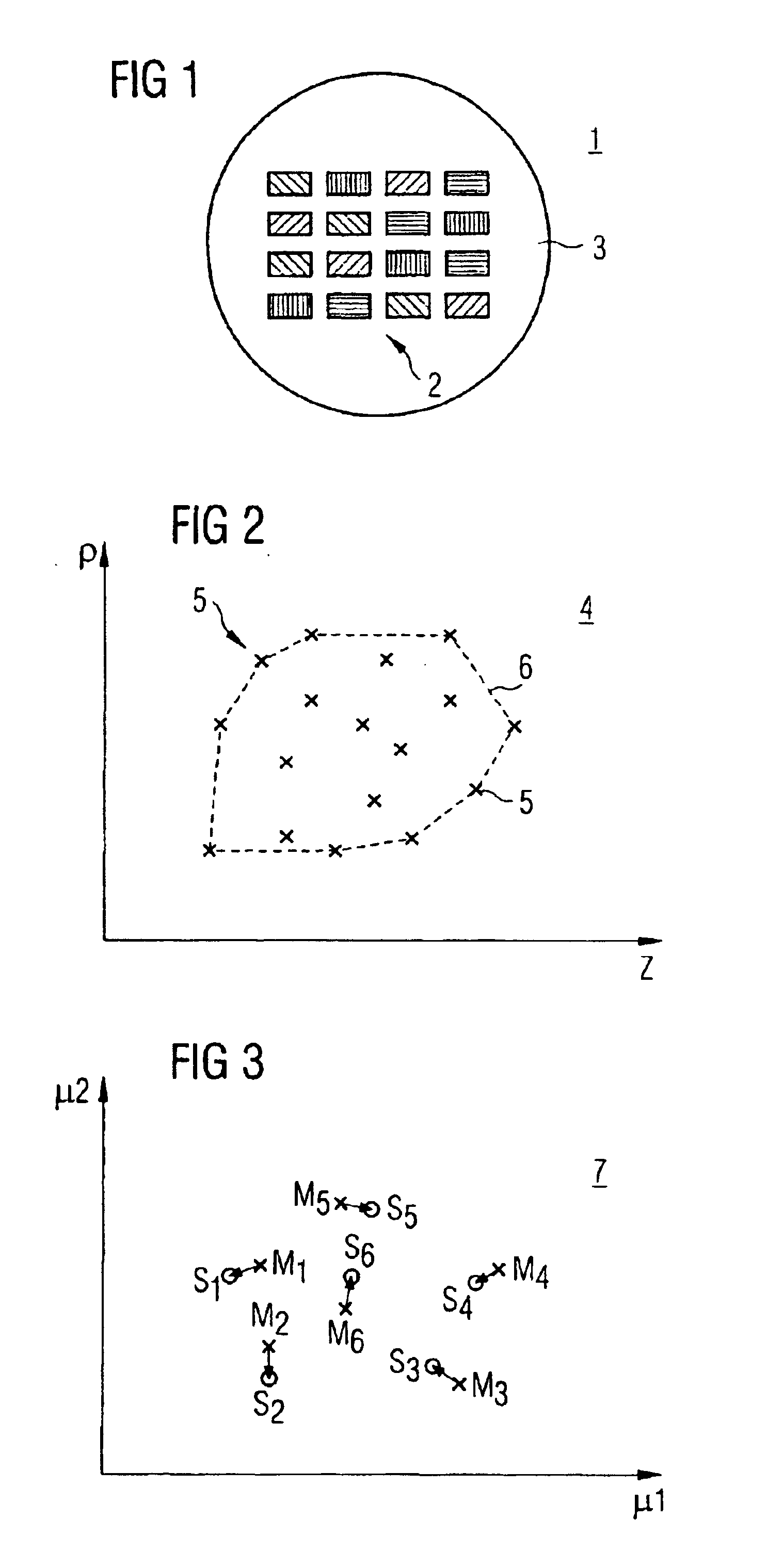
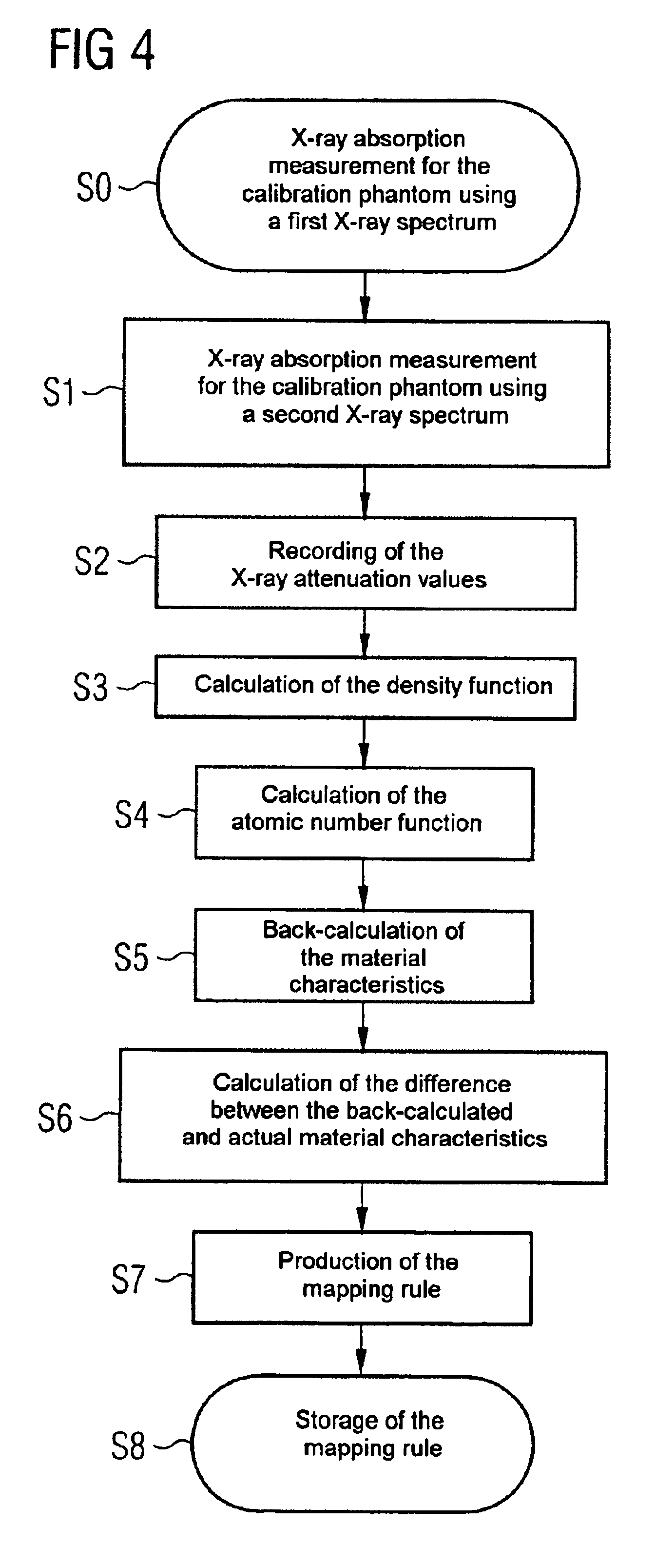
- A method involving a calibration phantom with multiple samples of varying densities and atomic numbers, using two X-ray spectra to record attenuation values, and creating density and atomic number functions to correct discrepancies, allowing for reliable transformation of attenuation values into accurate density and atomic number values without requiring spectral parameter recording.

Regulatory Standards for Radiopaque Calibration
Radiopaque density quantification for calibration purposes operates within a comprehensive regulatory framework designed to ensure patient safety and diagnostic accuracy across medical imaging applications. The International Organization for Standardization (ISO) has established fundamental standards, particularly ISO 5832 and ISO 25539 series, which define minimum radiopacity requirements for implantable medical devices. These standards mandate that radiopaque markers must demonstrate sufficient contrast against surrounding tissues to enable precise visualization during fluoroscopic procedures. The United States Food and Drug Administration (FDA) enforces stringent guidelines through 21 CFR Part 820 Quality System Regulation, requiring manufacturers to validate their radiopaque calibration methodologies through documented testing protocols and traceability to recognized measurement standards.
The European Medical Device Regulation (MDR 2017/745) imposes additional requirements for radiopaque materials, emphasizing the need for quantitative assessment methods that can be reproduced across different imaging systems and clinical settings. Compliance necessitates establishing calibration curves using reference phantoms with known densities, typically measured in Hounsfield Units (HU) for CT imaging or aluminum equivalent thickness for conventional radiography. Regulatory bodies require that calibration standards maintain stability over time and demonstrate minimal variation across batch productions.
The American Society for Testing and Materials (ASTM) provides complementary standards, particularly ASTM F640 for radiopacity testing of polymeric materials, which specifies standardized exposure conditions, phantom configurations, and measurement protocols. These standards define acceptable ranges for radiopaque density based on device type and intended anatomical location, with cardiovascular devices typically requiring higher radiopacity levels than orthopedic applications. Manufacturers must demonstrate compliance through comprehensive technical documentation, including calibration certificates, measurement uncertainty analyses, and validation studies comparing their quantification methods against internationally recognized reference standards.
Emerging regulatory trends emphasize harmonization across global markets, with the International Medical Device Regulators Forum (IMDRF) working to establish unified calibration protocols. Recent guidance documents stress the importance of digital imaging standardization and the adoption of quantitative metrics that remain consistent across evolving imaging technologies, ensuring long-term regulatory compliance and clinical reliability.
Quality Assurance in Medical Imaging Calibration
Quality assurance protocols in medical imaging calibration represent a critical framework for ensuring measurement accuracy and clinical reliability when quantifying radiopaque density. Establishing robust QA procedures requires systematic validation of calibration phantoms, imaging equipment performance, and measurement reproducibility across different operational conditions. The implementation of standardized QA programs directly impacts diagnostic confidence and treatment planning precision in radiological applications.
Calibration verification procedures must incorporate regular phantom scanning protocols using reference materials with known attenuation coefficients. These phantoms typically contain multiple inserts of varying densities, enabling comprehensive assessment of the imaging system's linearity and dynamic range. Daily, weekly, and monthly QA schedules should be established to monitor system stability, with acceptance criteria defined for density measurement deviations. Automated QA software solutions increasingly facilitate this process by comparing current measurements against baseline values and generating alerts when parameters drift beyond tolerance thresholds.
Traceability to international standards constitutes another essential component of quality assurance frameworks. Calibration procedures should reference established measurement standards from organizations such as NIST or equivalent national metrology institutes. This ensures that density quantification remains consistent across different institutions and imaging platforms. Documentation of calibration coefficients, environmental conditions, and equipment specifications provides an auditable trail supporting regulatory compliance and inter-institutional data comparison.
Operator training and competency assessment form integral elements of comprehensive QA programs. Standardized protocols for phantom positioning, scan parameter selection, and region-of-interest placement minimize inter-operator variability. Regular proficiency testing using blinded phantom studies helps identify systematic errors and training needs. Additionally, participation in external quality assessment schemes enables benchmarking against peer institutions and identification of potential systematic biases.
Advanced QA methodologies increasingly incorporate statistical process control techniques to detect subtle performance degradation before clinical impact occurs. Control charts tracking density measurement precision over time enable predictive maintenance strategies and optimize equipment replacement decisions. Integration of QA data with picture archiving and communication systems facilitates longitudinal analysis and supports continuous quality improvement initiatives essential for maintaining calibration accuracy throughout equipment lifecycles.
Unlock deeper insights with PatSnap Eureka Quick Research — get a full tech report to explore trends and direct your research. Try now!
Generate Your Research Report Instantly with AI Agent
Supercharge your innovation with PatSnap Eureka AI Agent Platform!