What Is Medical Image Analysis? Techniques and Challenges in MRI & CT
JUL 10, 2025 |
Understanding Medical Image Analysis
Medical image analysis involves the use of algorithms and computational techniques to extract meaningful information from medical images. It aims to enhance the interpretation of complex data, allowing healthcare professionals to make more informed decisions. This process involves several steps, including image acquisition, preprocessing, segmentation, feature extraction, and analysis. By transforming raw images into quantitative data, medical image analysis aids in identifying pathological abnormalities, assessing disease progression, and planning treatments.
Techniques in MRI and CT Image Analysis
In MRI image analysis, various techniques are employed to enhance the visualization and interpretation of soft tissues. One common approach is image segmentation, which involves partitioning the image into different regions or structures. This helps in isolating specific areas of interest, such as tumors or lesions, for further analysis. Additionally, techniques like registration align images from different time points or modalities, allowing for comparative studies and longitudinal assessments.
CT image analysis, on the other hand, is particularly useful for examining bone structures and detecting fractures or abnormalities. Techniques such as 3D reconstruction provide a detailed view of anatomical structures, facilitating surgical planning and intervention. Moreover, advanced algorithms can automatically detect and quantify features like nodules or calcifications, aiding in early disease detection and monitoring.
Challenges in Medical Image Analysis
Despite the advancements in medical image analysis, several challenges persist. One major hurdle is the variability in image quality and resolution, which can affect the accuracy of analysis. Factors such as noise, artifacts, and patient movement during image acquisition can compromise the clarity of images, necessitating robust preprocessing techniques to mitigate these issues.
Another challenge is the integration of multimodal data. Combining information from different imaging techniques, such as MRI and CT, can provide a more comprehensive understanding of a patient's condition. However, differences in image resolution, orientation, and scaling pose significant obstacles in achieving seamless integration.
Furthermore, the development of reliable and efficient algorithms is an ongoing challenge. The complexity and diversity of medical images require sophisticated computational models capable of handling large datasets and varying imaging conditions. Ensuring the generalizability and robustness of these algorithms across different patient populations and clinical settings remains a critical area of research.
Future Directions and Innovations
The field of medical image analysis is rapidly evolving, with ongoing research aimed at overcoming existing challenges and exploring new possibilities. Artificial intelligence (AI) and machine learning are playing an increasingly important role in this domain. AI-driven techniques, such as deep learning, are being harnessed to improve image interpretation, automate segmentation processes, and enhance diagnostic accuracy.
Moreover, the integration of big data analytics is opening new avenues for personalized medicine. By analyzing large-scale imaging data in conjunction with clinical and genomic information, healthcare providers can tailor treatments to individual patients, improving outcomes and reducing unnecessary interventions.
In conclusion, medical image analysis is a dynamic and essential field that continues to transform healthcare. As techniques in MRI and CT image analysis advance, and challenges are addressed, the potential for more precise diagnoses and targeted treatments becomes increasingly promising. Through ongoing research and innovation, the future of medical image analysis holds great promise in improving patient care and advancing medical science.
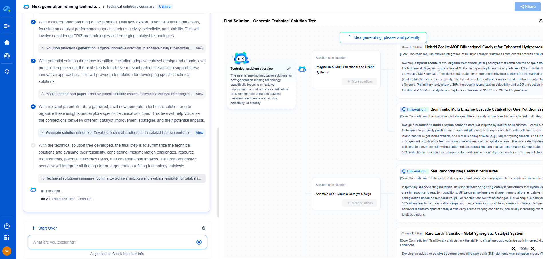
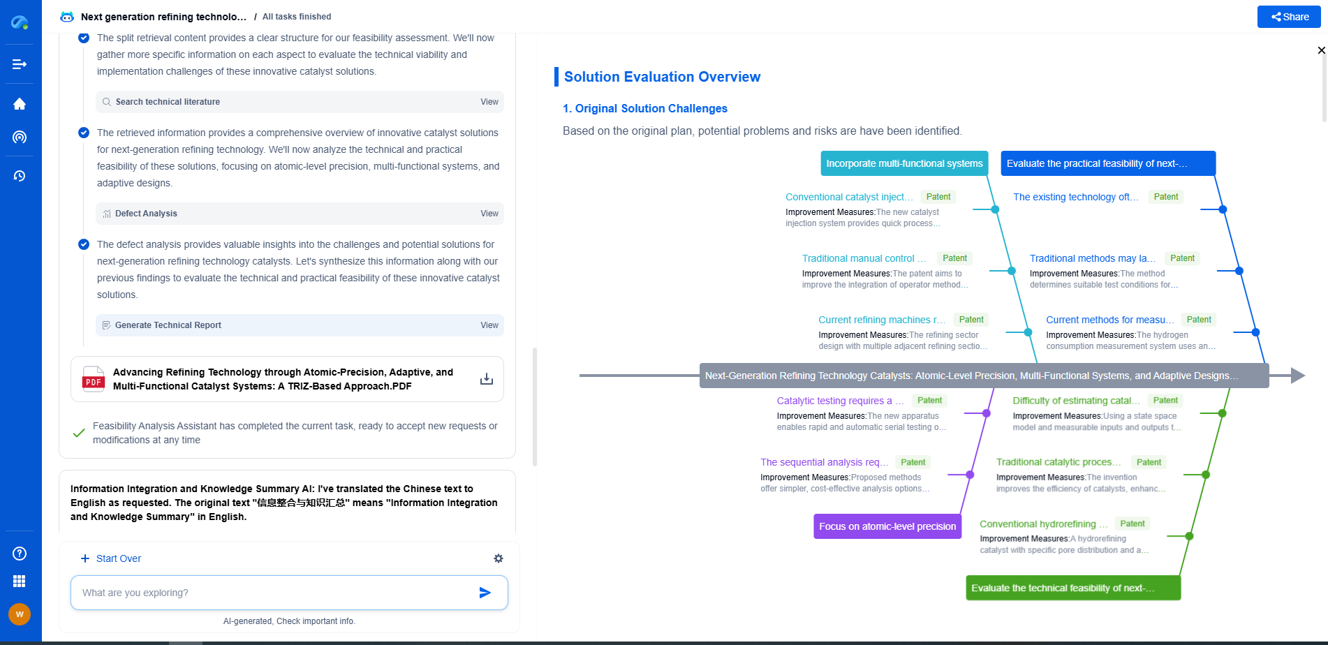
Image processing technologies—from semantic segmentation to photorealistic rendering—are driving the next generation of intelligent systems. For IP analysts and innovation scouts, identifying novel ideas before they go mainstream is essential.
Patsnap Eureka, our intelligent AI assistant built for R&D professionals in high-tech sectors, empowers you with real-time expert-level analysis, technology roadmap exploration, and strategic mapping of core patents—all within a seamless, user-friendly interface.
🎯 Try Patsnap Eureka now to explore the next wave of breakthroughs in image processing, before anyone else does.
- R&D
- Intellectual Property
- Life Sciences
- Materials
- Tech Scout
- Unparalleled Data Quality
- Higher Quality Content
- 60% Fewer Hallucinations
Browse by: Latest US Patents, China's latest patents, Technical Efficacy Thesaurus, Application Domain, Technology Topic, Popular Technical Reports.
© 2025 PatSnap. All rights reserved.Legal|Privacy policy|Modern Slavery Act Transparency Statement|Sitemap|About US| Contact US: help@patsnap.com

