Compare Cell Viability Under Different Bioprinting Cures
MAR 5, 20269 MIN READ
Generate Your Research Report Instantly with AI Agent
PatSnap Eureka helps you evaluate technical feasibility & market potential.
Bioprinting Cell Viability Background and Objectives
Bioprinting technology has emerged as a revolutionary approach in tissue engineering and regenerative medicine, offering unprecedented capabilities to fabricate three-dimensional biological constructs with precise spatial control of cells, biomaterials, and bioactive molecules. The field has evolved from simple cell deposition techniques to sophisticated multi-material printing systems capable of creating complex tissue architectures that mimic native biological structures.
The fundamental challenge in bioprinting lies in maintaining cell viability throughout the printing process and subsequent culture periods. Various curing mechanisms have been developed to solidify bioinks and provide structural integrity to printed constructs, including thermal gelation, photopolymerization, ionic crosslinking, and enzymatic crosslinking. Each curing method presents distinct advantages and limitations regarding processing conditions, mechanical properties, and most critically, their impact on encapsulated cell survival and functionality.
Current bioprinting applications span across multiple medical fields, from creating simple tissue models for drug testing to developing complex organ-on-chip systems and potential transplantable tissues. The success of these applications fundamentally depends on achieving high cell viability rates post-printing, as compromised cellular health directly translates to reduced tissue functionality and therapeutic efficacy.
The primary objective of comparing cell viability under different bioprinting cures is to establish evidence-based guidelines for selecting optimal curing strategies based on specific cell types, intended applications, and desired tissue properties. This comparative analysis aims to quantify the relationship between curing parameters and cellular outcomes, providing critical data for bioprinting protocol optimization.
Secondary objectives include identifying the underlying mechanisms responsible for cell death or stress during different curing processes, establishing standardized viability assessment protocols for bioprinted constructs, and developing predictive models that can guide bioink formulation and printing parameter selection. These insights will accelerate the translation of bioprinting technologies from laboratory research to clinical applications.
Furthermore, this comparative study seeks to bridge the gap between material science and cell biology perspectives in bioprinting, ensuring that advances in curing chemistry align with biological requirements for cell survival and tissue development. The ultimate goal is to enable the production of bioprinted tissues with viability rates approaching those of native tissues, thereby unlocking the full therapeutic potential of bioprinting technology.
Market Demand for Viable Bioprinted Tissues
The global bioprinting market is experiencing unprecedented growth driven by the critical need for viable tissue constructs that can address the severe shortage of transplantable organs and tissues. Current statistics indicate that over 100,000 patients in the United States alone are waiting for organ transplants, with only a fraction receiving suitable matches annually. This massive unmet medical need creates substantial market opportunities for bioprinting technologies that can produce viable, functional tissues.
Regenerative medicine applications represent the largest market segment for viable bioprinted tissues, encompassing skin grafts for burn victims, cartilage replacements for joint disorders, and cardiac patches for heart disease patients. The pharmaceutical industry demonstrates increasing interest in bioprinted tissue models for drug testing and toxicity screening, seeking alternatives to animal testing that provide more accurate human-relevant results. These applications require tissues with high cell viability to ensure proper biological responses and reliable experimental outcomes.
The cosmetics and personal care industry has emerged as another significant market driver, with companies seeking bioprinted skin models for product safety testing and efficacy evaluation. European regulations restricting animal testing have accelerated adoption of alternative testing methods, creating sustained demand for viable tissue constructs that can replicate human skin responses accurately.
Healthcare systems worldwide are recognizing the potential cost savings associated with bioprinted tissues compared to traditional treatment approaches. Chronic wound care, which currently costs healthcare systems billions annually, could benefit significantly from bioprinted skin grafts with high cell viability that promote faster healing and reduce infection rates.
Research institutions and academic medical centers constitute a growing market segment, requiring viable bioprinted tissues for fundamental research into disease mechanisms, tissue development, and therapeutic interventions. The increasing availability of research funding for bioprinting projects reflects the scientific community's recognition of this technology's transformative potential.
Market demand is further intensified by the aging global population, which faces higher incidences of degenerative diseases requiring tissue replacement therapies. The convergence of advancing bioprinting technologies, increasing healthcare expenditure, and growing awareness of personalized medicine approaches continues to expand market opportunities for viable bioprinted tissue solutions across multiple therapeutic areas.
Current Cell Survival Challenges in Bioprinting
Cell viability preservation during bioprinting processes represents one of the most critical technical barriers limiting the widespread adoption of three-dimensional bioprinting technologies in tissue engineering and regenerative medicine applications. The fundamental challenge stems from the inherent conflict between achieving precise structural control and maintaining optimal cellular health throughout the printing workflow.
Mechanical stress-induced cell damage constitutes a primary concern across all bioprinting modalities. Extrusion-based bioprinting subjects cells to significant shear forces as bioink passes through narrow nozzles, with stress levels often exceeding cellular tolerance thresholds. The magnitude of mechanical stress correlates directly with printing parameters including nozzle diameter, extrusion pressure, and bioink viscosity, creating complex optimization challenges for maintaining cell integrity.
Thermal fluctuations during printing processes pose additional survival challenges, particularly in systems requiring temperature-controlled bioink handling. Inkjet bioprinting technologies expose cells to rapid thermal cycling, while laser-assisted bioprinting generates localized heat that can denature cellular proteins and compromise membrane integrity. These temperature variations disrupt cellular homeostasis and trigger apoptotic pathways.
Chemical toxicity from crosslinking agents and photoinitiators represents another significant survival impediment. Many bioprinting applications rely on chemical or photochemical curing mechanisms that introduce cytotoxic compounds into the cellular microenvironment. Photoinitiators used in stereolithography-based bioprinting can generate reactive oxygen species that damage cellular components and DNA structures.
Osmotic stress emerges from bioink formulation incompatibilities with physiological conditions. Cells experience osmotic shock when bioink osmolarity deviates from optimal ranges, leading to cellular swelling, membrane rupture, or dehydration. This challenge is particularly pronounced when incorporating synthetic polymers or high-concentration natural hydrogels.
Temporal exposure to non-physiological conditions during extended printing procedures compounds cellular stress accumulation. Prolonged exposure to ambient atmospheric conditions, altered pH levels, and nutrient depletion progressively degrades cellular viability. Multi-layer printing processes exacerbate this issue as initial cell populations endure extended exposure periods before final construct completion.
Inadequate oxygen and nutrient diffusion within printed constructs creates survival challenges post-printing. Dense bioink formulations and thick construct geometries limit mass transport, establishing hypoxic conditions that compromise cellular metabolism and long-term viability in printed tissue constructs.
Existing Cell Viability Assessment Methods
01 Bioink composition optimization for cell viability
The formulation of bioinks with specific compositions including hydrogels, growth factors, and biocompatible materials can significantly enhance cell viability during and after the bioprinting process. These compositions provide appropriate mechanical properties, nutrient delivery, and cellular microenvironment to support cell survival and function. The optimization of bioink viscosity, crosslinking mechanisms, and degradation rates are critical factors in maintaining high cell viability throughout the printing process.- Bioink formulation and composition for enhanced cell viability: Development of specialized bioink formulations that incorporate biocompatible materials, hydrogels, and growth factors to maintain and enhance cell viability during and after the bioprinting process. These formulations are designed to provide optimal viscosity, crosslinking properties, and nutrient delivery to support cellular functions and minimize mechanical stress on cells during extrusion or deposition.
- Temperature and environmental control during bioprinting: Methods and systems for controlling temperature, humidity, and atmospheric conditions during the bioprinting process to maintain optimal cell viability. This includes maintaining physiological temperature ranges, controlling oxygen levels, and minimizing exposure time to non-physiological conditions. Temperature-controlled print heads and chambers are utilized to prevent thermal stress on cells.
- Post-printing culture and maturation techniques: Protocols and systems for culturing and maturing bioprinted constructs after the printing process to improve cell viability and tissue functionality. These techniques include perfusion bioreactors, dynamic culture conditions, and controlled delivery of nutrients and growth factors to support cell proliferation, differentiation, and tissue integration.
- Real-time monitoring and assessment of cell viability: Integration of sensors and imaging systems for real-time monitoring of cell viability during and after bioprinting. These systems employ various techniques including fluorescence imaging, impedance measurements, and metabolic activity assays to assess cell health and enable immediate adjustments to printing parameters or post-processing conditions.
- Optimization of printing parameters and mechanical forces: Methods for optimizing bioprinting parameters such as nozzle diameter, extrusion pressure, printing speed, and droplet size to minimize mechanical stress and shear forces on cells. This includes computational modeling and experimental validation to determine optimal parameter ranges that balance printing resolution with cell viability maintenance.
02 Temperature and environmental control during bioprinting
Maintaining optimal temperature and environmental conditions during the bioprinting process is essential for preserving cell viability. Controlled temperature systems, humidity regulation, and sterile environments help minimize cellular stress and damage. The implementation of real-time monitoring systems and adaptive control mechanisms ensures that cells remain within physiological parameters throughout the printing procedure, reducing apoptosis and maintaining cellular functionality.Expand Specific Solutions03 Nozzle design and printing parameter optimization
The design of printing nozzles and optimization of printing parameters such as extrusion pressure, printing speed, and nozzle diameter directly impact cell viability. Advanced nozzle geometries that reduce shear stress, along with optimized flow rates and deposition patterns, minimize mechanical damage to cells during extrusion. Precise control of these parameters ensures that cells experience minimal stress while maintaining printing accuracy and resolution.Expand Specific Solutions04 Post-printing culture and maturation protocols
Post-printing protocols including culture medium composition, incubation conditions, and maturation strategies are crucial for maintaining and improving cell viability after bioprinting. These protocols involve the use of specific growth factors, nutrients, and mechanical stimulation to promote cell recovery, proliferation, and tissue maturation. The implementation of perfusion systems and dynamic culture conditions further enhances long-term cell survival and tissue functionality.Expand Specific Solutions05 Cell viability assessment and monitoring methods
Various assessment methods and monitoring techniques are employed to evaluate cell viability during and after bioprinting. These include fluorescence-based assays, metabolic activity measurements, and real-time imaging systems that provide quantitative data on cell survival rates. Advanced monitoring technologies enable continuous assessment of cellular health, allowing for immediate adjustments to printing parameters and post-processing conditions to optimize viability outcomes.Expand Specific Solutions
Key Players in Bioprinting and Cell Analysis
The bioprinting cell viability comparison field represents an emerging yet rapidly evolving sector within the broader bioprinting market, currently valued at approximately $2.8 billion and projected to reach $8.3 billion by 2030. The industry is transitioning from early research phases to clinical applications, with technology maturity varying significantly across different approaches. Leading companies like BICO Group AB and Aspect Biosystems Ltd. have established sophisticated bioprinting platforms, while research institutions including Tsinghua University, Zhejiang University, and Osaka University are advancing fundamental cell viability assessment methodologies. Clinical-stage companies such as Aleph Farms Ltd. and Carcinotech Ltd. demonstrate practical applications in food technology and oncology respectively. The competitive landscape shows a convergence of established biotechnology firms, specialized bioprinting companies, and academic institutions, indicating a maturing ecosystem where standardized cell viability protocols are becoming critical for regulatory approval and commercial success.
Aspect Biosystems Ltd.
Technical Solution: Aspect Biosystems has developed advanced microfluidic bioprinting technology that enables precise control over cell placement and viability assessment. Their platform utilizes lab-on-a-chip technology to create controlled microenvironments for bioprinting, allowing real-time monitoring of cell viability under different curing conditions. The company's proprietary bioinks are formulated to maintain high cell viability rates exceeding 85% post-printing, with specialized crosslinking mechanisms that minimize cellular stress during the curing process. Their system incorporates automated viability assessment tools using fluorescent markers and live/dead staining protocols.
Strengths: High precision control, real-time monitoring capabilities, proven high viability rates. Weaknesses: Limited scalability for large tissue constructs, high equipment costs.
Aleph Farms Ltd.
Technical Solution: Aleph Farms has developed specialized bioprinting techniques for cultivated meat production, with particular focus on maintaining high cell viability during the printing and curing processes. Their proprietary technology utilizes gentle crosslinking methods that preserve cellular function while providing structural integrity to printed constructs. The company has established protocols for comparing cell viability under different curing conditions specific to muscle and fat cell types, employing advanced imaging techniques and metabolic assays to assess cellular health. Their approach emphasizes minimizing mechanical stress and optimizing nutrient delivery during the curing phase to maximize cell survival rates.
Strengths: Specialized expertise in cellular agriculture, optimized protocols for specific cell types. Weaknesses: Limited to food industry applications, narrow cell type focus compared to medical bioprinting.
Core Innovations in Bioprinting Curing Processes
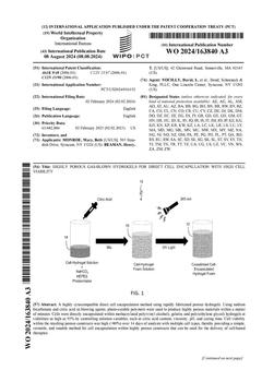
Highly porous gas-blown hydrogels for direct cell encapsulation with high cell viability
PatentWO2024163840A3
Innovation
- Novel gas-blowing technique using sodium bicarbonate and citric acid as blowing agents to create highly porous hydrogels within minutes, enabling rapid fabrication compared to traditional slow gelation methods.
- Direct cell encapsulation method achieving exceptionally high cell viability (up to 93%) by precisely controlling solution parameters including citric acid content, viscosity, pH, and curing time.
- Versatile platform compatible with multiple photo-curable polymers (methacrylated PVA, gelatin, and PEG) maintaining high cell viability (>80%) over 14 days across various cell types.

Cell viability apparatus, system, and methods thereof
PatentActiveUS12428619B2
Innovation
- A non-destructive oxygen imaging system using Electron Paramagnetic Resonance Oxygen Imaging (EPROI) with a trityl OX071 contrast agent to create oxygen maps, allowing for the assessment of cell viability and functionality without destroying cells or scaffolds.


Regulatory Framework for Bioprinted Products
The regulatory landscape for bioprinted products represents one of the most complex and evolving areas in biotechnology governance. Current frameworks primarily rely on existing medical device and tissue engineering regulations, which were not specifically designed to address the unique challenges posed by bioprinting technologies. The FDA has established preliminary guidelines through its Center for Biologics Evaluation and Research (CBER) and Center for Devices and Radiological Health (CDRH), treating bioprinted products as combination products requiring multi-disciplinary review processes.
Cell viability assessment under different bioprinting cures falls under stringent regulatory scrutiny, as it directly impacts patient safety and therapeutic efficacy. Regulatory bodies require comprehensive documentation of cell survival rates, functionality maintenance, and long-term stability across various curing conditions. The validation protocols must demonstrate consistent cell viability metrics regardless of the bioprinting parameters employed, including temperature variations, UV exposure duration, and chemical crosslinking agents.
International regulatory harmonization remains a significant challenge, with the European Medicines Agency (EMA) following different pathways compared to FDA approaches. The EMA's Advanced Therapy Medicinal Products (ATMP) regulation provides a framework that encompasses bioprinted tissues, requiring extensive preclinical data on cell viability and biocompatibility. Japan's Pharmaceuticals and Medical Devices Agency (PMDA) has developed parallel guidelines emphasizing regenerative medicine applications.
Quality control standards mandate rigorous testing protocols for cell viability assessment, including standardized assays such as MTT, Live/Dead staining, and metabolic activity measurements. These requirements ensure that bioprinted products maintain consistent therapeutic potential across different manufacturing conditions and curing methodologies.
The regulatory approval pathway typically involves phased clinical trials, with particular emphasis on demonstrating that cell viability outcomes translate to clinical efficacy. Manufacturers must establish clear correlations between in vitro cell survival data and in vivo performance, creating comprehensive risk-benefit profiles that satisfy regulatory requirements for market authorization.
Standardization of Cell Viability Metrics
The standardization of cell viability metrics represents a critical foundation for advancing bioprinting technology and ensuring reproducible research outcomes across different laboratories and applications. Currently, the field faces significant challenges due to the lack of universally accepted protocols and measurement standards, which hampers meaningful comparison of results between different bioprinting systems and research groups.
Existing cell viability assessment methods vary considerably in their approaches, timing, and interpretation criteria. Common techniques include live/dead staining assays, metabolic activity measurements such as MTT or alamarBlue assays, and flow cytometry-based analyses. However, these methods often employ different threshold values for determining cell viability, varying incubation periods, and inconsistent sample preparation protocols, leading to substantial variability in reported results.
The temporal aspect of viability assessment presents another standardization challenge. Different studies measure cell viability at varying time points post-printing, ranging from immediate assessment to extended culture periods of several weeks. This inconsistency makes it difficult to establish baseline viability expectations and compare the performance of different bioprinting techniques or bioink formulations.
International standardization organizations and research consortiums are beginning to address these challenges by developing comprehensive guidelines for cell viability assessment in bioprinting applications. These efforts focus on establishing standardized protocols for sample preparation, measurement techniques, data analysis methods, and reporting formats. Key parameters being standardized include cell counting methodologies, viability threshold definitions, statistical analysis requirements, and quality control measures.
The development of reference materials and control standards is also gaining momentum, with efforts to create standardized cell lines, bioink formulations, and printing parameters that can serve as benchmarks for viability assessments. These reference standards would enable researchers to validate their measurement systems and ensure consistency across different experimental setups.
Furthermore, emerging digital pathology and automated image analysis technologies are being integrated into standardization efforts, offering the potential for more objective and reproducible viability measurements. These advanced analytical tools can reduce human bias and improve the precision of cell viability quantification, supporting the establishment of more robust standardized protocols.
Unlock deeper insights with PatSnap Eureka Quick Research — get a full tech report to explore trends and direct your research. Try now!
Generate Your Research Report Instantly with AI Agent
Supercharge your innovation with PatSnap Eureka AI Agent Platform!