

How to Capture Rapid Dynamic Changes With PET Scans
MAR 2, 20269 MIN READ
Generate Your Research Report Instantly with AI Agent
PatSnap Eureka helps you evaluate technical feasibility & market potential.
PET Dynamic Imaging Background and Objectives
Positron Emission Tomography (PET) has evolved from a primarily static imaging modality to a sophisticated dynamic imaging technique capable of capturing temporal physiological processes. Traditional PET imaging typically acquires data over extended periods to achieve adequate signal-to-noise ratios, often resulting in static representations that average biological activity over time. However, many critical physiological processes occur on timescales of seconds to minutes, necessitating the development of dynamic PET imaging capabilities.
The fundamental challenge in dynamic PET imaging lies in the inherent trade-off between temporal resolution and image quality. Rapid data acquisition requires shorter frame durations, which inevitably reduces photon counts per frame and increases noise levels. This limitation has historically constrained the ability to visualize fast biological processes such as neurotransmitter release, cardiac perfusion changes, or rapid metabolic shifts during physiological stress.
Recent technological advances have created new opportunities to overcome these limitations. Modern PET scanners equipped with advanced detector technologies, including silicon photomultipliers and time-of-flight capabilities, offer improved sensitivity and temporal resolution. Additionally, sophisticated reconstruction algorithms and artificial intelligence-based denoising techniques have emerged as powerful tools to enhance image quality in low-count scenarios.
The primary objective of advancing dynamic PET imaging is to enable real-time visualization of biological processes with sufficient temporal resolution to capture rapid physiological changes while maintaining diagnostic image quality. This capability would revolutionize applications in cardiology, neurology, and oncology by providing unprecedented insights into dynamic biological processes.
Specific technical goals include achieving sub-second temporal resolution for cardiac applications, enabling real-time neurotransmitter tracking in brain studies, and developing robust motion correction algorithms for dynamic acquisitions. Furthermore, the integration of machine learning approaches aims to predict and compensate for the inherent noise limitations in rapid dynamic imaging.
The successful development of rapid dynamic PET imaging would establish new clinical protocols for time-sensitive diagnoses, enable personalized medicine approaches based on individual physiological response patterns, and facilitate the development of novel radiopharmaceuticals designed specifically for dynamic studies. This technological advancement represents a critical step toward transforming PET from a static diagnostic tool into a dynamic physiological monitoring system.
Market Demand for Rapid PET Dynamic Scanning
The healthcare industry is experiencing unprecedented demand for advanced imaging technologies that can capture physiological processes in real-time, with rapid dynamic PET scanning emerging as a critical diagnostic tool. This growing market demand stems from the increasing need for precise, time-sensitive medical imaging that can track metabolic changes, drug distribution, and biological processes as they occur within the human body.
Oncology represents the largest market segment driving demand for rapid dynamic PET scanning capabilities. Cancer treatment protocols increasingly require real-time monitoring of tumor metabolism and treatment response, necessitating imaging systems that can capture rapid physiological changes during therapy administration. The ability to visualize how tumors respond to treatment within minutes rather than hours provides oncologists with invaluable data for treatment optimization and patient management.
Cardiology applications constitute another significant market driver, particularly for myocardial perfusion studies and cardiac metabolism assessment. Cardiologists require imaging systems capable of tracking blood flow dynamics and metabolic changes in cardiac tissue during stress tests and pharmaceutical interventions. The demand for rapid dynamic scanning in this field has intensified due to the growing prevalence of cardiovascular diseases and the need for more precise diagnostic tools.
Neurological applications present substantial market opportunities, especially in neurodegenerative disease research and brain function studies. Neurologists and researchers demand imaging capabilities that can capture rapid neurotransmitter dynamics, glucose metabolism changes, and drug uptake patterns in the brain. The increasing focus on early detection and monitoring of conditions such as Alzheimer's disease and Parkinson's disease has amplified the need for high-temporal-resolution PET imaging.
The pharmaceutical industry represents a rapidly expanding market segment for dynamic PET scanning technologies. Drug development companies require sophisticated imaging tools to track pharmacokinetics and pharmacodynamics in real-time during clinical trials. The ability to visualize drug distribution, target engagement, and metabolic effects as they occur provides pharmaceutical companies with critical data for drug development and regulatory approval processes.
Research institutions and academic medical centers constitute a specialized but influential market segment. These organizations drive demand for cutting-edge dynamic PET scanning capabilities to support translational research and clinical studies. Their requirements often push the boundaries of current technology, creating demand for more advanced rapid scanning solutions.
Market growth is further accelerated by regulatory pressures for more precise diagnostic tools and the healthcare industry's shift toward personalized medicine approaches that require detailed, real-time physiological monitoring capabilities.
Current PET Temporal Resolution Limitations
Current PET imaging systems face significant temporal resolution constraints that fundamentally limit their ability to capture rapid physiological processes. The temporal resolution of conventional PET scanners typically ranges from 30 seconds to several minutes per frame, which is inadequate for monitoring dynamic biological events that occur on timescales of seconds or milliseconds. This limitation stems from the inherent physics of positron emission detection and the statistical requirements for generating meaningful images.
The primary bottleneck lies in the photon counting statistics required for acceptable image quality. PET imaging relies on detecting coincident gamma ray pairs produced by positron-electron annihilation events. To achieve sufficient signal-to-noise ratios, current systems must accumulate millions of coincidence events over extended time periods. This statistical requirement creates a fundamental trade-off between temporal resolution and image quality, where shorter acquisition times result in noisy, clinically unusable images.
Hardware limitations further compound these challenges. Traditional photomultiplier tubes and early-generation silicon photomultipliers exhibit timing uncertainties in the nanosecond range, contributing to coincidence timing windows that reduce system sensitivity. The detector crystal response time, typically 200-400 nanoseconds for commonly used materials like LSO and LYSO, adds additional temporal blur to the detection process.
Data processing and reconstruction algorithms represent another significant constraint. Current iterative reconstruction methods require substantial computational time, often taking several minutes to hours for complex dynamic studies. Real-time reconstruction capabilities remain limited, preventing immediate visualization of rapid physiological changes during scanning procedures.
Geometric factors also impose temporal limitations. The physical arrangement of detector rings and the need for sufficient angular sampling to avoid artifacts necessitate longer acquisition times. Limited-angle reconstructions, while faster, suffer from reduced image quality and spatial resolution that may obscure subtle dynamic changes.
Motion artifacts present additional challenges for high temporal resolution imaging. Patient movement during rapid sequential acquisitions can introduce significant image degradation, requiring either motion correction algorithms that further slow processing or gating techniques that effectively reduce temporal sampling rates.
These combined limitations create a technological ceiling that prevents PET from effectively monitoring rapid biological processes such as neurotransmitter release, cardiac perfusion changes during stress, or real-time drug distribution kinetics, highlighting the critical need for innovative solutions to overcome current temporal resolution barriers.
Current Solutions for Fast Dynamic PET Acquisition
01 Dynamic PET imaging protocols and rapid acquisition methods
Advanced PET scanning techniques focus on rapid dynamic imaging protocols that enable faster data acquisition while maintaining image quality. These methods involve optimized scanning sequences, reduced acquisition times, and improved temporal resolution to capture dynamic physiological changes. The protocols are designed to minimize patient discomfort and motion artifacts while maximizing diagnostic information through accelerated imaging processes.- Dynamic PET imaging protocols and rapid acquisition methods: Advanced PET scanning techniques focus on rapid dynamic imaging protocols that enable faster data acquisition while maintaining image quality. These methods involve optimized scanning sequences, reduced acquisition times, and improved temporal resolution to capture dynamic physiological changes. The protocols are designed to minimize patient discomfort and motion artifacts while maximizing diagnostic information through accelerated imaging processes.
- Real-time image reconstruction and processing algorithms: Sophisticated algorithms enable real-time reconstruction of PET scan data to visualize rapid dynamic changes in metabolic activity. These computational methods employ advanced mathematical models and machine learning techniques to process imaging data quickly and accurately. The algorithms enhance image quality, reduce noise, and provide immediate feedback during scanning procedures, allowing for dynamic assessment of physiological processes.
- Motion correction and compensation techniques: Technologies for detecting and correcting patient motion during dynamic PET scans ensure accurate imaging of rapid physiological changes. These techniques utilize tracking systems, gating methods, and computational algorithms to compensate for respiratory, cardiac, and voluntary movements. The correction methods improve image quality and quantitative accuracy by aligning sequential scan data and reducing motion-induced artifacts.
- Tracer kinetic modeling for dynamic studies: Mathematical modeling approaches analyze the temporal distribution and uptake of radiotracers to characterize rapid dynamic changes in tissue metabolism. These models quantify physiological parameters by analyzing time-activity curves and tracer kinetics. The modeling techniques provide detailed information about blood flow, metabolic rates, and receptor binding dynamics, enabling comprehensive assessment of biological processes over time.
- Multi-parametric and hybrid imaging systems: Integrated imaging systems combine PET with other modalities to capture comprehensive dynamic information about physiological changes. These hybrid approaches synchronize multiple imaging techniques to provide complementary anatomical and functional data. The systems enable simultaneous acquisition of different types of information, improving the detection and characterization of rapid dynamic processes through multi-parametric analysis.
02 Real-time image reconstruction and processing algorithms
Sophisticated algorithms enable real-time reconstruction of PET scan data to visualize rapid dynamic changes in metabolic activity. These computational methods employ advanced mathematical models and machine learning techniques to process imaging data quickly and accurately. The algorithms can handle large datasets and provide immediate feedback during scanning procedures, allowing for dynamic assessment of physiological processes.Expand Specific Solutions03 Motion correction and compensation techniques
Technologies for detecting and correcting patient motion during PET scans are essential for capturing accurate dynamic changes. These techniques utilize tracking systems, gating methods, and computational corrections to account for respiratory, cardiac, and voluntary movements. The approaches ensure that rapid physiological changes are accurately captured without artifacts from patient motion, improving the reliability of dynamic PET studies.Expand Specific Solutions04 Tracer kinetic modeling for dynamic PET analysis
Mathematical modeling approaches analyze the temporal distribution and uptake of radiotracers to quantify rapid dynamic changes in tissue metabolism. These models calculate kinetic parameters that describe tracer behavior over time, providing insights into physiological processes such as blood flow, glucose metabolism, and receptor binding. The modeling techniques enable quantitative assessment of dynamic biological processes captured in PET imaging.Expand Specific Solutions05 Multi-parametric and time-resolved PET imaging systems
Integrated imaging systems combine multiple detection parameters and time-resolved capabilities to capture comprehensive dynamic information. These systems feature enhanced detector sensitivity, improved timing resolution, and simultaneous acquisition of multiple imaging parameters. The technology enables detailed characterization of rapid physiological changes through continuous monitoring and multi-dimensional data analysis, providing superior temporal and spatial information for clinical and research applications.Expand Specific Solutions
Key Players in Advanced PET Scanner Development
The competitive landscape for capturing rapid dynamic changes with PET scans reflects a mature but evolving market driven by technological advancement needs. The industry is in a growth phase, with increasing demand for real-time imaging capabilities in oncology and neurology applications. Market size is substantial, dominated by established players like Siemens Healthineers AG, Koninklijke Philips NV, and Canon Medical Systems Corp., who control significant market share through comprehensive imaging portfolios. Technology maturity varies across segments, with companies like Shanghai United Imaging Healthcare Co., Ltd. and MinFound Medical Systems Co., Ltd. emerging as innovative challengers, particularly in AI-enhanced dynamic imaging solutions. Research institutions including Washington University in St. Louis and Zhejiang University are advancing fundamental technologies, while specialized firms like ADM Diagnostics focus on brain imaging applications, indicating a competitive ecosystem balancing established infrastructure with cutting-edge innovation.
Shanghai United Imaging Healthcare Co., Ltd.
Technical Solution: United Imaging has developed the uEXPLORER total-body PET scanner with unprecedented sensitivity for dynamic imaging applications. Their technology enables simultaneous imaging of the entire body with temporal resolution down to seconds, allowing capture of rapid tracer distribution and clearance patterns. The system utilizes advanced coincidence detection algorithms and real-time data processing to track dynamic changes across multiple organ systems simultaneously, particularly valuable for pharmacokinetic studies and rapid metabolic assessments.
Strengths: Total-body imaging capability with exceptional sensitivity for dynamic studies. Weaknesses: Very high cost and limited global availability outside of major research centers.
Koninklijke Philips NV
Technical Solution: Philips has developed advanced PET/CT systems with continuous bed motion (CBM) technology that enables dynamic whole-body imaging for capturing rapid physiological changes. Their Vereos Digital PET technology incorporates digital photon counting with improved temporal resolution, allowing for faster acquisition times and enhanced sensitivity for dynamic studies. The system features advanced reconstruction algorithms that can process rapid sequential imaging data to track tracer kinetics in real-time, particularly useful for cardiac and oncological applications where metabolic changes occur within minutes.
Strengths: Market-leading digital PET technology with superior temporal resolution and sensitivity. Weaknesses: High cost and complex calibration requirements for dynamic protocols.
Core Innovations in Rapid PET Data Reconstruction
Ultrafast tracer imaging for positron emission tomography
PatentActiveUS20220304596A1
Innovation
- A high-temporal-resolution PET scanning system that performs subsecond total-body dynamic scans using a kernel-regularized reconstruction technique to produce 100 millisecond temporal frames, enabling visualization of tracer propagation during individual heart beats and extracting motion signals for motion gating without external devices.


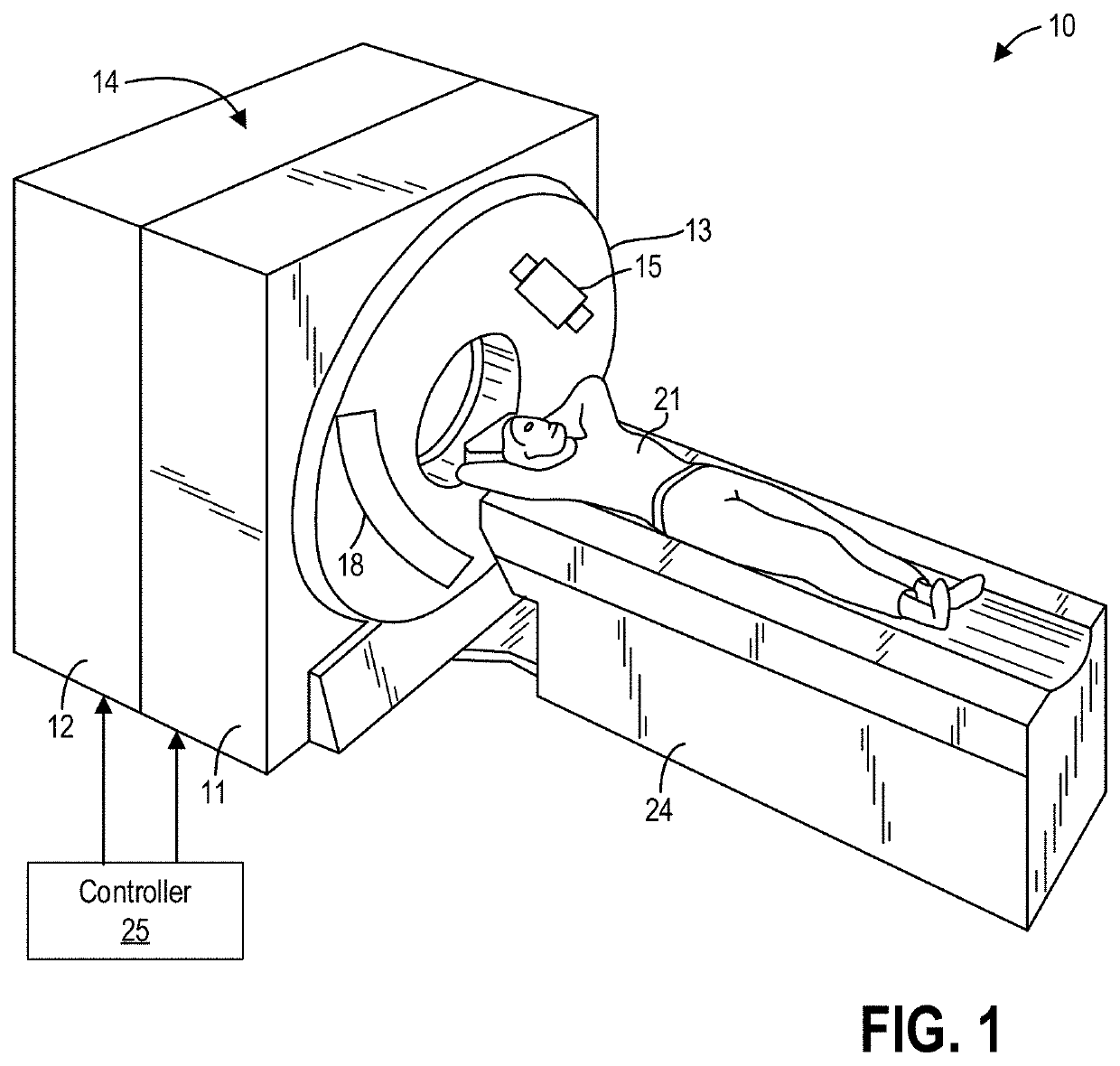
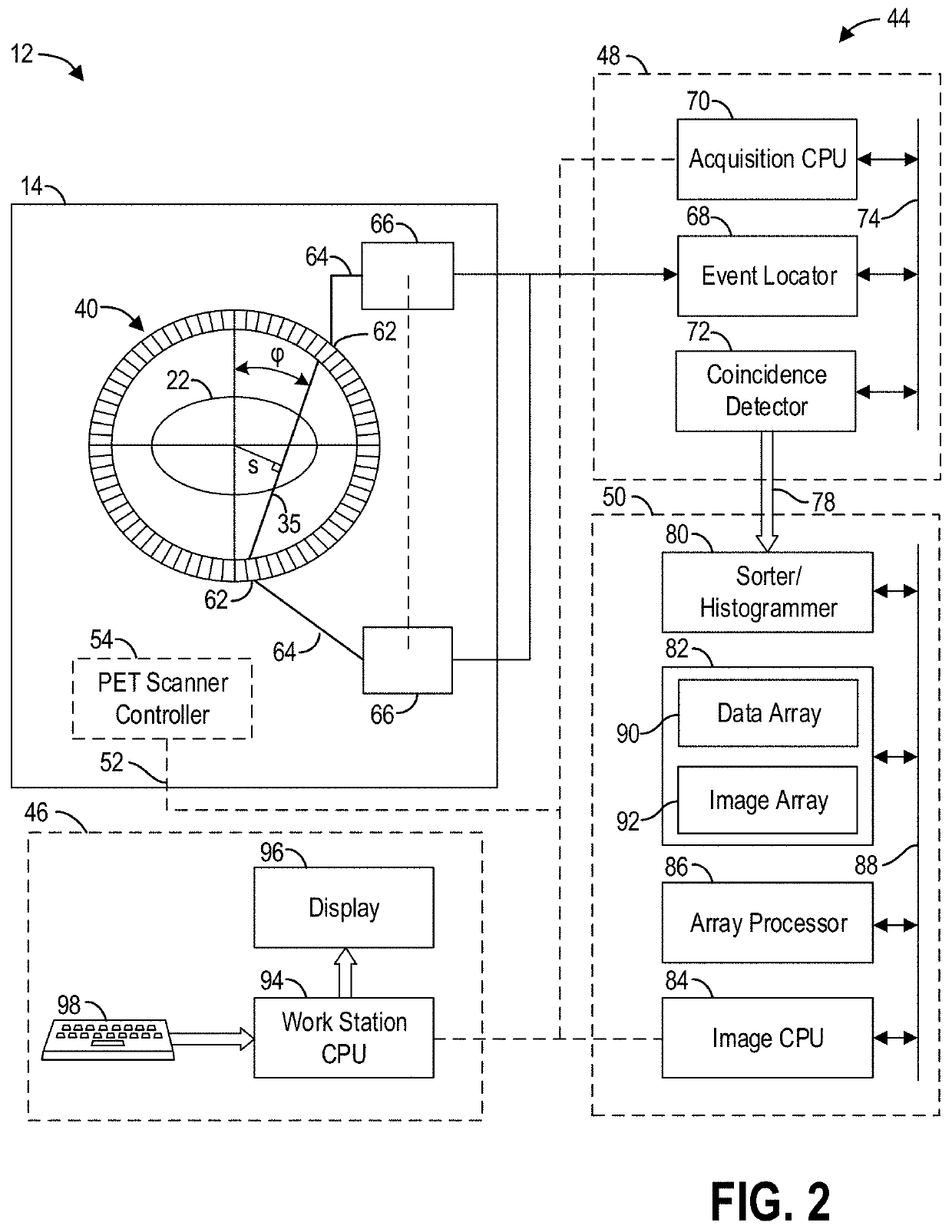
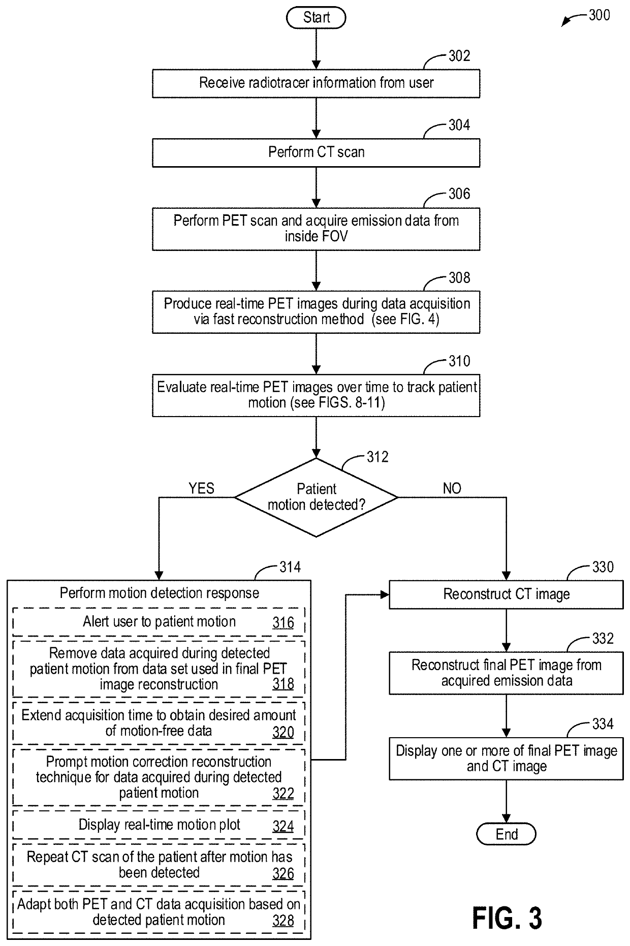
Methods and systems for motion detection in positron emission tomography
PatentActiveUS20220047227A1
Innovation
- A method for real-time PET image reconstruction during data acquisition, which tracks patient motion by analyzing voxel-wise variations and outputs motion indicators, allowing for immediate adjustments to the scan protocol to minimize motion artifacts and enhance image quality.

Radiation Safety Standards for Dynamic PET
Dynamic PET imaging presents unique radiation safety challenges that require specialized regulatory frameworks and safety protocols. The rapid acquisition sequences and extended scanning durations inherent to dynamic studies necessitate careful consideration of radiation exposure limits for both patients and healthcare personnel. Current international standards, including those established by the International Commission on Radiological Protection (ICRP) and national regulatory bodies, provide foundational guidelines that must be adapted for dynamic PET applications.
Patient radiation exposure in dynamic PET studies typically exceeds that of conventional static imaging due to prolonged acquisition periods and potentially higher tracer doses required for adequate temporal resolution. The effective dose calculations must account for the extended biological half-life of tracers during continuous monitoring, with particular attention to pediatric populations where dose optimization becomes critical. Regulatory frameworks mandate that cumulative exposure remains within acceptable diagnostic reference levels while maintaining image quality sufficient for clinical decision-making.
Healthcare worker protection requires enhanced protocols during dynamic PET procedures, as extended patient contact time and repeated tracer administrations increase occupational exposure risks. Radiation safety standards mandate continuous monitoring of personnel doses, implementation of time-distance-shielding principles, and establishment of controlled access zones during dynamic acquisitions. Special consideration must be given to nursing staff and technologists who may require prolonged patient interaction during stress protocols or pharmacological interventions.
Facility design standards for dynamic PET installations must incorporate additional shielding calculations based on increased workload factors and extended occupancy times. Regulatory requirements specify minimum wall thickness, door specifications, and ventilation systems capable of managing increased radioactive waste generation from dynamic studies. Emergency protocols must address potential equipment failures during extended acquisitions and procedures for managing patients with retained activity.
Quality assurance programs mandated by radiation safety standards include regular calibration of dose calibrators, survey meters, and imaging equipment to ensure accurate dose measurements throughout dynamic acquisitions. Documentation requirements encompass detailed exposure records, protocol justifications, and regular safety training for all personnel involved in dynamic PET procedures, ensuring compliance with evolving regulatory standards while maintaining optimal patient care.
Clinical Validation Requirements for Dynamic PET
Clinical validation of dynamic PET imaging systems requires adherence to stringent regulatory frameworks and standardized protocols to ensure patient safety and diagnostic accuracy. The validation process must demonstrate that rapid dynamic imaging capabilities maintain image quality standards while capturing temporal changes in tracer kinetics. Regulatory bodies such as the FDA and EMA have established specific guidelines for medical imaging devices, requiring comprehensive documentation of system performance characteristics, radiation dose optimization, and clinical efficacy data.
The validation framework encompasses multiple phases of clinical trials, beginning with phantom studies to establish baseline performance metrics. These studies must demonstrate that accelerated acquisition protocols maintain adequate signal-to-noise ratios and spatial resolution while achieving the temporal resolution necessary for dynamic imaging. Subsequent human studies require careful patient selection criteria, with particular attention to motion artifacts and patient comfort during extended imaging sessions.
Quality assurance protocols for dynamic PET systems must address unique challenges associated with rapid data acquisition. Validation studies must establish acceptable limits for image noise, uniformity, and quantitative accuracy across different temporal sampling rates. Cross-calibration procedures between different scanner models and manufacturers become critical when multi-center trials are conducted, requiring standardized phantom protocols and data analysis methodologies.
Patient safety considerations extend beyond traditional radiation exposure limits to include psychological factors associated with longer scan durations. Validation protocols must demonstrate that dynamic imaging procedures can be completed safely across diverse patient populations, including pediatric and elderly patients who may have difficulty remaining motionless during extended acquisitions.
The clinical validation process must also establish standardized metrics for evaluating dynamic PET data quality and diagnostic performance. This includes defining acceptable thresholds for kinetic parameter estimation accuracy, test-retest reproducibility, and inter-observer variability in image interpretation. Validation studies should demonstrate clinical utility through comparison with established diagnostic methods and correlation with patient outcomes.
Regulatory approval pathways for dynamic PET systems often require demonstration of substantial equivalence to existing imaging modalities while providing evidence of clinical benefit from enhanced temporal information. The validation process must address software validation requirements for kinetic modeling algorithms and ensure compliance with medical device quality management systems throughout the product lifecycle.
Unlock deeper insights with PatSnap Eureka Quick Research — get a full tech report to explore trends and direct your research. Try now!
Generate Your Research Report Instantly with AI Agent
Supercharge your innovation with PatSnap Eureka AI Agent Platform!