

PET Scan Vs Contrast-Enhanced CT: Visualization Infections
MAR 2, 20269 MIN READ
Generate Your Research Report Instantly with AI Agent
PatSnap Eureka helps you evaluate technical feasibility & market potential.
PET vs CT Imaging Background and Clinical Goals
Medical imaging has undergone revolutionary transformations over the past several decades, with computed tomography (CT) and positron emission tomography (PET) emerging as cornerstone technologies in diagnostic medicine. The evolution from basic radiographic techniques to sophisticated cross-sectional imaging has fundamentally changed how clinicians approach infection detection and characterization. CT technology, first introduced in the 1970s, provided unprecedented anatomical detail through cross-sectional imaging, while PET scanning, developed in the 1980s, offered unique metabolic and functional insights into tissue behavior.
The integration of contrast enhancement in CT imaging marked a significant milestone in infection visualization. Contrast-enhanced CT enables superior delineation of vascular structures, tissue perfusion patterns, and inflammatory processes that are characteristic of infectious conditions. This advancement allowed radiologists to identify subtle changes in tissue density, enhancement patterns, and morphological alterations that indicate active infection or inflammatory response.
PET imaging represents a paradigm shift from purely anatomical to functional imaging approaches. By utilizing radiolabeled glucose analogs, particularly fluorodeoxyglucose (F-18 FDG), PET scanners can detect areas of increased metabolic activity that often correlate with infectious processes. This metabolic imaging capability provides unique advantages in identifying infections that may not yet manifest significant anatomical changes visible on conventional imaging modalities.
The clinical objectives driving the comparison between PET and contrast-enhanced CT for infection visualization center on improving diagnostic accuracy, reducing time to diagnosis, and optimizing patient management strategies. Healthcare systems worldwide face increasing pressure to provide rapid, accurate diagnoses while minimizing patient exposure to unnecessary procedures and radiation. The ability to differentiate between active infection, sterile inflammation, and post-treatment changes has become increasingly critical in clinical decision-making.
Contemporary imaging goals emphasize the need for comprehensive evaluation protocols that can distinguish between various infectious etiologies, assess treatment response, and guide therapeutic interventions. The development of hybrid imaging systems, such as PET-CT, reflects the growing recognition that combining anatomical and functional information provides superior diagnostic capabilities compared to either modality alone. This technological convergence aims to leverage the strengths of both imaging approaches while mitigating their individual limitations in infection detection and characterization.
Market Demand for Advanced Infection Imaging Solutions
The global medical imaging market is experiencing unprecedented growth driven by the increasing prevalence of infectious diseases and the critical need for accurate diagnostic tools. Healthcare systems worldwide are grappling with complex infection cases that require sophisticated imaging solutions beyond conventional radiography. The demand for advanced infection imaging technologies has intensified following recent global health challenges, highlighting the limitations of traditional diagnostic approaches.
Hospital networks and diagnostic centers are actively seeking imaging modalities that can provide superior soft tissue contrast and metabolic information for infection detection. The market shows particularly strong demand for technologies capable of distinguishing between active infections and post-inflammatory changes, a clinical challenge that significantly impacts treatment decisions. Emergency departments and intensive care units represent high-volume segments requiring rapid, accurate infection imaging capabilities.
Oncology departments constitute another substantial market segment, where differentiating between malignant lesions and infectious processes remains a persistent clinical need. The growing cancer patient population, often immunocompromised and susceptible to opportunistic infections, drives demand for imaging solutions that can reliably distinguish between these conditions. This dual-purpose imaging capability represents a significant market opportunity.
The pediatric imaging market presents unique demands for infection visualization, requiring technologies that minimize radiation exposure while maintaining diagnostic accuracy. Healthcare providers increasingly prioritize imaging modalities that can deliver comprehensive infection assessment with reduced patient risk, particularly for vulnerable populations including children and pregnant women.
Emerging markets in developing countries show accelerating adoption of advanced imaging technologies as healthcare infrastructure expands. These regions face significant infectious disease burdens, creating substantial demand for cost-effective yet sophisticated imaging solutions. The market trend indicates preference for versatile imaging platforms that can address multiple clinical scenarios within resource-constrained environments.
Private imaging centers and outpatient facilities represent rapidly growing market segments, driven by healthcare decentralization trends and patient preference for accessible diagnostic services. These facilities require imaging technologies that combine clinical excellence with operational efficiency, supporting high patient throughput while maintaining diagnostic quality standards.
Current State of PET and Contrast-Enhanced CT Technologies
Positron Emission Tomography (PET) scanning has evolved significantly since its clinical introduction in the 1970s, with current systems achieving spatial resolutions of 4-6mm and incorporating advanced reconstruction algorithms. Modern PET scanners utilize fluorine-18 fluorodeoxyglucose (FDG) as the primary tracer for infection imaging, leveraging the increased glucose metabolism in inflammatory tissues. Recent technological advances include digital PET detectors, time-of-flight capabilities, and hybrid PET/CT systems that provide simultaneous metabolic and anatomical information.
Contemporary contrast-enhanced CT technology has reached remarkable sophistication with multi-detector row scanners offering submillimeter spatial resolution and rapid acquisition times. Current systems feature dual-energy CT capabilities, iterative reconstruction techniques, and advanced contrast protocols optimized for infection detection. The integration of artificial intelligence algorithms has enhanced image quality while reducing radiation exposure, with some systems achieving dose reductions of up to 80% compared to previous generations.
PET imaging demonstrates exceptional sensitivity for detecting metabolically active infections, particularly in cases where anatomical changes are minimal or absent. Current FDG-PET protocols achieve sensitivity rates of 85-95% for various infectious processes, with the ability to detect inflammation within hours of onset. The technology excels in differentiating between active infection and sterile inflammation, though specificity can be challenged by concurrent inflammatory conditions or malignancies.
Contrast-enhanced CT provides superior anatomical detail and remains the gold standard for visualizing structural changes associated with infections. Modern protocols utilize optimized contrast timing and multi-phase acquisitions to enhance lesion conspicuity. The technology demonstrates particular strength in detecting abscesses, fluid collections, and complications such as perforation or vascular involvement, with spatial resolution capabilities that far exceed those of PET imaging.
Current limitations in PET technology include relatively high costs, limited availability, and the need for specialized radiopharmaceutical production facilities. The technology also faces challenges in distinguishing between infectious and non-infectious inflammatory processes, particularly in immunocompromised patients or those with autoimmune conditions.
Contrast-enhanced CT faces constraints related to iodinated contrast agent contraindications, including renal dysfunction and allergic reactions. Additionally, the technique may miss early-stage infections that have not yet produced significant anatomical changes, and radiation exposure considerations limit repeated examinations in certain patient populations.
Existing Solutions for Infection Visualization Techniques
01 Integrated PET-CT imaging systems and methods
Systems and methods that combine positron emission tomography (PET) and computed tomography (CT) imaging capabilities into a single integrated device. These systems enable simultaneous or sequential acquisition of both functional metabolic information from PET and anatomical structural information from contrast-enhanced CT, providing comprehensive diagnostic imaging. The integration allows for precise spatial registration and fusion of the two imaging modalities, improving diagnostic accuracy and workflow efficiency.- Integrated PET-CT imaging systems and methods: Systems and methods that combine positron emission tomography (PET) and computed tomography (CT) imaging capabilities into a single integrated platform. These systems enable simultaneous or sequential acquisition of both functional metabolic information from PET and anatomical structural information from contrast-enhanced CT, providing comprehensive diagnostic imaging. The integration allows for precise spatial registration and fusion of the two imaging modalities, improving diagnostic accuracy and workflow efficiency.
- Image registration and fusion techniques for multi-modality visualization: Advanced algorithms and techniques for aligning, registering, and fusing images from different imaging modalities. These methods enable accurate overlay and correlation of functional PET data with anatomical CT data, compensating for patient motion and anatomical variations. The fusion techniques enhance visualization by combining complementary information from both modalities, allowing clinicians to precisely localize metabolic abnormalities within anatomical structures.
- Contrast agent optimization and timing protocols: Methods and protocols for optimizing the administration and timing of contrast agents in combined imaging procedures. These techniques ensure optimal contrast enhancement during CT acquisition while coordinating with PET imaging sequences. The protocols address challenges such as determining appropriate contrast agent dosages, injection timing, and scan delays to maximize image quality and diagnostic value in combined examinations.
- Attenuation correction using CT data for PET imaging: Techniques that utilize CT imaging data to perform attenuation correction for PET images. The CT scan provides accurate electron density maps that can be converted to attenuation coefficients for correcting photon attenuation in PET data. This approach improves the quantitative accuracy of PET imaging and eliminates the need for separate transmission scans, reducing overall scan time and radiation exposure.
- Advanced visualization and display methods for combined imaging data: Sophisticated visualization tools and display methods specifically designed for presenting combined functional and anatomical imaging data. These methods include multi-planar reconstruction, three-dimensional rendering, color-coded overlay displays, and interactive viewing interfaces that allow clinicians to navigate through fused datasets. The visualization techniques facilitate interpretation by presenting complex multi-modal information in an intuitive and clinically useful manner.
02 Image registration and fusion techniques for multi-modality imaging
Advanced algorithms and methods for aligning and combining images from different imaging modalities. These techniques enable accurate spatial correspondence between PET functional data and CT anatomical data, accounting for patient motion, breathing artifacts, and differences in acquisition timing. The registration process ensures that metabolic abnormalities detected in PET scans can be precisely localized on the corresponding anatomical structures visible in contrast-enhanced CT images.Expand Specific Solutions03 Contrast agent optimization for combined PET-CT imaging
Methods and compositions for optimizing contrast agents used in combined imaging protocols. This includes timing protocols for contrast administration relative to PET tracer injection, selection of appropriate contrast concentrations that enhance CT visualization without interfering with PET signal detection, and development of dual-purpose agents that can provide both CT contrast enhancement and metabolic information. These optimizations improve the quality and diagnostic value of both imaging modalities when performed together.Expand Specific Solutions04 Attenuation correction methods using CT data for PET imaging
Techniques that utilize CT imaging data to correct for photon attenuation in PET imaging, improving quantitative accuracy of PET measurements. The CT scan provides detailed tissue density information that can be used to calculate attenuation coefficients, eliminating the need for separate transmission scans. This approach is particularly important when contrast agents are present, requiring specialized algorithms to account for the altered attenuation properties of contrast-enhanced tissues.Expand Specific Solutions05 Visualization and display systems for combined PET-CT data
Advanced visualization platforms and display methods for presenting combined PET and contrast-enhanced CT imaging data to clinicians. These systems provide tools for simultaneous viewing of fused images, side-by-side comparisons, and interactive manipulation of overlay parameters. Features include color-coded metabolic activity maps superimposed on anatomical CT images, three-dimensional rendering capabilities, and quantitative analysis tools that leverage information from both modalities to enhance diagnostic interpretation and treatment planning.Expand Specific Solutions
Key Players in PET and CT Imaging Industry
The medical imaging sector for PET scan versus contrast-enhanced CT in infection visualization represents a mature, multi-billion-dollar market experiencing steady technological advancement. The industry has evolved from early adoption to widespread clinical integration, with established players dominating through continuous innovation. Market leaders include Siemens Healthineers AG and Koninklijke Philips NV, who leverage decades of expertise in both PET and CT technologies. Emerging competitors like Shanghai United Imaging Healthcare Co., Ltd. and Beijing Arrays Medical Imaging Corp. are challenging traditional hierarchies with cost-effective solutions. Technology maturity varies significantly - while CT imaging has reached high standardization, PET technology continues evolving with enhanced radiopharmaceuticals and hybrid systems. Companies like GE Precision Healthcare LLC and Neusoft Medical Systems Co., Ltd. are driving integration of AI-powered diagnostic capabilities, positioning the sector for next-generation precision medicine applications in infectious disease detection.
Koninklijke Philips NV
Technical Solution: Philips Healthcare develops integrated PET/CT imaging solutions specifically optimized for infection and inflammation imaging. Their Vereos PET/CT systems feature digital photon counting technology that enhances sensitivity for detecting subtle infectious processes. The platform supports comprehensive contrast-enhanced CT protocols combined with FDG-PET and specialized tracers like Ga-68 citrate for infection imaging. Their IntelliSpace Portal provides advanced visualization tools including automated lesion detection, SUV quantification, and multi-parametric analysis comparing metabolic activity with contrast enhancement patterns. The system includes dedicated infection imaging protocols that optimize scan parameters for different clinical scenarios, from acute infections to chronic inflammatory conditions, enabling precise localization and characterization of infectious processes.
Strengths: Digital PET technology provides superior image quality and sensitivity for infection detection. Weaknesses: Premium pricing and complex maintenance requirements may limit adoption in smaller healthcare facilities.
Shanghai United Imaging Healthcare Co., Ltd.
Technical Solution: United Imaging Healthcare offers cost-effective PET/CT solutions combining molecular and anatomical imaging for infection visualization. Their uMI Panorama PET/CT systems integrate high-resolution PET detectors with advanced CT imaging capabilities, supporting comprehensive infection imaging protocols. The platform features proprietary reconstruction algorithms that enhance image quality while reducing radiation dose, particularly important for infection imaging that may require follow-up studies. Their uWS-MI workstation includes specialized infection imaging tools that facilitate comparison between PET metabolic activity and CT contrast enhancement patterns. The system supports various PET tracers including FDG and emerging infection-specific radiopharmaceuticals, providing flexible imaging options for different infectious conditions and clinical requirements.
Strengths: Competitive pricing with good image quality makes advanced imaging more accessible globally. Weaknesses: Limited global service network and newer market presence compared to established competitors.
Core Innovations in PET and CT Contrast Enhancement
PET image and CT image fusion device and method
PatentActiveCN118172252A
Innovation
- Adopting a method based on soft and hard computing, through the Fourier image adjustment system, Fourier image fusion system and Fourier image enhancement system, FPGA is used to adjust, fuse and enhance the image in real time to avoid the impact of noise in the post-processing process. , realizing real-time fusion of images and detection of lesion locations.


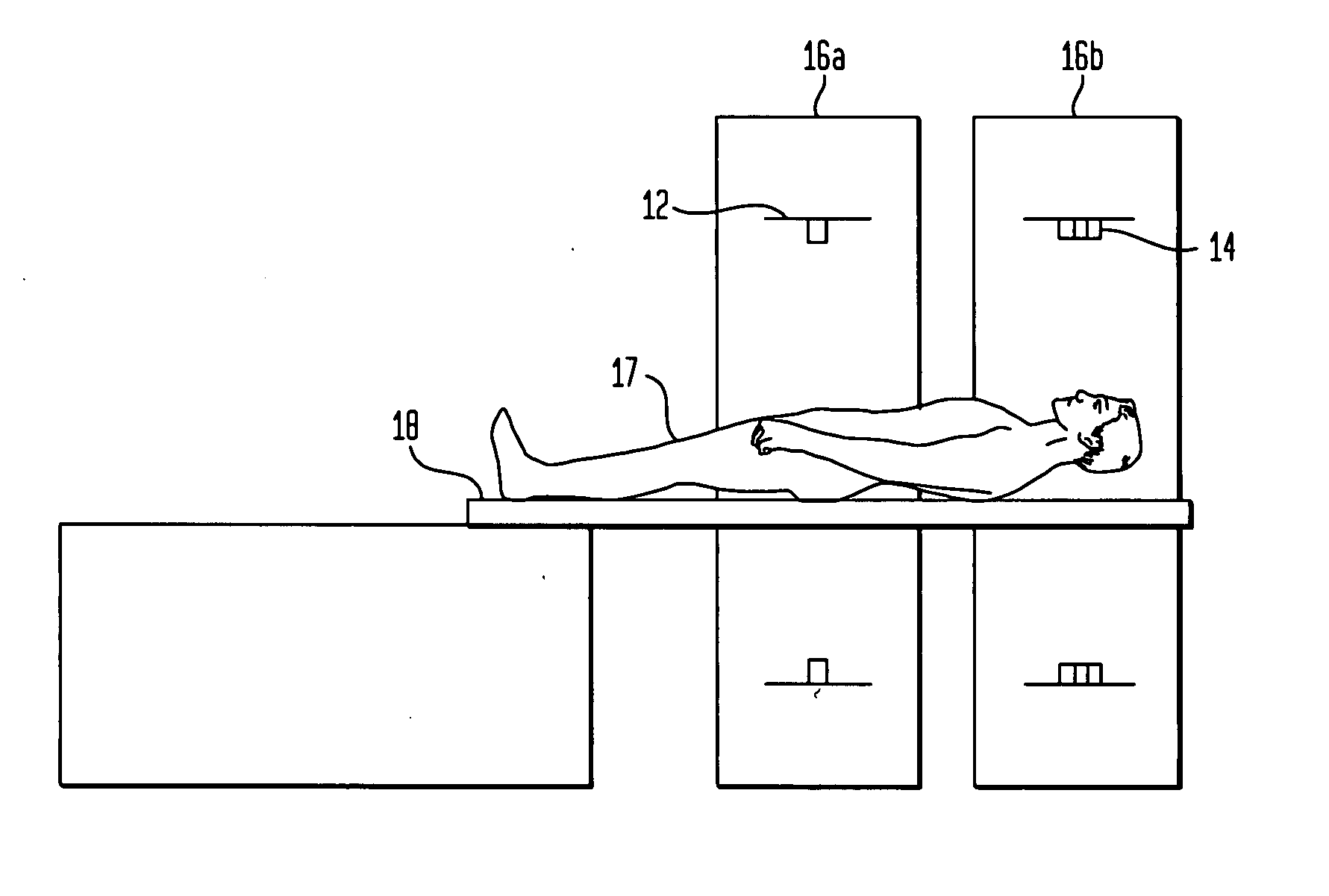
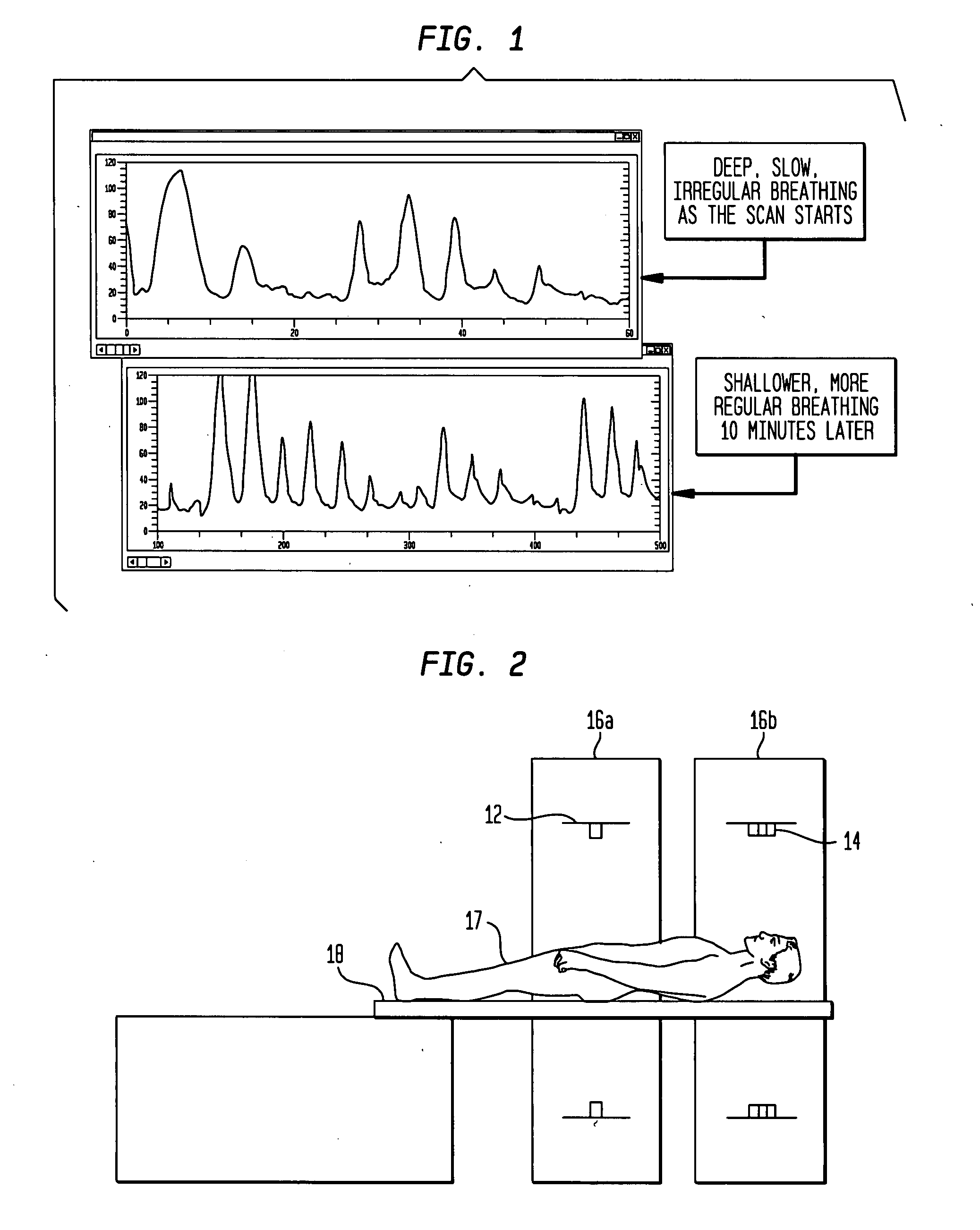
Registration of computed tomography (CT) and positron emission tomography (PET) image scans with automatic patient motion correction
PatentActiveUS20070232903A1
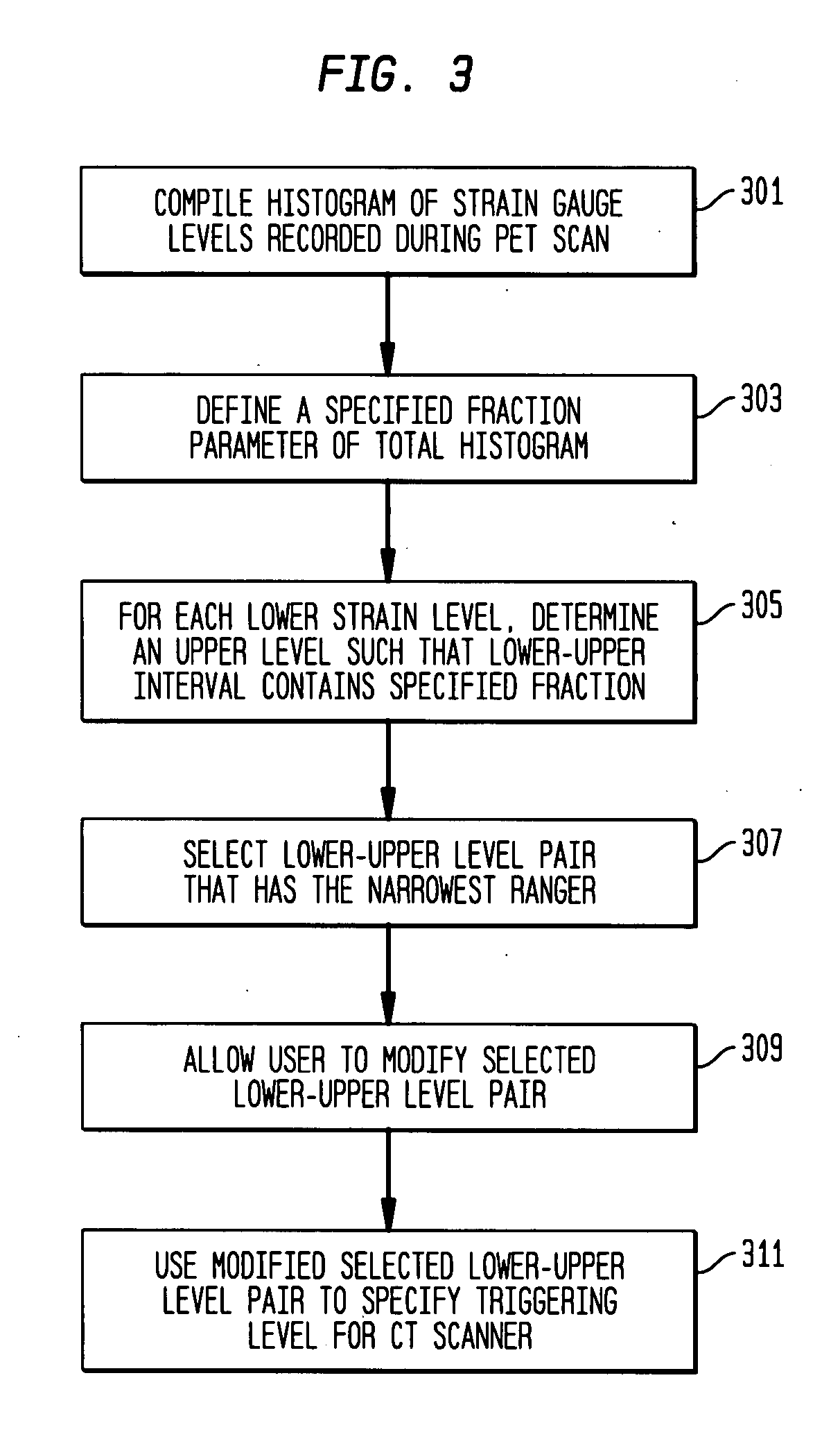
Innovation
- Determine optimal CT scan gating criteria based on strain gauge levels recorded during a PET scan, and perform a second CT scan with triggering that minimizes respiratory motion, allowing for improved registration between CT and PET images.

Radiation Safety Standards for Medical Imaging Devices
Medical imaging devices utilizing ionizing radiation, including PET scanners and contrast-enhanced CT systems, operate under stringent regulatory frameworks designed to minimize radiation exposure while maintaining diagnostic efficacy. The International Commission on Radiological Protection (ICRP) establishes fundamental dose limits, with occupational exposure capped at 20 mSv annually averaged over five years, and public exposure limited to 1 mSv per year. These standards form the foundation for national regulatory bodies such as the FDA, European Medicines Agency, and various nuclear regulatory commissions worldwide.
PET scanning systems must comply with specific radiation safety protocols due to their use of radioactive tracers, typically fluorine-18 labeled compounds. The effective dose from a standard FDG-PET scan ranges from 5-7 mSv, requiring careful justification for infection imaging applications. Facilities must implement comprehensive radiation protection programs including proper shielding design, contamination control procedures, and radioactive waste management systems. Staff handling radiopharmaceuticals require specialized training and regular dosimetry monitoring to ensure compliance with occupational exposure limits.
Contrast-enhanced CT systems present different radiation safety considerations, with typical effective doses ranging from 2-10 mSv depending on scan parameters and anatomical coverage. Modern CT scanners incorporate dose optimization technologies such as automatic exposure control, iterative reconstruction algorithms, and organ-based tube current modulation. Regulatory standards mandate regular quality assurance testing, including dose verification measurements and image quality assessments to maintain the delicate balance between diagnostic image quality and radiation exposure minimization.
Quality assurance protocols for both modalities require systematic monitoring of radiation output, image quality metrics, and safety system functionality. Annual inspections by qualified medical physicists ensure continued compliance with established safety standards. Emergency procedures must be documented and regularly practiced, particularly for PET facilities where radioactive contamination scenarios require specialized response protocols.
Emerging regulatory trends focus on dose tracking and reporting systems, with increasing emphasis on patient-specific dose monitoring and cumulative exposure tracking across multiple imaging studies. These evolving standards reflect the growing recognition of radiation protection as a critical component of infection imaging protocols, ensuring that diagnostic benefits consistently outweigh potential radiation risks while maintaining optimal image quality for accurate infection detection and monitoring.
Cost-Effectiveness Analysis of PET vs CT Modalities
The economic evaluation of PET versus contrast-enhanced CT for infection visualization reveals significant disparities in cost structures and clinical outcomes. PET imaging typically incurs higher upfront costs, ranging from $3,000 to $5,000 per scan, compared to contrast-enhanced CT at approximately $1,200 to $2,500. However, the superior diagnostic accuracy of PET, particularly in detecting occult infections and inflammatory processes, often translates to reduced downstream healthcare expenditures through more precise treatment targeting and shorter diagnostic workups.
Healthcare systems implementing PET for infection detection report 15-25% reduction in unnecessary antibiotic treatments and surgical interventions. This improvement stems from PET's enhanced specificity in differentiating active infection from sterile inflammation, particularly in post-surgical patients and those with chronic conditions. The metabolic information provided by FDG-PET enables clinicians to avoid exploratory procedures that frequently follow inconclusive CT results.
Cost-effectiveness ratios demonstrate favorable outcomes for PET in complex cases, with incremental cost-effectiveness ratios ranging from $12,000 to $18,000 per quality-adjusted life year gained. These figures become more compelling when considering patient populations with recurrent infections, immunocompromised states, or prosthetic device-related complications, where diagnostic precision significantly impacts treatment success rates.
The economic burden analysis reveals that while CT maintains advantages in screening scenarios and acute care settings due to rapid acquisition and lower costs, PET demonstrates superior value in cases requiring definitive infection localization. Healthcare institutions report 20-30% reduction in readmission rates when PET guides infection management protocols, offsetting initial imaging costs through improved patient outcomes and reduced length of stay.
Reimbursement patterns increasingly favor PET utilization for specific infection indications, with Medicare and major insurers expanding coverage criteria. This trend reflects growing recognition of PET's role in reducing overall healthcare costs through enhanced diagnostic confidence and treatment optimization in challenging infectious disease cases.
Unlock deeper insights with PatSnap Eureka Quick Research — get a full tech report to explore trends and direct your research. Try now!
Generate Your Research Report Instantly with AI Agent
Supercharge your innovation with PatSnap Eureka AI Agent Platform!