

How to Divide Tissue Sections While Preserving Transcriptomic Layers
JUN 3, 20269 MIN READ
Generate Your Research Report Instantly with AI Agent
Patsnap Eureka helps you evaluate technical feasibility & market potential.
Spatial Transcriptomics Tissue Division Background and Goals
Spatial transcriptomics represents a revolutionary advancement in molecular biology, emerging from the convergence of traditional transcriptomics and spatial biology. This field addresses the fundamental limitation of conventional RNA sequencing methods, which lose crucial spatial context information during tissue dissociation. The technology enables researchers to map gene expression patterns while maintaining the precise spatial coordinates of cells within their native tissue architecture.
The historical development of spatial transcriptomics began with early in situ hybridization techniques in the 1980s, evolved through fluorescence-based methods in the 1990s, and culminated in the breakthrough technologies of the 2010s. Key milestones include the development of spatial transcriptomics arrays, slide-seq technology, and high-resolution imaging-based approaches. These innovations have transformed our understanding of tissue organization and cellular communication networks.
Current technological evolution focuses on achieving single-cell resolution while maintaining spatial fidelity across entire tissue sections. The field has progressed from initial 100-micrometer resolution spots to sub-cellular precision, enabling detailed analysis of tissue microenvironments. Advanced computational algorithms now integrate spatial coordinates with transcriptomic data, creating comprehensive molecular atlases of complex tissues.
The primary technical objective centers on developing methodologies that can accurately divide tissue sections into meaningful biological units while preserving the integrity of transcriptomic layers. This involves maintaining RNA quality throughout sectioning procedures, ensuring consistent capture efficiency across different tissue regions, and preserving spatial relationships between adjacent cellular populations.
Strategic goals encompass establishing standardized protocols for tissue preparation, sectioning, and spatial coordinate mapping. The technology aims to achieve reproducible results across different laboratories and tissue types, while minimizing technical artifacts that could compromise data interpretation. Long-term objectives include developing automated systems capable of processing large-scale tissue collections with minimal human intervention.
Future aspirations involve creating three-dimensional spatial transcriptomic reconstructions from serial tissue sections, enabling comprehensive analysis of organ-level gene expression patterns. The ultimate goal is establishing spatial transcriptomics as a routine diagnostic and research tool, comparable to current histopathological methods but with unprecedented molecular resolution and quantitative precision.
Market Demand for Spatial Transcriptomics Solutions
The spatial transcriptomics market has experienced unprecedented growth driven by the increasing recognition that understanding gene expression patterns within their native tissue architecture is crucial for advancing biomedical research and clinical applications. Traditional bulk RNA sequencing methods, while valuable, fail to capture the spatial heterogeneity of tissues, creating a significant gap in our understanding of cellular interactions and tissue organization.
Research institutions and pharmaceutical companies are increasingly demanding solutions that can preserve transcriptomic information while enabling precise tissue sectioning for spatial analysis. This demand stems from the need to understand disease mechanisms, drug responses, and developmental processes at the cellular and subcellular levels within their spatial context. Cancer research represents a particularly strong market driver, as tumor heterogeneity and microenvironment interactions are critical factors in treatment efficacy and resistance mechanisms.
The clinical diagnostics sector is emerging as a major growth area, with pathologists and clinicians seeking tools that can provide molecular insights while maintaining morphological information. This dual requirement for molecular and spatial data is driving demand for advanced tissue processing techniques that can preserve RNA integrity during sectioning procedures. Hospitals and diagnostic laboratories are investing in spatial transcriptomics capabilities to enhance precision medicine approaches.
Academic research institutions constitute the largest current market segment, with increasing funding allocated to spatial biology projects. Government initiatives and research grants specifically targeting spatial omics technologies have accelerated adoption rates. Core facilities at major universities are establishing spatial transcriptomics platforms, creating sustained demand for reliable tissue processing solutions.
The pharmaceutical industry represents a high-value market segment, utilizing spatial transcriptomics for drug discovery, target validation, and biomarker identification. Companies are particularly interested in understanding how therapeutic compounds affect different cell populations within tissues and how spatial organization influences drug efficacy. This application requires robust tissue processing methods that maintain both spatial integrity and transcriptomic fidelity.
Biotechnology companies developing spatial analysis platforms are driving demand for complementary tissue processing technologies. These companies require standardized, reproducible methods for tissue sectioning that can integrate seamlessly with their analytical workflows. The market is characterized by a need for solutions that can handle diverse tissue types while maintaining consistent quality across different experimental conditions.
Current Challenges in Tissue Section Processing
Tissue section processing for transcriptomic analysis faces significant technical barriers that compromise data quality and experimental reproducibility. The primary challenge lies in maintaining RNA integrity during the sectioning process, as mechanical stress from microtome blades generates heat and physical disruption that degrades transcriptomic material. Traditional sectioning methods often result in RNA fragmentation rates exceeding 15-20%, particularly affecting longer transcripts and low-abundance species.
Spatial resolution preservation represents another critical bottleneck in current workflows. Conventional sectioning techniques struggle to maintain precise cellular boundaries, leading to cross-contamination between adjacent tissue layers. This issue becomes particularly pronounced when processing heterogeneous tissues with distinct transcriptomic profiles, where even minimal displacement can confound downstream spatial transcriptomics analyses.
Temperature control during sectioning remains inadequately addressed by existing protocols. Most standard procedures operate at suboptimal temperatures that fail to prevent enzymatic degradation while simultaneously causing ice crystal formation in frozen specimens. These temperature fluctuations create artifacts that distort native transcriptomic landscapes and introduce systematic biases in gene expression measurements.
Sample preparation inconsistencies plague current methodologies, with significant variability in fixation protocols, embedding procedures, and storage conditions. These inconsistencies result in batch effects that can overshadow biological signals, making it difficult to achieve reproducible results across different laboratories or experimental timepoints. The lack of standardized protocols particularly affects multi-institutional studies requiring consistent tissue processing approaches.
Automation limitations in existing sectioning equipment present scalability challenges for high-throughput transcriptomic studies. Current microtomes lack the precision control necessary for consistent section thickness and uniform cutting speeds, leading to variable tissue quality that impacts downstream molecular analyses. Manual intervention requirements further introduce operator-dependent variability that compromises experimental standardization.
Contamination prevention remains insufficiently addressed in standard protocols, with inadequate measures to prevent cross-sample contamination during sequential sectioning procedures. This challenge becomes critical when processing multiple specimens with different transcriptomic profiles, where even trace contamination can generate false-positive signals in sensitive detection methods.
Existing Tissue Division Methods for Transcriptomic Analysis
01 Spatial transcriptomic analysis methods for tissue sections
Advanced methodologies for analyzing gene expression patterns across different spatial locations within tissue sections. These techniques enable researchers to map transcriptomic data to specific anatomical regions and cellular layers, providing insights into tissue organization and function. The methods involve sophisticated sample preparation, sequencing protocols, and computational analysis to generate spatially resolved transcriptomic maps.- Spatial transcriptomic analysis methods for tissue sections: Advanced methodologies for analyzing gene expression patterns across different spatial locations within tissue sections. These techniques enable researchers to map transcriptomic data to specific anatomical regions and cellular layers, providing insights into tissue organization and function. The methods involve sophisticated sample preparation, sequencing protocols, and computational analysis to generate spatially resolved transcriptomic maps.
- Multi-layer tissue sectioning and processing techniques: Specialized approaches for preparing and processing tissue samples to preserve multiple transcriptomic layers while maintaining spatial integrity. These techniques focus on optimal sectioning methods, fixation protocols, and preservation strategies that allow for accurate analysis of different tissue depths and cellular compartments. The methods ensure minimal degradation of genetic material while preserving morphological structure.
- Computational algorithms for transcriptomic layer reconstruction: Software tools and computational methods designed to reconstruct and analyze transcriptomic data from tissue sections across multiple layers. These algorithms process complex datasets to identify gene expression patterns, perform layer-specific analysis, and generate three-dimensional transcriptomic models. The computational approaches integrate imaging data with sequencing results to create comprehensive tissue maps.
- Imaging and visualization systems for tissue transcriptomics: Integrated imaging platforms and visualization technologies specifically designed for capturing and displaying transcriptomic information from tissue sections. These systems combine high-resolution microscopy with molecular detection methods to visualize gene expression patterns across tissue layers. The platforms provide real-time analysis capabilities and support various staining and labeling techniques for enhanced visualization.
- Sample preparation and preservation methods for layered analysis: Specialized protocols for preparing tissue samples to maintain the integrity of transcriptomic information across different tissue layers. These methods include novel fixation techniques, cryopreservation approaches, and embedding procedures that preserve both morphological structure and genetic material. The preparation methods are optimized to prevent cross-contamination between layers while ensuring high-quality sequencing results.
02 Multi-layer tissue sectioning and processing techniques
Specialized approaches for preparing and processing tissue samples to preserve multiple transcriptomic layers while maintaining spatial integrity. These techniques focus on optimal sectioning thickness, preservation methods, and handling protocols that ensure high-quality RNA extraction from different tissue depths. The methods enable comprehensive analysis of layered tissue architecture and corresponding gene expression profiles.Expand Specific Solutions03 Computational algorithms for transcriptomic layer reconstruction
Software tools and algorithms designed to reconstruct and analyze transcriptomic data from sectioned tissues. These computational methods integrate data from multiple tissue layers to create comprehensive three-dimensional transcriptomic maps. The algorithms handle data alignment, layer registration, and statistical analysis to identify layer-specific gene expression patterns and cellular interactions.Expand Specific Solutions04 Imaging and visualization systems for tissue transcriptomics
Integrated imaging platforms that combine microscopy with transcriptomic analysis to visualize gene expression patterns within tissue sections. These systems provide real-time visualization of transcriptomic layers and enable correlation between morphological features and gene expression data. The technology supports both automated and manual analysis workflows for comprehensive tissue characterization.Expand Specific Solutions05 Sample preparation and RNA preservation protocols
Specialized protocols for preparing tissue sections while maintaining RNA integrity across different tissue layers. These methods include fixation techniques, cryopreservation approaches, and embedding procedures that optimize RNA quality for downstream transcriptomic analysis. The protocols ensure consistent and reproducible results across multiple tissue sections and experimental conditions.Expand Specific Solutions
Key Players in Spatial Omics and Tissue Processing
The tissue sectioning technology for preserving transcriptomic layers represents an emerging field at the intersection of spatial biology and precision medicine, currently in early development stages with significant growth potential. The market is driven by increasing demand for spatial transcriptomics and single-cell analysis, with estimated values reaching billions globally as research institutions and pharmaceutical companies invest heavily in these capabilities. Technology maturity varies considerably across players, with specialized companies like Singular Genomics Systems and Clarapath leading in automated laboratory solutions and next-generation sequencing platforms, while major technology corporations such as Huawei Technologies and Siemens Healthineers contribute advanced imaging and computational infrastructure. Academic institutions including Max Planck Society, University of Oslo, and various Chinese universities provide foundational research, though commercial applications remain limited. The competitive landscape shows fragmentation between pure-play biotechnology firms developing specialized instruments and established healthcare technology companies integrating spatial analysis into broader diagnostic platforms.
Max Planck Gesellschaft zur Förderung der Wissenschaften eV
Technical Solution: Max Planck Institute has pioneered research-based approaches to tissue sectioning that preserve transcriptomic layers through innovative cryosectioning methodologies and spatial transcriptomics protocols. Their research focuses on developing novel embedding media that maintain RNA integrity while allowing for precise mechanical sectioning. The institute has created specialized protocols that combine optimized tissue orientation strategies with controlled sectioning environments to preserve cellular architecture and gene expression gradients. Their approach includes the development of custom sectioning equipment with enhanced precision controls and real-time monitoring systems. The methodology emphasizes understanding the fundamental principles of tissue mechanics and RNA preservation, leading to protocols that can be adapted for various tissue types while maintaining spatial transcriptomic information across different cellular layers and anatomical structures.
Strengths: Strong research foundation, innovative methodologies, adaptable protocols for various tissue types. Weaknesses: Research-focused rather than commercial solutions, may require significant technical expertise, limited scalability for high-throughput applications.
Singular Genomics Systems, Inc.
Technical Solution: Singular Genomics has developed advanced sequencing platforms that integrate spatial transcriptomics capabilities for tissue analysis. Their G4 sequencing system incorporates novel flow cell technology and real-time imaging to enable high-resolution mapping of gene expression patterns within tissue sections. The platform utilizes proprietary barcoding methods to maintain spatial coordinates while preserving RNA integrity during tissue processing. Their approach combines automated sectioning protocols with temperature-controlled environments to minimize RNA degradation. The system features specialized reagents and protocols designed specifically for spatial transcriptomics applications, allowing researchers to divide tissue sections into precise regions while maintaining the molecular signatures of different cellular populations and tissue layers.
Strengths: High-resolution spatial mapping, automated processing reduces human error, specialized reagents for RNA preservation. Weaknesses: High equipment costs, requires specialized training, limited to specific tissue types.
Core Innovations in Layer-Preserving Sectioning Techniques
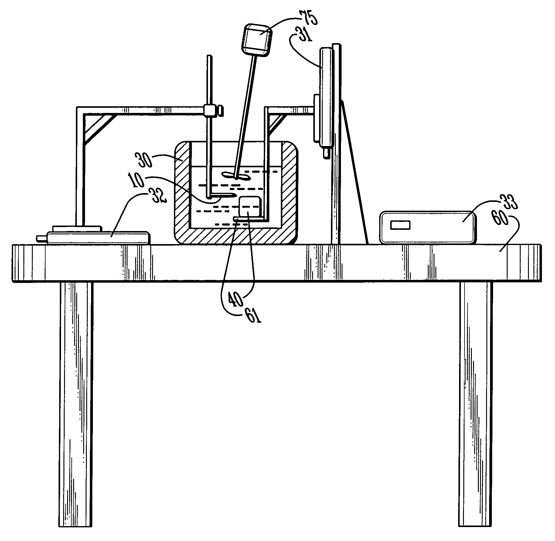
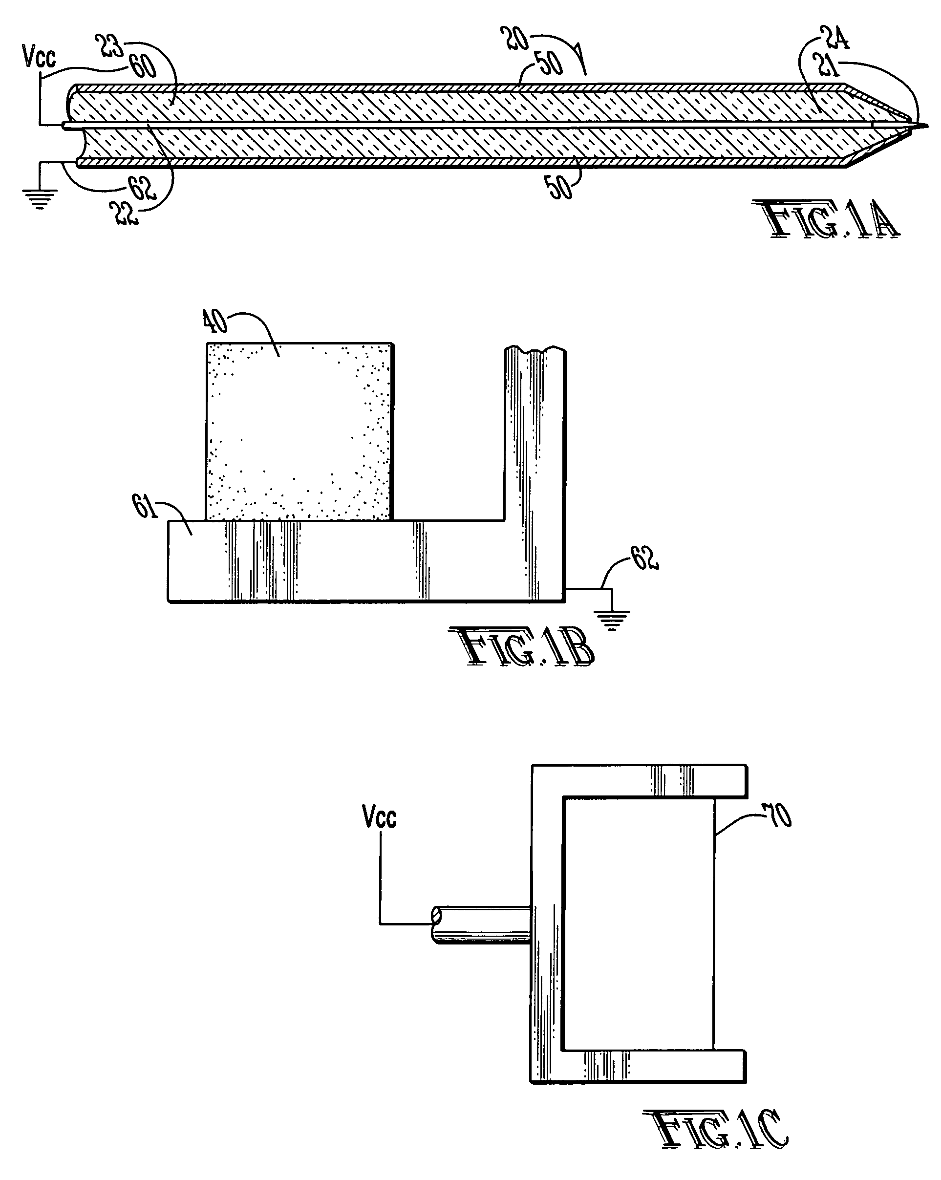
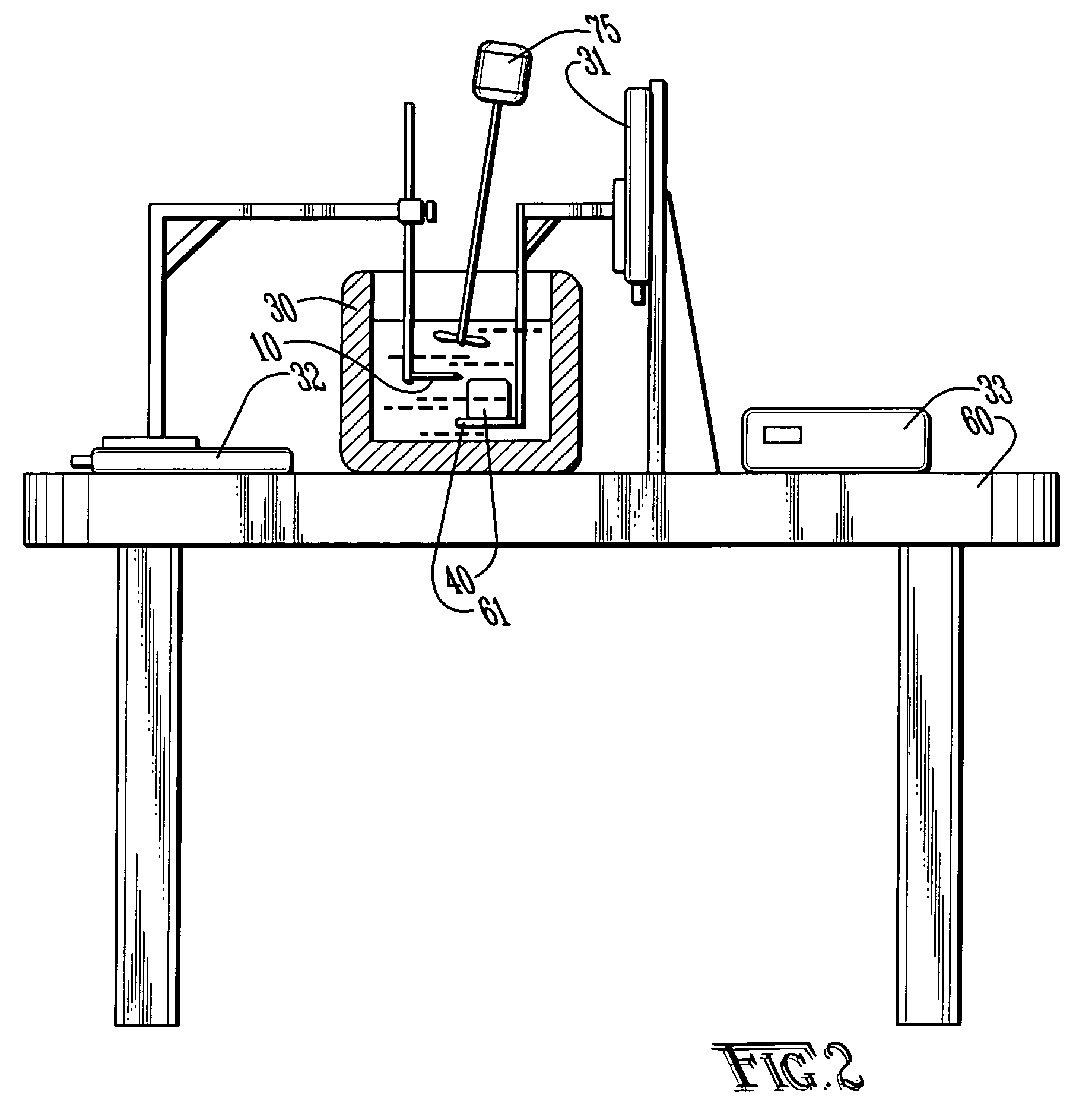
Tissue electro-sectioning apparatus
PatentInactiveUS20050220674A1
Innovation
- The use of electro-dissociation with a focused electromagnetic field to section fresh, unfixed tissues without mechanical or thermal damage, allowing for the preservation of tissue architecture and mRNA content, enabling high-resolution 2-D or 3-D reconstruction.

Analysis of nucleic acid molecules distributed on a surface or within a layer by sequencing with position identification
PatentWO2013150083A1
Innovation
- The method involves labeling nucleic acid molecules with two-dimensionally distributed oligonucleotide markers, allowing for their identification and analysis by hybridization, which enables the preservation of their original spatial distribution during transfer from a surface to another surface or into solution, using hybridization-based binding and subsequent covalent linking.


Quality Control Standards for Spatial Transcriptomics
Quality control standards for spatial transcriptomics represent a critical framework for ensuring data reliability and reproducibility when dividing tissue sections while preserving transcriptomic layers. These standards encompass multiple validation checkpoints throughout the experimental workflow, from initial tissue preparation to final data analysis.
RNA integrity assessment forms the cornerstone of quality control protocols. The RNA Integrity Number (RIN) should exceed 7.0 for optimal spatial transcriptomic analysis, with additional metrics including DV200 values above 50% to ensure sufficient transcript preservation. Post-sectioning RNA quality must be monitored through quantitative PCR of housekeeping genes and tissue-specific markers to verify that transcriptomic layers remain intact during the division process.
Morphological preservation standards require comprehensive histological evaluation using standard H&E staining protocols. Tissue architecture must maintain cellular boundaries, nuclear morphology, and extracellular matrix organization. Specific criteria include absence of ice crystal artifacts, minimal tissue folding, and preservation of tissue-specific structural features that correlate with expected transcriptomic patterns.
Spatial resolution validation involves establishing minimum spot-to-spot correlation thresholds and maximum acceptable cross-contamination rates between adjacent tissue regions. Current industry standards mandate correlation coefficients above 0.8 for technical replicates and contamination rates below 5% for neighboring spots. These metrics ensure that tissue division procedures do not compromise the spatial integrity of transcriptomic data.
Technical reproducibility standards encompass inter-batch variation coefficients, typically requiring less than 20% variation for highly expressed genes and 30% for moderately expressed transcripts. Quality control also includes assessment of capture efficiency, with acceptable rates ranging from 10-50% depending on the specific spatial transcriptomics platform employed.
Data completeness metrics establish minimum thresholds for gene detection rates, typically requiring identification of at least 1,000 unique genes per spot or region, with total UMI counts exceeding platform-specific benchmarks. These standards ensure sufficient transcriptomic depth for meaningful biological interpretation while maintaining spatial context integrity.
Integration Strategies for Multi-Modal Spatial Analysis
The integration of multi-modal spatial analysis represents a critical advancement in tissue section analysis, enabling researchers to combine transcriptomic data with complementary analytical modalities while maintaining spatial resolution. This approach addresses the fundamental challenge of preserving transcriptomic layer integrity during tissue division by establishing comprehensive data fusion frameworks that capture both molecular and morphological information simultaneously.
Contemporary integration strategies primarily focus on combining spatial transcriptomics with histological imaging, immunofluorescence microscopy, and mass spectrometry imaging. These multi-modal approaches create synergistic datasets that provide enhanced biological insights compared to single-modality analyses. The integration process typically involves sophisticated computational pipelines that align different data types spatially and temporally, ensuring that transcriptomic information remains accurately mapped to specific tissue regions throughout the sectioning process.
Advanced registration algorithms form the backbone of successful multi-modal integration, utilizing fiducial markers, anatomical landmarks, and computational image alignment techniques. These methods enable precise overlay of transcriptomic data with complementary modalities, creating comprehensive spatial maps that preserve the original tissue architecture. Machine learning approaches, particularly deep learning networks, have emerged as powerful tools for automated registration and data fusion, significantly improving accuracy and reducing processing time.
Cross-platform data standardization presents both opportunities and challenges in multi-modal integration. Standardized data formats and metadata schemas facilitate seamless integration across different analytical platforms, while normalization techniques ensure compatibility between diverse measurement scales and detection sensitivities. The development of universal coordinate systems and reference frameworks enables consistent spatial mapping across multiple analytical sessions and tissue sections.
Quality control mechanisms are essential for maintaining data integrity throughout the integration process. These include spatial correlation assessments, signal-to-noise ratio evaluations, and cross-modal validation procedures that verify the accuracy of integrated datasets. Automated quality metrics help identify potential artifacts or misalignments that could compromise the preservation of transcriptomic layers during tissue division and subsequent analysis.
Future integration strategies are evolving toward real-time multi-modal analysis platforms that can simultaneously capture and process multiple data streams during tissue sectioning procedures. These emerging approaches promise to further enhance the preservation of transcriptomic information while expanding the analytical capabilities available for comprehensive tissue characterization.
Unlock deeper insights with Patsnap Eureka Quick Research — get a full tech report to explore trends and direct your research. Try now!
Generate Your Research Report Instantly with AI Agent
Supercharge your innovation with Patsnap Eureka AI Agent Platform!