
Method for automatic sorting and representation of medical data sets
a medical data and automatic sorting technology, applied in the field of automatic sorting and representation of medical data sets, can solve the problems of inability to quickly change between the two views, time-consuming procedure for the physician, etc., and achieve the effect of convenient sorting and display of medical data sets
- Summary
- Abstract
- Description
- Claims
- Application Information
AI Technical Summary
Benefits of technology
Problems solved by technology
Method used
Image

Examples
Embodiment Construction
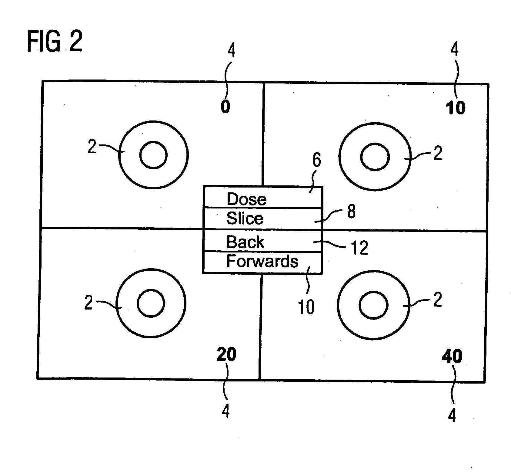
[0011] In a magnetic resonance stress examination, various doses of a stress agent are typically administered and magnetic resonance exposures are typically measured at various points of the heart. Slices are typically acquired at four different positions with each dose. A sequence of various images is acquired in succession at each slice position so that a time curve is documented. The corresponding image file denoting which slice belongs to which dose is stored in the header. For the physician, for generation of a finding it is now important either to show the same slice multiple times on the display medium for various doses of the stress means, or to display a number of slices for a dose on the display medium. Easy access to the slices measured in temporal succession is thereby desirable.
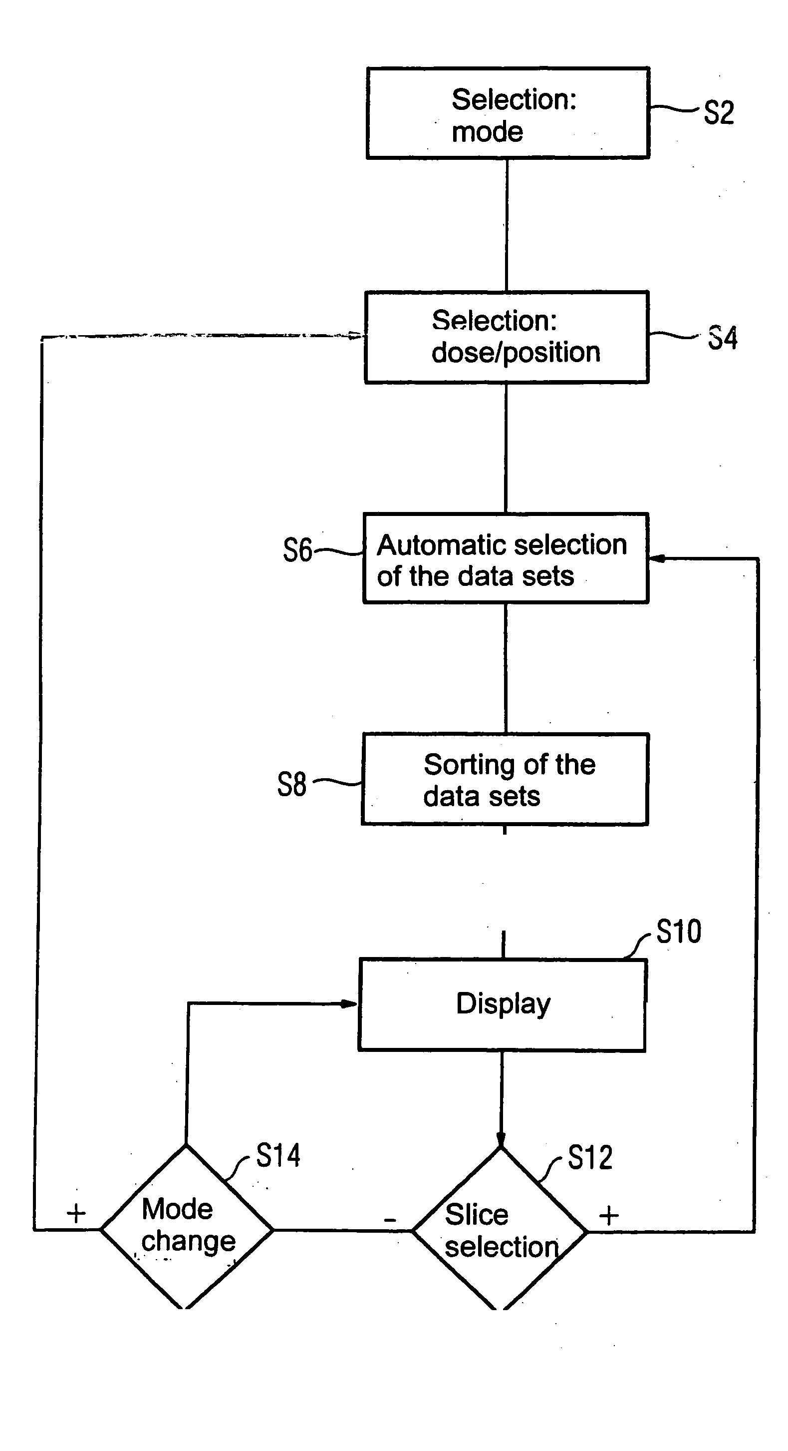
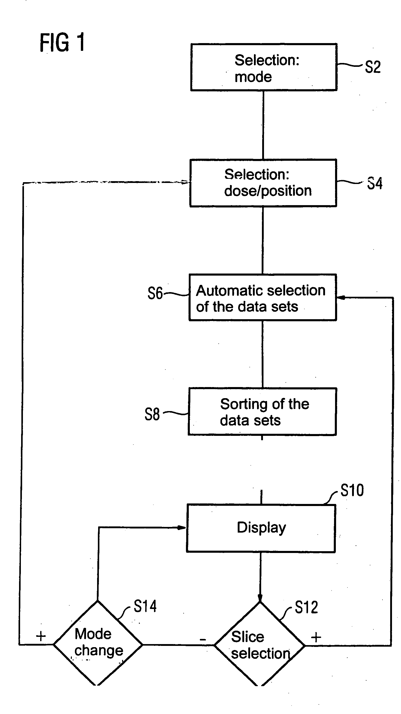
[0012] According to FIG. 1, the representation mode is selected by the physician in a first method step S2. This establishes whether various slice positions of various doses of the stress agent ...
PUM
 Abstract
Description
Claims
Application Information