
Method and apparatus for analysis of tissue microarrays
a tissue microarray and analysis method technology, applied in the field of image processing and image analysis, can solve the problems of loss of correspondence between serial sections, difficult alignment of tmas made from serial sections of blocks, and inconvenient imaging system
Patent Information
- Authority / Receiving Office
- US · United States
- Patent Type
- Patents(United States)
- Current Assignee / Owner
- Publication Date
- 2014-11-04
Smart Images
Figure 1 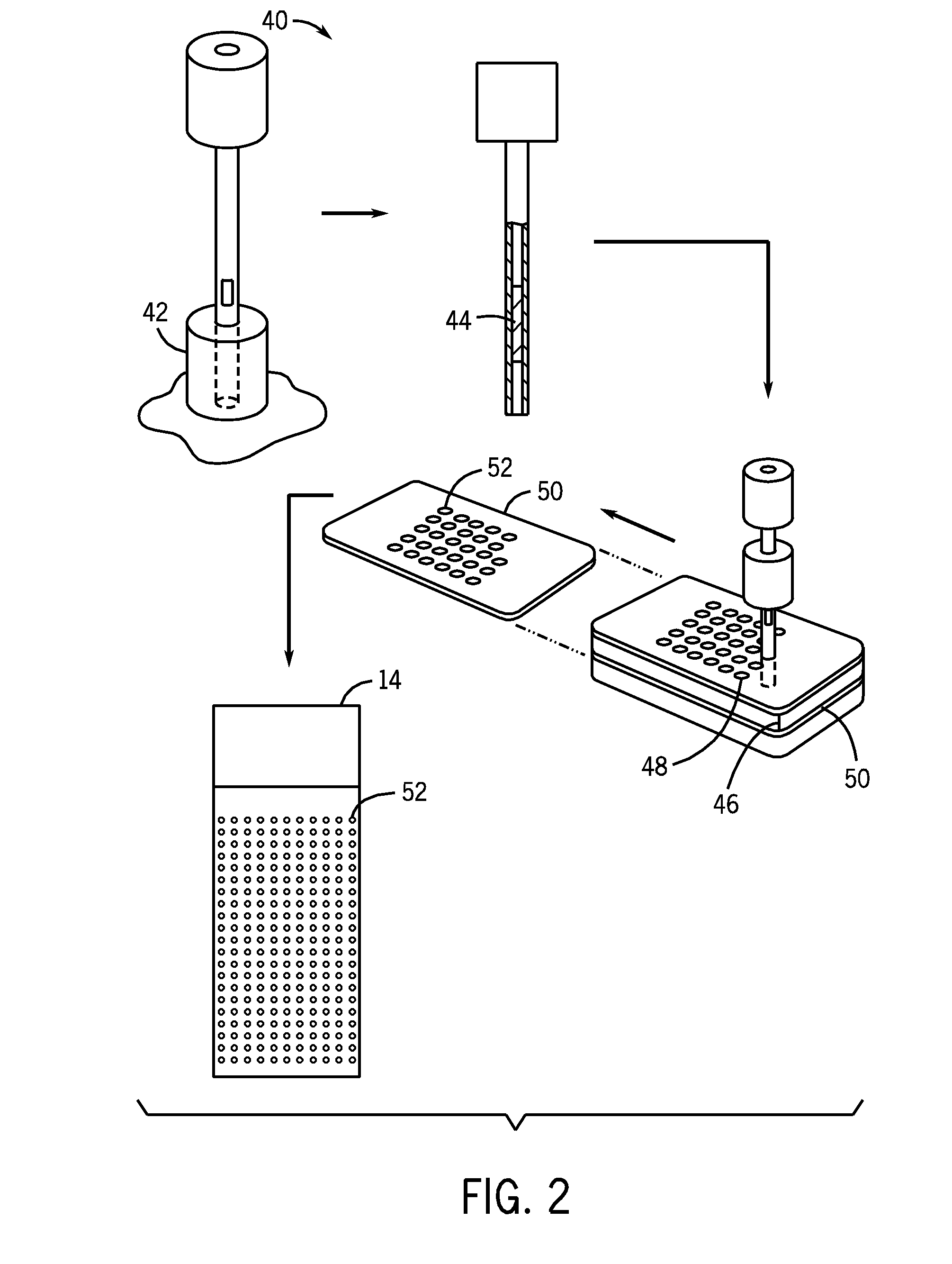
Figure 2 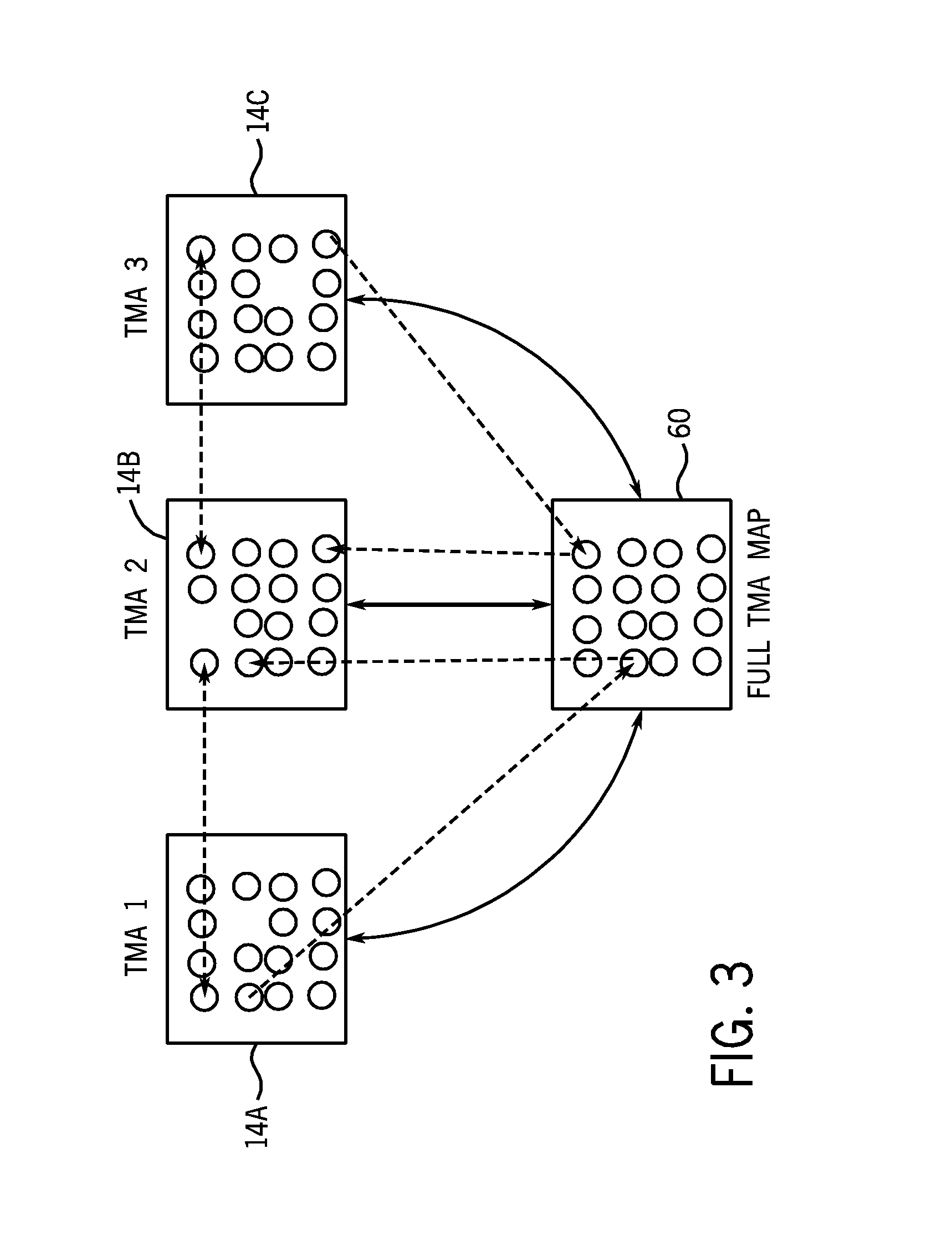
Figure 3
Abstract
Description
BACKGROUND
[0001] The invention relates generally to image processing and image analysis. More specifically, the present techniques relate to analysis of tissue microarrays made from serial sections of a multiple-tissue sample block.
[0002] Tissue microarrray (TMA) technology has become the standard in large-scale immunohistochemistry (IHC), fluorescent in situ hybridization (FISH), and mRNA in situ hybridization (RNA-ISH) studies for protein, DNA and RNA expression. To prepare the TMA slides, a tissue core is typically obtained from the patient tissue and inserted in a paraffin recipient block. The resulting recipient block typically has hundreds of tissue cores from multiple patients. This block may then be cut into sections that may be placed on glass slides for examination and imaging. In each of the serial sections, corresponding tissue cores belong to the same patient, and it is advantageous to be able to relate each patient core in one TMA slide to the corresponding one belonging...
Examples
examples
[0046]The following examples illustrate embodiments of the present techniques. In one example, ten TMA slides from a recipient tissue block, originally consisting of 217 breast tissue cores from 55 patients, were analyzed. Six of the slides were conjugated with immuno-fluorescent (IF) dyes and examined under fluorescent microscopy. The remaining four slides were examined with bright field microscopy after diaminobenzidine tetrahydrochloride (DAB) staining. The whole slide scan was not available for the DAB slides and one of the IF slides. Pathologists provided a TMA-map (shown in FIG. 7D) for the block. Serial numbers were automatically assigned to the images during scanning for identification. The tissue spots on four of the IF slides were labeled with AR, ER, p53, and Her2 biomarkers respectively and cores on the remaining two were used for multiplexing with multiple biomarkers. The DAB stains were also used to test for the same four biomarkers under bright field microscopy.
[0047]...