

Pediatric implications of P wave morphology changes
AUG 19, 20259 MIN READ
Generate Your Research Report Instantly with AI Agent
PatSnap Eureka helps you evaluate technical feasibility & market potential.
P Wave Morphology in Pediatrics: Background and Objectives
P wave morphology analysis in pediatric electrocardiography has emerged as a crucial area of study, offering valuable insights into cardiac function and potential abnormalities in children. The evolution of this field has been marked by significant advancements in technology and understanding of pediatric cardiac physiology. Historically, P wave analysis was primarily focused on adult populations, but recent decades have seen a shift towards recognizing its unique implications in pediatric cardiology.
The primary objective of studying P wave morphology changes in pediatrics is to enhance early detection and diagnosis of cardiac abnormalities specific to children. This includes identifying atrial enlargement, conduction disorders, and other structural or functional cardiac issues that may manifest differently in pediatric patients compared to adults. By understanding these changes, clinicians aim to improve risk stratification and guide more targeted therapeutic interventions in young patients.
Recent technological advancements have significantly contributed to the precision and reliability of P wave analysis in pediatric populations. High-resolution electrocardiography and advanced signal processing techniques have enabled more detailed examination of P wave characteristics, including duration, amplitude, and morphology. These improvements have led to a better understanding of normal P wave variations in different age groups within the pediatric population, from neonates to adolescents.
The growing interest in pediatric P wave morphology is driven by the recognition that early cardiac changes can have long-term implications for a child's health. Researchers and clinicians are increasingly focusing on how alterations in P wave characteristics might predict future cardiovascular risks or indicate the need for early interventions. This proactive approach aligns with the broader trend in pediatric medicine towards preventive care and early disease management.
Furthermore, the study of P wave morphology in pediatrics intersects with other areas of cardiac research, including genetics and developmental biology. There is a growing body of evidence suggesting that certain P wave abnormalities may be linked to genetic factors or developmental processes unique to childhood. This interdisciplinary approach is opening new avenues for understanding the complex interplay between genetic predisposition, cardiac development, and electrophysiological manifestations in children.
As research in this field progresses, there is an increasing emphasis on establishing standardized criteria for P wave analysis specific to pediatric populations. This standardization is crucial for accurate interpretation of electrocardiographic findings across different age groups and clinical settings. The ultimate goal is to develop robust, age-specific reference values and diagnostic criteria that can be universally applied in pediatric cardiology practice.
The primary objective of studying P wave morphology changes in pediatrics is to enhance early detection and diagnosis of cardiac abnormalities specific to children. This includes identifying atrial enlargement, conduction disorders, and other structural or functional cardiac issues that may manifest differently in pediatric patients compared to adults. By understanding these changes, clinicians aim to improve risk stratification and guide more targeted therapeutic interventions in young patients.
Recent technological advancements have significantly contributed to the precision and reliability of P wave analysis in pediatric populations. High-resolution electrocardiography and advanced signal processing techniques have enabled more detailed examination of P wave characteristics, including duration, amplitude, and morphology. These improvements have led to a better understanding of normal P wave variations in different age groups within the pediatric population, from neonates to adolescents.
The growing interest in pediatric P wave morphology is driven by the recognition that early cardiac changes can have long-term implications for a child's health. Researchers and clinicians are increasingly focusing on how alterations in P wave characteristics might predict future cardiovascular risks or indicate the need for early interventions. This proactive approach aligns with the broader trend in pediatric medicine towards preventive care and early disease management.
Furthermore, the study of P wave morphology in pediatrics intersects with other areas of cardiac research, including genetics and developmental biology. There is a growing body of evidence suggesting that certain P wave abnormalities may be linked to genetic factors or developmental processes unique to childhood. This interdisciplinary approach is opening new avenues for understanding the complex interplay between genetic predisposition, cardiac development, and electrophysiological manifestations in children.
As research in this field progresses, there is an increasing emphasis on establishing standardized criteria for P wave analysis specific to pediatric populations. This standardization is crucial for accurate interpretation of electrocardiographic findings across different age groups and clinical settings. The ultimate goal is to develop robust, age-specific reference values and diagnostic criteria that can be universally applied in pediatric cardiology practice.
Clinical Significance and Diagnostic Value
P wave morphology changes in pediatric patients hold significant clinical importance and diagnostic value in the field of pediatric cardiology. These alterations in the P wave, which represents atrial depolarization, can provide crucial insights into various cardiac conditions affecting children.
The clinical significance of P wave morphology changes in pediatrics lies in their ability to indicate underlying atrial abnormalities or conduction disturbances. These changes can be early markers of structural or functional cardiac issues that may not be immediately apparent through other diagnostic methods. For instance, P wave abnormalities may suggest atrial enlargement, which could be associated with conditions such as congenital heart defects or cardiomyopathies.
In terms of diagnostic value, P wave morphology analysis offers a non-invasive and cost-effective method for initial cardiac assessment in children. It can be readily obtained from standard 12-lead electrocardiograms (ECGs), making it an accessible tool for pediatric cardiologists and general practitioners alike. The ability to detect subtle changes in P wave characteristics can aid in early identification of potential cardiac issues, allowing for timely intervention and management.
P wave morphology changes can also provide valuable information about the progression of certain cardiac conditions in pediatric patients. For example, serial ECG evaluations focusing on P wave changes can help monitor the development of atrial remodeling in children with chronic cardiac conditions, such as hypertension or valvular heart disease.
Furthermore, the diagnostic value of P wave morphology extends to the differentiation of various arrhythmias in pediatric populations. Certain P wave patterns can be indicative of specific types of supraventricular tachycardias, helping clinicians distinguish between different arrhythmia mechanisms and guide appropriate treatment strategies.
In the context of congenital heart defects, P wave morphology analysis can contribute to the assessment of atrial situs and the identification of atrial isomerism. This information is crucial for accurate diagnosis and surgical planning in complex congenital heart anomalies.
The integration of P wave morphology analysis with other diagnostic modalities, such as echocardiography and cardiac MRI, can enhance the overall diagnostic accuracy in pediatric cardiology. This multimodal approach allows for a more comprehensive evaluation of cardiac structure and function, particularly in cases where P wave changes may be the first indication of an underlying cardiac abnormality.
The clinical significance of P wave morphology changes in pediatrics lies in their ability to indicate underlying atrial abnormalities or conduction disturbances. These changes can be early markers of structural or functional cardiac issues that may not be immediately apparent through other diagnostic methods. For instance, P wave abnormalities may suggest atrial enlargement, which could be associated with conditions such as congenital heart defects or cardiomyopathies.
In terms of diagnostic value, P wave morphology analysis offers a non-invasive and cost-effective method for initial cardiac assessment in children. It can be readily obtained from standard 12-lead electrocardiograms (ECGs), making it an accessible tool for pediatric cardiologists and general practitioners alike. The ability to detect subtle changes in P wave characteristics can aid in early identification of potential cardiac issues, allowing for timely intervention and management.
P wave morphology changes can also provide valuable information about the progression of certain cardiac conditions in pediatric patients. For example, serial ECG evaluations focusing on P wave changes can help monitor the development of atrial remodeling in children with chronic cardiac conditions, such as hypertension or valvular heart disease.
Furthermore, the diagnostic value of P wave morphology extends to the differentiation of various arrhythmias in pediatric populations. Certain P wave patterns can be indicative of specific types of supraventricular tachycardias, helping clinicians distinguish between different arrhythmia mechanisms and guide appropriate treatment strategies.
In the context of congenital heart defects, P wave morphology analysis can contribute to the assessment of atrial situs and the identification of atrial isomerism. This information is crucial for accurate diagnosis and surgical planning in complex congenital heart anomalies.
The integration of P wave morphology analysis with other diagnostic modalities, such as echocardiography and cardiac MRI, can enhance the overall diagnostic accuracy in pediatric cardiology. This multimodal approach allows for a more comprehensive evaluation of cardiac structure and function, particularly in cases where P wave changes may be the first indication of an underlying cardiac abnormality.
Current Challenges in Pediatric ECG Interpretation
Pediatric ECG interpretation presents unique challenges due to the dynamic nature of cardiac development in children. One of the primary difficulties lies in the wide range of normal variations observed in pediatric ECGs, which can often mimic pathological conditions in adults. This variability is particularly evident in P wave morphology, which undergoes significant changes throughout childhood and adolescence.
The interpretation of P wave morphology in pediatric patients is complicated by the fact that normal ranges for P wave amplitude, duration, and axis shift with age. Neonates and infants typically exhibit higher P wave amplitudes and shorter durations compared to older children and adults. As the child grows, these parameters gradually transition towards adult values, creating a moving target for what constitutes "normal" at any given age.
Another challenge is the lack of standardized, age-specific criteria for P wave abnormalities in children. While adult ECG criteria are well-established, applying these same standards to pediatric patients can lead to misdiagnosis or overdiagnosis of atrial abnormalities. This is particularly problematic when assessing conditions such as atrial enlargement or intra-atrial conduction delays, where adult criteria may not be directly applicable to younger patients.
The presence of physiological right ventricular dominance in infants and young children further complicates P wave interpretation. This normal developmental feature can result in rightward P wave axis and increased amplitude in right precordial leads, potentially mimicking right atrial enlargement or other pathological conditions when viewed through an adult lens.
Technological limitations also contribute to the challenges in pediatric ECG interpretation. Many ECG machines and automated interpretation algorithms are primarily designed and validated for adult populations. These systems may not accurately account for age-related variations in P wave morphology, leading to potential errors in automated measurements and interpretations when applied to pediatric ECGs.
The impact of respiratory variations on P wave morphology is more pronounced in children, particularly in infants and young children with higher respiratory rates. This can lead to beat-to-beat variability in P wave appearance, making consistent interpretation challenging and potentially masking subtle abnormalities.
Lastly, the scarcity of large-scale, longitudinal studies on P wave morphology changes throughout childhood limits our understanding of the full spectrum of normal variations. This knowledge gap hampers the development of robust, age-specific criteria for identifying truly pathological P wave changes in pediatric patients.
Addressing these challenges requires a multifaceted approach, including the development of age-specific normative data, refinement of ECG acquisition and analysis techniques for pediatric patients, and improved education for clinicians on the nuances of pediatric ECG interpretation, particularly with respect to P wave morphology changes.
The interpretation of P wave morphology in pediatric patients is complicated by the fact that normal ranges for P wave amplitude, duration, and axis shift with age. Neonates and infants typically exhibit higher P wave amplitudes and shorter durations compared to older children and adults. As the child grows, these parameters gradually transition towards adult values, creating a moving target for what constitutes "normal" at any given age.
Another challenge is the lack of standardized, age-specific criteria for P wave abnormalities in children. While adult ECG criteria are well-established, applying these same standards to pediatric patients can lead to misdiagnosis or overdiagnosis of atrial abnormalities. This is particularly problematic when assessing conditions such as atrial enlargement or intra-atrial conduction delays, where adult criteria may not be directly applicable to younger patients.
The presence of physiological right ventricular dominance in infants and young children further complicates P wave interpretation. This normal developmental feature can result in rightward P wave axis and increased amplitude in right precordial leads, potentially mimicking right atrial enlargement or other pathological conditions when viewed through an adult lens.
Technological limitations also contribute to the challenges in pediatric ECG interpretation. Many ECG machines and automated interpretation algorithms are primarily designed and validated for adult populations. These systems may not accurately account for age-related variations in P wave morphology, leading to potential errors in automated measurements and interpretations when applied to pediatric ECGs.
The impact of respiratory variations on P wave morphology is more pronounced in children, particularly in infants and young children with higher respiratory rates. This can lead to beat-to-beat variability in P wave appearance, making consistent interpretation challenging and potentially masking subtle abnormalities.
Lastly, the scarcity of large-scale, longitudinal studies on P wave morphology changes throughout childhood limits our understanding of the full spectrum of normal variations. This knowledge gap hampers the development of robust, age-specific criteria for identifying truly pathological P wave changes in pediatric patients.
Addressing these challenges requires a multifaceted approach, including the development of age-specific normative data, refinement of ECG acquisition and analysis techniques for pediatric patients, and improved education for clinicians on the nuances of pediatric ECG interpretation, particularly with respect to P wave morphology changes.
Existing Methods for P Wave Morphology Assessment
01 Analysis of P wave morphology changes
Methods and systems for analyzing changes in P wave morphology to detect and diagnose cardiac conditions. This involves examining variations in the shape, duration, and amplitude of P waves over time, which can indicate atrial abnormalities or other cardiac issues.- Analysis of P wave morphology changes: Methods and systems for analyzing changes in P wave morphology to detect and diagnose cardiac conditions. This involves examining variations in the shape, duration, and amplitude of P waves over time, which can indicate atrial abnormalities or other cardiac issues.
- Machine learning for P wave analysis: Application of machine learning algorithms to analyze P wave morphology changes. These techniques can improve the accuracy of detecting subtle alterations in P wave characteristics, potentially leading to earlier diagnosis of cardiac conditions.
- Real-time monitoring of P wave changes: Development of systems for continuous, real-time monitoring of P wave morphology changes. These systems can provide immediate alerts to healthcare providers when significant alterations in P wave characteristics are detected, enabling prompt intervention.
- P wave morphology in atrial fibrillation prediction: Utilization of P wave morphology changes as a predictor for atrial fibrillation. By analyzing specific alterations in P wave characteristics, these methods aim to identify patients at higher risk of developing atrial fibrillation before its onset.
- Integration of P wave analysis in ECG devices: Incorporation of advanced P wave morphology analysis capabilities into ECG devices and wearable health monitors. This integration allows for more comprehensive cardiac assessments in both clinical and home settings, improving the detection of various cardiac abnormalities.
02 Machine learning for P wave analysis
Application of machine learning algorithms to analyze P wave morphology changes. These techniques can improve the accuracy of detecting subtle alterations in P wave characteristics, potentially leading to earlier diagnosis of cardiac conditions.Expand Specific Solutions03 Real-time monitoring of P wave changes
Development of systems for continuous, real-time monitoring of P wave morphology changes. These systems can provide immediate alerts to healthcare providers when significant alterations in P wave characteristics are detected, enabling prompt intervention.Expand Specific Solutions04 P wave morphology in specific cardiac conditions
Studies focusing on P wave morphology changes associated with specific cardiac conditions such as atrial fibrillation, atrial flutter, and other arrhythmias. This research aims to identify unique P wave patterns that can serve as early indicators of these conditions.Expand Specific Solutions05 Integration of P wave analysis in ECG devices
Incorporation of advanced P wave morphology analysis capabilities into ECG devices and monitoring systems. This integration allows for more comprehensive cardiac assessments and improved detection of subtle changes in atrial activity.Expand Specific Solutions
Key Players in Pediatric Cardiology Research
The pediatric implications of P wave morphology changes represent an evolving field in cardiac electrophysiology. The competitive landscape is characterized by a mix of established pharmaceutical companies, innovative biotechnology firms, and academic research institutions. The market is in its early growth stage, with increasing research focus but limited commercialization. Key players like Merck Patent GmbH, Vertex Pharmaceuticals, and Bardy Diagnostics are investing in R&D to develop advanced diagnostic tools and therapies. Academic institutions such as the University of Copenhagen and Cincinnati Children's Hospital Medical Center are contributing significant research. While the technology is still maturing, there is growing interest due to its potential impact on pediatric cardiac care and early disease detection.
Pacesetter, Inc.
Technical Solution: Pacesetter, Inc. has developed advanced algorithms for analyzing P wave morphology changes in pediatric patients. Their technology utilizes machine learning techniques to process electrocardiogram (ECG) data and detect subtle alterations in P wave characteristics. The system can identify changes in P wave amplitude, duration, and axis, which may indicate atrial abnormalities or conduction disorders in children[1]. The algorithm incorporates age-specific normative data to account for the natural variations in P wave morphology throughout childhood development[3]. This allows for more accurate interpretation of ECG findings in pediatric populations.
Strengths: Specialized focus on pediatric ECG analysis, incorporation of age-specific data. Weaknesses: May require extensive clinical validation in diverse pediatric populations.
Bardy Diagnostics, Inc.
Technical Solution: Bardy Diagnostics has developed a novel wearable ECG monitor specifically designed for pediatric patients. The device utilizes advanced signal processing algorithms to capture and analyze P wave morphology changes in children. The monitor's unique placement on the sternum allows for optimal P wave detection, even in active pediatric patients[2]. The system employs cloud-based analytics to process the collected data and identify subtle P wave alterations that may indicate atrial abnormalities or conduction disorders. The technology also incorporates machine learning algorithms to improve detection accuracy over time, adapting to individual patient characteristics[4].
Strengths: Pediatric-specific wearable design, continuous monitoring capability. Weaknesses: May be limited by patient compliance and comfort issues in long-term use.
Innovative Approaches in P Wave Analysis
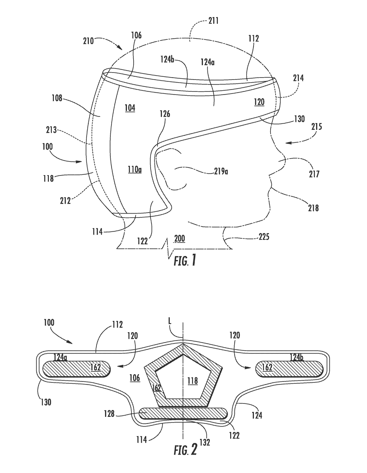
Force Distribution Method and Apparatus for Neonates at Risk of Cranial Molding
PatentActiveUS20160038332A1
Innovation
- A force-distributing cranial support apparatus that disperses external forces across a larger area, reducing pressure on the infant's head and promoting proper head development, designed to be secured on the occipital region and forehead, using a gel assembly and padded layers to prevent and treat cranial molding and skin breakdown.


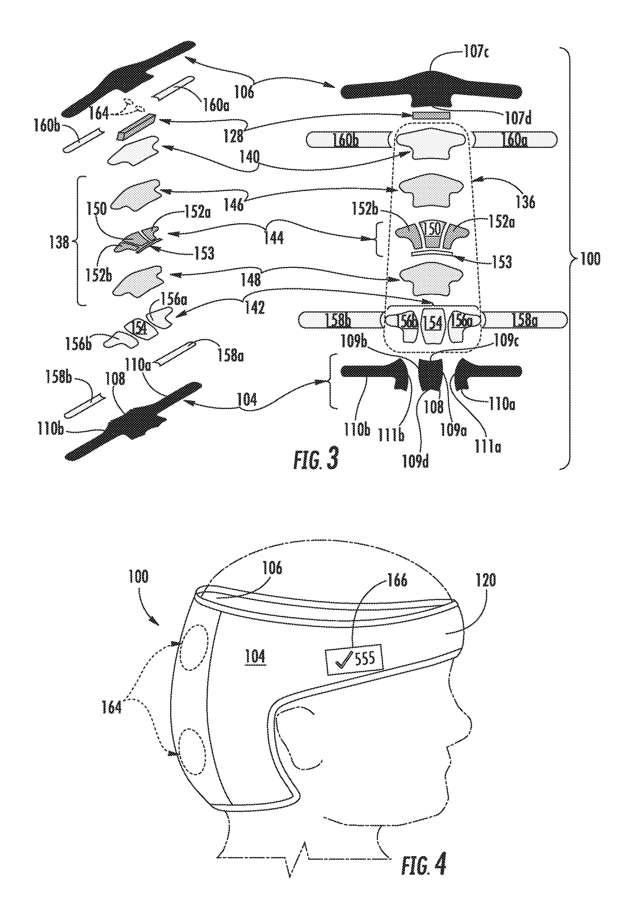
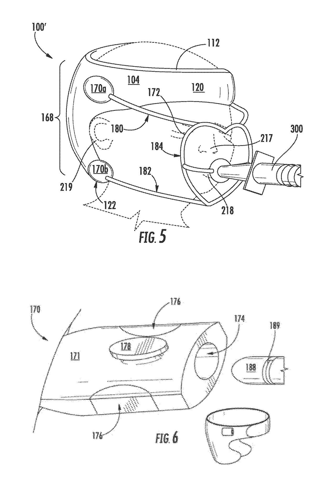
Force distribution method and apparatus for neonates at risk of cranial molding
PatentActiveUS20180344505A1
Innovation
- A force-distributing cranial support that disperses external forces across a larger area, reducing pressure on the infant's head and promoting proper head development, designed to fit various head sizes and shapes, with a gel assembly and adjustable straps to secure the device.

Age-Specific P Wave Morphology Standards
The establishment of age-specific P wave morphology standards is crucial for accurately interpreting electrocardiograms (ECGs) in pediatric populations. These standards account for the developmental changes in cardiac anatomy and physiology that occur throughout childhood and adolescence, which significantly impact P wave characteristics.
In infants and young children, the P wave typically appears more prominent and peaked due to the relative right atrial dominance. As children grow, the P wave morphology gradually evolves, reflecting the changing balance between right and left atrial contributions to atrial depolarization. By adolescence, the P wave morphology begins to resemble that of adults, with a more balanced contribution from both atria.
Age-specific standards for P wave amplitude, duration, and axis have been developed through extensive research and analysis of large pediatric ECG databases. These standards are typically presented as percentile charts or tables, stratified by age groups and gender. For instance, P wave amplitude tends to be higher in younger children and gradually decreases with age, while P wave duration generally increases as children grow older.
The normal range for P wave axis also shifts with age, typically starting more rightward in infants and gradually moving leftward as children mature. This shift reflects the changing orientation of the heart within the chest cavity as children grow. Understanding these age-related changes is essential for accurately identifying abnormal P wave morphologies that may indicate underlying cardiac pathologies.
Importantly, these age-specific standards must account for variations in lead placement and chest wall thickness, which can significantly affect P wave measurements in pediatric patients. Standardized lead placement techniques and adjustment factors have been developed to ensure consistent and accurate P wave analysis across different age groups.
Clinicians and researchers have also established criteria for identifying abnormal P wave morphologies in pediatric populations, such as P pulmonale, P mitrale, and interatrial conduction delays. These criteria are tailored to different age groups, recognizing that what may be considered abnormal in an adolescent might be within normal limits for an infant or toddler.
The implementation of age-specific P wave morphology standards in clinical practice and research settings has significantly improved the accuracy of ECG interpretation in pediatric cardiology. These standards serve as a crucial reference point for identifying potential atrial abnormalities, conduction disorders, and other cardiac conditions in children of all ages.
In infants and young children, the P wave typically appears more prominent and peaked due to the relative right atrial dominance. As children grow, the P wave morphology gradually evolves, reflecting the changing balance between right and left atrial contributions to atrial depolarization. By adolescence, the P wave morphology begins to resemble that of adults, with a more balanced contribution from both atria.
Age-specific standards for P wave amplitude, duration, and axis have been developed through extensive research and analysis of large pediatric ECG databases. These standards are typically presented as percentile charts or tables, stratified by age groups and gender. For instance, P wave amplitude tends to be higher in younger children and gradually decreases with age, while P wave duration generally increases as children grow older.
The normal range for P wave axis also shifts with age, typically starting more rightward in infants and gradually moving leftward as children mature. This shift reflects the changing orientation of the heart within the chest cavity as children grow. Understanding these age-related changes is essential for accurately identifying abnormal P wave morphologies that may indicate underlying cardiac pathologies.
Importantly, these age-specific standards must account for variations in lead placement and chest wall thickness, which can significantly affect P wave measurements in pediatric patients. Standardized lead placement techniques and adjustment factors have been developed to ensure consistent and accurate P wave analysis across different age groups.
Clinicians and researchers have also established criteria for identifying abnormal P wave morphologies in pediatric populations, such as P pulmonale, P mitrale, and interatrial conduction delays. These criteria are tailored to different age groups, recognizing that what may be considered abnormal in an adolescent might be within normal limits for an infant or toddler.
The implementation of age-specific P wave morphology standards in clinical practice and research settings has significantly improved the accuracy of ECG interpretation in pediatric cardiology. These standards serve as a crucial reference point for identifying potential atrial abnormalities, conduction disorders, and other cardiac conditions in children of all ages.
Ethical Considerations in Pediatric Cardiac Research
Ethical considerations in pediatric cardiac research involving P wave morphology changes are of paramount importance due to the vulnerability of the pediatric population and the potential long-term implications of cardiac interventions. The primary ethical principle guiding such research is the protection of children's rights and well-being, which must be balanced against the potential benefits of advancing medical knowledge and improving cardiac care for pediatric patients.
One of the key ethical challenges in this field is obtaining informed consent. Given that the subjects are minors, researchers must navigate the complex process of obtaining parental or guardian consent while also considering the child's assent when appropriate. This process requires clear communication of the research objectives, potential risks, and benefits in a manner that is understandable to both parents and children of varying ages and cognitive abilities.
The principle of minimal risk is particularly crucial in pediatric cardiac research. Investigators must ensure that any interventions or procedures related to studying P wave morphology changes do not expose children to risks that are greater than those encountered in daily life or during routine medical examinations. This principle may limit the types of studies that can be ethically conducted, potentially slowing the pace of research but safeguarding the well-being of young participants.
Confidentiality and privacy protection are also significant ethical concerns, especially given the sensitive nature of cardiac data and the potential for long-term implications of early-life cardiac findings. Researchers must implement robust data protection measures and consider the implications of storing and sharing pediatric cardiac data over extended periods.
The ethical framework for pediatric cardiac research must also address the issue of equitable subject selection. Researchers should strive to include a diverse range of pediatric participants to ensure that the benefits and risks of research are distributed fairly across different demographic groups. However, this must be balanced against the need to protect vulnerable populations from exploitation or undue burden.
Long-term follow-up considerations present another ethical challenge. Given that P wave morphology changes in childhood may have implications for adult cardiac health, researchers have an ethical obligation to consider how findings will be communicated and managed over time. This includes planning for the transition of care as pediatric subjects reach adulthood and ensuring that important health information is not lost in the process.
Lastly, the ethical conduct of pediatric cardiac research requires careful consideration of the risk-benefit ratio. While the potential for improving cardiac care for children is significant, researchers must continually assess whether the anticipated benefits justify the risks and burdens placed on young participants and their families. This ongoing evaluation should inform study design, recruitment practices, and the overall ethical framework of the research endeavor.
One of the key ethical challenges in this field is obtaining informed consent. Given that the subjects are minors, researchers must navigate the complex process of obtaining parental or guardian consent while also considering the child's assent when appropriate. This process requires clear communication of the research objectives, potential risks, and benefits in a manner that is understandable to both parents and children of varying ages and cognitive abilities.
The principle of minimal risk is particularly crucial in pediatric cardiac research. Investigators must ensure that any interventions or procedures related to studying P wave morphology changes do not expose children to risks that are greater than those encountered in daily life or during routine medical examinations. This principle may limit the types of studies that can be ethically conducted, potentially slowing the pace of research but safeguarding the well-being of young participants.
Confidentiality and privacy protection are also significant ethical concerns, especially given the sensitive nature of cardiac data and the potential for long-term implications of early-life cardiac findings. Researchers must implement robust data protection measures and consider the implications of storing and sharing pediatric cardiac data over extended periods.
The ethical framework for pediatric cardiac research must also address the issue of equitable subject selection. Researchers should strive to include a diverse range of pediatric participants to ensure that the benefits and risks of research are distributed fairly across different demographic groups. However, this must be balanced against the need to protect vulnerable populations from exploitation or undue burden.
Long-term follow-up considerations present another ethical challenge. Given that P wave morphology changes in childhood may have implications for adult cardiac health, researchers have an ethical obligation to consider how findings will be communicated and managed over time. This includes planning for the transition of care as pediatric subjects reach adulthood and ensuring that important health information is not lost in the process.
Lastly, the ethical conduct of pediatric cardiac research requires careful consideration of the risk-benefit ratio. While the potential for improving cardiac care for children is significant, researchers must continually assess whether the anticipated benefits justify the risks and burdens placed on young participants and their families. This ongoing evaluation should inform study design, recruitment practices, and the overall ethical framework of the research endeavor.
Unlock deeper insights with PatSnap Eureka Quick Research — get a full tech report to explore trends and direct your research. Try now!
Generate Your Research Report Instantly with AI Agent
Supercharge your innovation with PatSnap Eureka AI Agent Platform!