
Method and apparatus for repairing the mid-food region via an intramedullary nail
a technology of intramedullary nail and midfoot, which is applied in the direction of non-surgical orthopedic devices, osteosynthesis devices, fasteners, etc., can solve the problems of deteriorating the bones in the foot, limiting diabetic foot treatment to date, and collapse of the foo
- Summary
- Abstract
- Description
- Claims
- Application Information
AI Technical Summary
Benefits of technology
Problems solved by technology
Method used
Image

Examples
Embodiment Construction
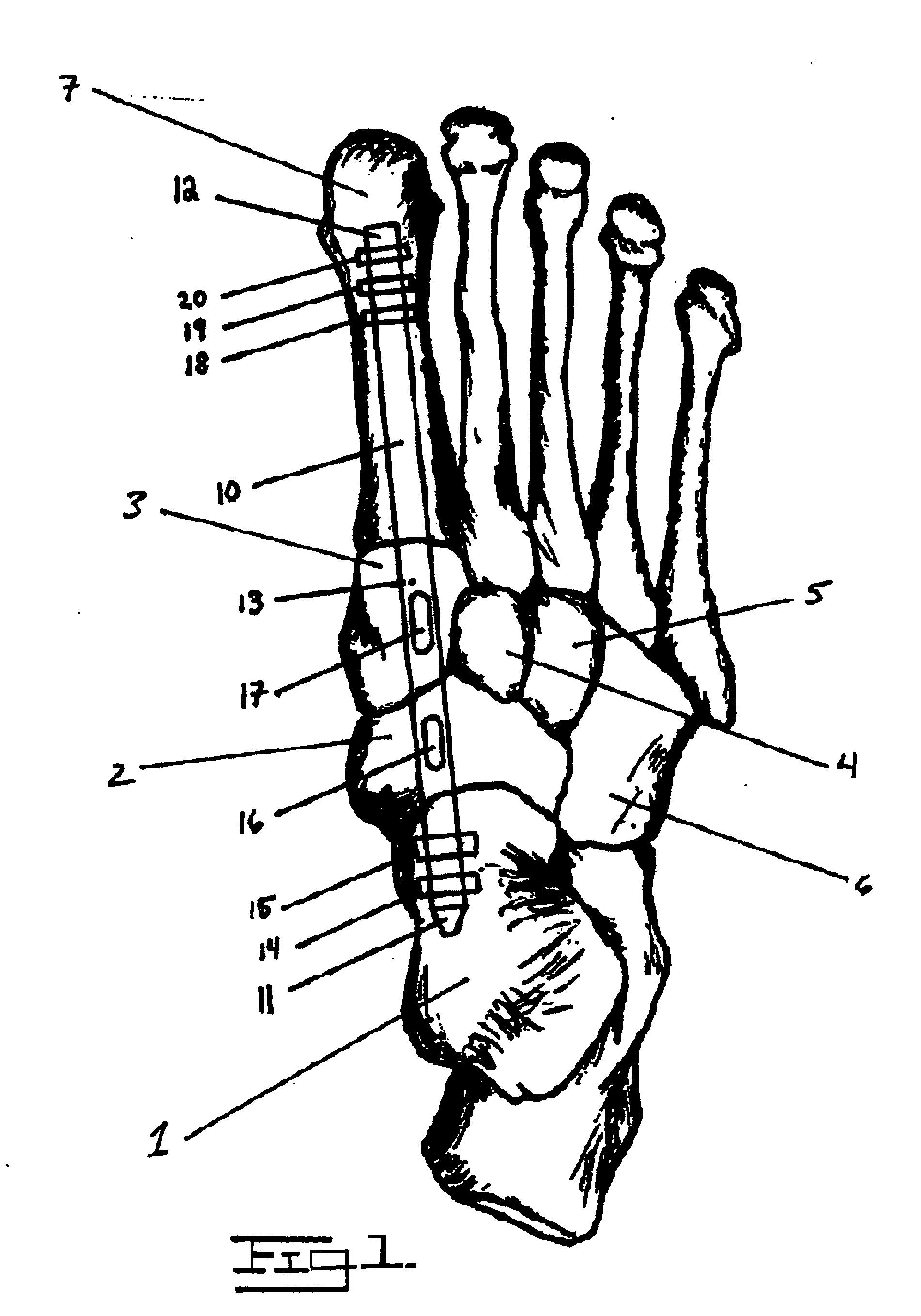
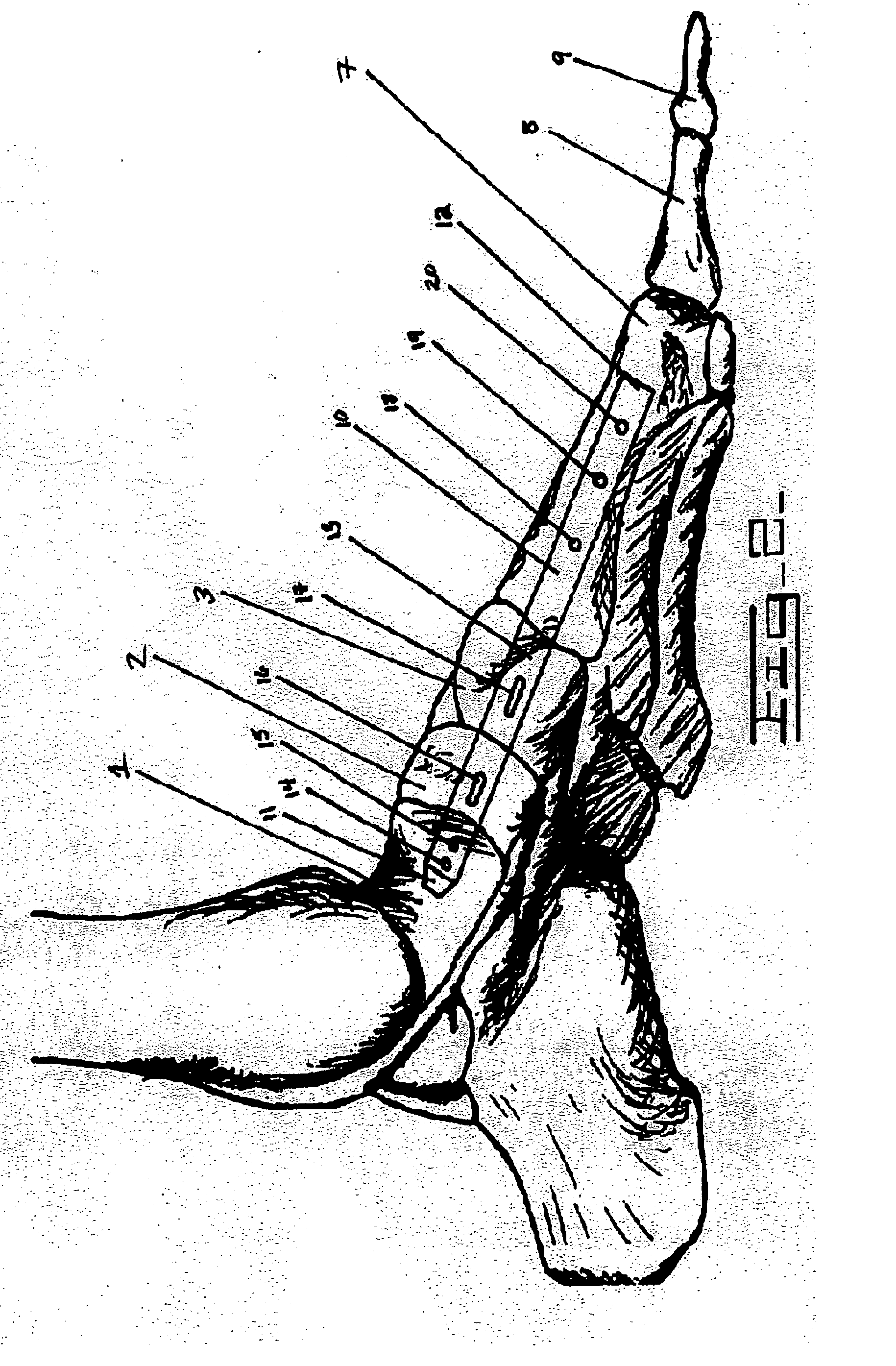
[0052] The following description will explain the embodiments of the invention, beginning with a device used for treating deteriorating or damaged bones in the medial column of a human foot, and concluding with the method for treating deteriorating or damaged bones in the medial column of a human foot. Both the first and second figures are skeletal outlines of a human foot, depicting the device therein and the resulting orientation of the bones by using the method specified below.
[0053]FIG. 1 is a top down view of a right human foot, depicting the bones therein and an implant or intramedullary nail 10 running through the mid-foot region. The implant or intramedullary nail 10 runs through the medullary canal of first metatarsal 7, medial cuneiform 3, navicular 2, and talus bone 1. The talus bone 1 makes up the lower part of the ankle joint where the proximal end 11 of the implant or intramedullary nail 10 is attached with at least one fastener (or locking screw, with two depicted in...
PUM
 Abstract
Description
Claims
Application Information