Echogenicity in Evaluating Embryonic Development Anomalies
JAN 20, 20269 MIN READ
Generate Your Research Report Instantly with AI Agent
PatSnap Eureka helps you evaluate technical feasibility & market potential.
Echogenicity in Embryonic Development: Background and Objectives
Echogenicity, defined as the ability of tissue to reflect ultrasound waves, has emerged as a fundamental parameter in prenatal diagnostic imaging since the widespread adoption of ultrasonography in obstetric practice during the 1970s. The varying acoustic properties of embryonic tissues create distinct echo patterns that enable clinicians to visualize and assess developmental structures non-invasively. Early observations revealed that abnormal echogenicity patterns often correlate with structural and chromosomal anomalies, establishing this parameter as a critical diagnostic marker in first-trimester screening protocols.
The evolution of ultrasound technology has significantly enhanced the detection capabilities of subtle echogenic variations. High-frequency transvaginal probes and advanced image processing algorithms now permit detailed visualization of embryonic structures as early as six weeks of gestation. This technological progression has transformed echogenicity assessment from a qualitative observation into a quantifiable diagnostic tool, enabling earlier detection of developmental abnormalities and improving clinical decision-making processes.
Current research objectives focus on establishing standardized echogenicity measurement protocols and defining normative reference ranges across different gestational ages. The primary goal is to develop reliable predictive models that can differentiate between physiological variations and pathological conditions. Particular emphasis is placed on characterizing echogenic patterns associated with neural tube defects, cardiac malformations, and chromosomal aneuploidies, which represent the most prevalent categories of embryonic anomalies.
Another critical objective involves integrating artificial intelligence and machine learning algorithms to enhance diagnostic accuracy. These computational approaches aim to identify subtle echogenic signatures that may escape human visual detection, potentially enabling earlier intervention and improved prognostic assessment. Additionally, research efforts are directed toward understanding the biological mechanisms underlying abnormal echogenicity, including cellular composition changes, tissue mineralization patterns, and vascular development alterations.
The ultimate goal is to establish echogenicity evaluation as a standardized, reproducible, and highly sensitive screening tool that can be seamlessly integrated into routine prenatal care protocols, thereby reducing the incidence of undetected developmental anomalies and improving maternal-fetal outcomes through timely clinical intervention.
Clinical Demand for Embryonic Anomaly Detection
The clinical demand for embryonic anomaly detection has intensified significantly in recent decades, driven by advances in prenatal care standards and evolving patient expectations. Early identification of developmental abnormalities during the first trimester has become a critical component of obstetric practice, enabling timely clinical decision-making and appropriate counseling for expectant parents. The ability to detect structural and chromosomal anomalies at earlier gestational ages directly impacts pregnancy management strategies and outcomes.
Echogenicity assessment represents a fundamental diagnostic approach in evaluating embryonic development, as variations in tissue acoustic properties often correlate with underlying pathological conditions. Healthcare providers increasingly rely on ultrasound imaging to screen for neural tube defects, cardiac malformations, skeletal dysplasias, and other congenital abnormalities. The non-invasive nature of ultrasound technology, combined with its real-time imaging capabilities, positions it as the primary modality for routine prenatal screening across diverse healthcare settings.
Current clinical protocols emphasize the importance of standardized echogenicity evaluation during critical developmental windows. However, significant challenges persist in achieving consistent diagnostic accuracy, particularly in distinguishing between normal physiological variations and pathological changes. The subjective nature of echogenicity interpretation, coupled with operator-dependent variability, creates substantial demand for improved assessment methodologies and objective quantification techniques.
The growing prevalence of advanced maternal age pregnancies has further amplified the need for reliable embryonic anomaly detection systems. Healthcare systems worldwide face increasing pressure to provide comprehensive screening services while managing resource constraints and maintaining cost-effectiveness. This demographic shift, combined with heightened awareness of prenatal diagnostic options, has created a substantial market for enhanced ultrasound technologies and decision-support tools.
Regulatory bodies and professional societies continue to refine guidelines for embryonic anomaly screening, establishing quality benchmarks and performance standards. These evolving requirements drive continuous innovation in imaging technologies and interpretation algorithms, creating sustained demand for research into echogenicity-based diagnostic approaches that can deliver higher sensitivity and specificity while reducing false-positive rates.
Current Echogenicity Assessment: Status and Challenges
Echogenicity assessment has become a fundamental component of prenatal ultrasound examination for detecting embryonic development anomalies. Current clinical practice relies primarily on B-mode ultrasound imaging, where tissue echogenicity patterns serve as critical indicators of normal versus abnormal embryonic structures. The grayscale variations in ultrasound images reflect differences in acoustic impedance between tissues, enabling clinicians to identify structural abnormalities, growth restrictions, and developmental defects during early pregnancy stages.
Despite widespread clinical adoption, echogenicity evaluation faces significant technical limitations that compromise diagnostic accuracy. The subjective nature of image interpretation remains a primary challenge, as echogenicity assessment depends heavily on operator experience and individual judgment. Different sonographers may interpret the same echogenic patterns differently, leading to inconsistent diagnostic outcomes. This variability is particularly problematic in borderline cases where subtle echogenicity changes may indicate early-stage developmental anomalies.
Image quality variability presents another substantial obstacle in current practice. Factors including maternal body habitus, gestational age, fetal position, and equipment specifications significantly influence echogenicity visualization. Adipose tissue in obese patients attenuates ultrasound waves, reducing image resolution and making it difficult to distinguish normal from abnormal echogenic patterns. Additionally, the limited penetration depth of high-frequency transducers restricts detailed assessment of deeper embryonic structures.
Standardization deficiencies further complicate echogenicity assessment across different clinical settings. The absence of universally accepted quantitative metrics for echogenicity measurement results in predominantly qualitative descriptions such as hyperechoic, hypoechoic, or isoechoic. These descriptive terms lack precision and reproducibility, hindering effective communication between healthcare providers and limiting the development of standardized diagnostic protocols.
Technological constraints in current ultrasound systems also impede comprehensive evaluation. Real-time imaging capabilities are limited by temporal resolution trade-offs, and three-dimensional reconstruction of echogenic structures remains computationally intensive. Furthermore, distinguishing between physiological variations and pathological changes in echogenicity patterns during rapid embryonic development stages requires sophisticated analytical capabilities that exceed current conventional ultrasound technology.
Mainstream Echogenicity Evaluation Solutions
01 Ultrasound contrast agents for enhanced echogenicity
Contrast agents containing microbubbles or nanoparticles are used to enhance echogenicity in ultrasound imaging. These agents improve visualization of blood flow, tissue perfusion, and organ structures by increasing the acoustic impedance difference between tissues. The contrast agents can be formulated with various shell materials and gas cores to optimize their acoustic properties and stability.- Ultrasound contrast agents for enhanced echogenicity: Contrast agents containing microbubbles or nanoparticles are used to enhance echogenicity in ultrasound imaging. These agents improve visualization of blood flow, tissue perfusion, and organ structures by increasing the acoustic impedance difference between tissues. The contrast agents can be formulated with various shell materials and gas cores to optimize their acoustic properties and stability.
- Echogenic medical devices and implants: Medical devices such as catheters, needles, and implants are designed with echogenic properties to improve their visibility during ultrasound-guided procedures. These devices incorporate materials or surface modifications that enhance ultrasound reflection, allowing clinicians to accurately track device placement and positioning in real-time imaging. Various coating techniques and material compositions are employed to achieve optimal echogenicity.
- Tissue characterization using echogenicity analysis: Methods and systems for analyzing tissue echogenicity patterns to characterize tissue properties and detect abnormalities. Image processing algorithms evaluate echo intensity, texture, and distribution patterns to differentiate between normal and pathological tissues. These techniques are applied in diagnostic applications for identifying lesions, tumors, and other tissue changes based on their echogenic characteristics.
- Echogenic drug delivery systems: Pharmaceutical formulations designed with echogenic properties for image-guided drug delivery and monitoring. These systems combine therapeutic agents with ultrasound-visible components, enabling real-time tracking of drug distribution and release. The echogenic carriers can be triggered by ultrasound energy to release their payload at targeted sites, improving treatment precision and efficacy.
- Echogenicity enhancement in diagnostic imaging systems: Advanced ultrasound imaging techniques and signal processing methods to optimize echogenicity detection and display. These innovations include harmonic imaging, adaptive filtering, and machine learning algorithms that enhance contrast resolution and reduce artifacts. System configurations and transducer designs are optimized to improve sensitivity to echogenic signals across different tissue types and imaging depths.
02 Echogenic medical devices and implants
Medical devices such as catheters, needles, and implants are designed with echogenic properties to improve their visibility during ultrasound-guided procedures. These devices incorporate materials or surface modifications that enhance ultrasound reflection, allowing clinicians to accurately track device placement and positioning in real-time imaging. Various coating techniques and material compositions are employed to achieve optimal echogenicity.Expand Specific Solutions03 Tissue characterization using echogenicity analysis
Methods and systems for analyzing tissue echogenicity patterns to diagnose and characterize pathological conditions are developed. These techniques involve quantitative assessment of echo intensity, texture analysis, and pattern recognition algorithms to differentiate between normal and abnormal tissues. The analysis can be applied to various organs and conditions including liver disease, thyroid nodules, and tumor detection.Expand Specific Solutions04 Echogenic drug delivery systems
Drug delivery formulations are designed with echogenic properties to enable ultrasound-guided targeting and monitoring of therapeutic agents. These systems combine pharmaceutical compounds with echogenic materials that allow real-time visualization of drug distribution and release. The formulations can be triggered or activated by ultrasound energy to achieve controlled and localized drug delivery.Expand Specific Solutions05 Image processing methods for echogenicity enhancement
Advanced signal processing and image enhancement algorithms are developed to improve the detection and interpretation of echogenic signals in ultrasound imaging. These methods include noise reduction, contrast enhancement, and automated detection algorithms that optimize the visualization of structures with varying echogenicity. Machine learning and artificial intelligence techniques are increasingly incorporated to improve diagnostic accuracy.Expand Specific Solutions
Key Players in Prenatal Ultrasound Diagnostics
The field of echogenicity research for evaluating embryonic development anomalies represents a maturing diagnostic technology sector within prenatal and reproductive medicine. The market demonstrates significant growth potential, driven by increasing demand for non-invasive prenatal testing and advanced ultrasound imaging capabilities. The competitive landscape features established medical device manufacturers like Koninklijke Philips NV, Shenzhen Mindray Bio-Medical Electronics, and Chison Medical Technologies providing ultrasound imaging systems, alongside specialized genetic testing companies such as Natera and Verinata Health offering complementary diagnostic solutions. Academic institutions including Yale University, University of Birmingham, and Katholieke Universiteit Leuven contribute foundational research advancing echogenicity assessment methodologies. Technology maturity varies across applications, with ultrasound imaging being well-established while AI-enhanced analysis and integration with genomic data remain emerging capabilities, positioning this sector at the intersection of traditional medical imaging and precision medicine innovation.
Shenzhen Mindray Bio-Medical Electronics Co., Ltd.
Technical Solution: Mindray has developed advanced ultrasound imaging systems with sophisticated echogenicity analysis capabilities for prenatal diagnosis. Their technology incorporates multi-frequency transducers (2-9 MHz) and tissue harmonic imaging to enhance visualization of embryonic structures. The system utilizes automated measurement algorithms that assess echogenicity patterns in gestational sacs, yolk sacs, and embryonic tissues to detect developmental anomalies such as anembryonic pregnancy, abnormal yolk sac morphology, and cardiac activity abnormalities. Their platforms feature real-time image optimization with speckle reduction and contrast enhancement, enabling clinicians to differentiate between normal hypoechoic embryonic tissue and pathological hyperechoic or heterogeneous patterns indicative of chromosomal abnormalities or structural defects.
Strengths: Cost-effective solutions with comprehensive imaging modes, strong market presence in emerging markets, integrated AI-assisted diagnostic tools. Weaknesses: Lower brand recognition compared to Western competitors in premium segments, limited advanced research collaborations in cutting-edge prenatal genomics integration.
Koninklijke Philips NV
Technical Solution: Philips offers premium ultrasound systems with advanced echogenicity assessment capabilities through their EPIQ and Affiniti platforms. Their technology employs PureWave crystal transducers and nSIGHT imaging architecture to provide superior resolution for early embryonic evaluation (5-12 weeks gestation). The systems feature automated biometry and echogenicity quantification tools that analyze grayscale distribution patterns to identify markers such as increased nuchal translucency, abnormal yolk sac echogenicity, and irregular gestational sac borders. Philips integrates AI-powered decision support systems that correlate echogenic findings with clinical databases to predict risks of conditions like triploidy, Turner syndrome, and neural tube defects. Their 3D/4D imaging capabilities enable volumetric assessment of embryonic structures with echogenicity mapping across multiple planes.
Strengths: Industry-leading image quality and resolution, extensive clinical validation studies, seamless integration with hospital information systems and AI analytics. Weaknesses: High equipment and maintenance costs, complex user interface requiring specialized training, limited penetration in cost-sensitive markets.
Core Technologies in Embryonic Echogenicity Interpretation
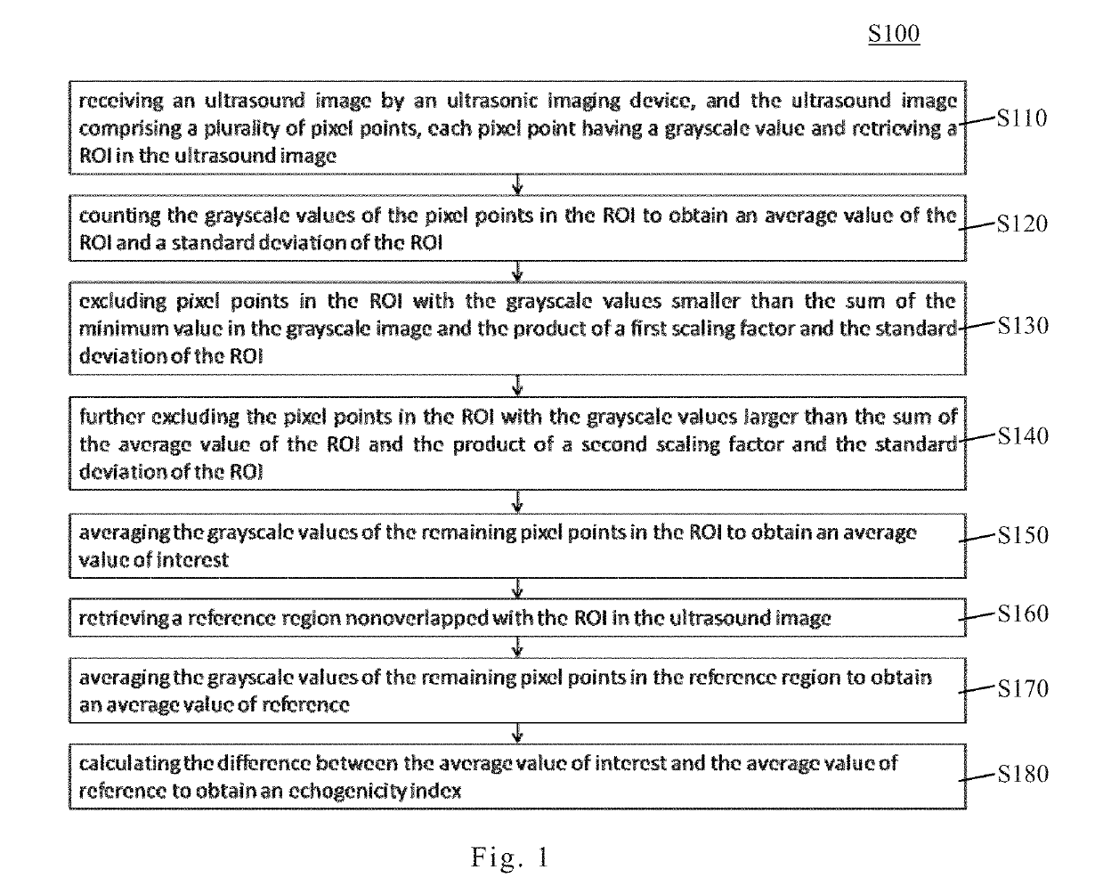
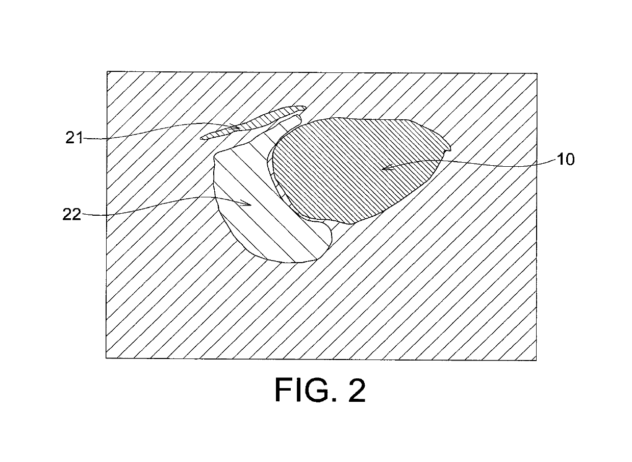
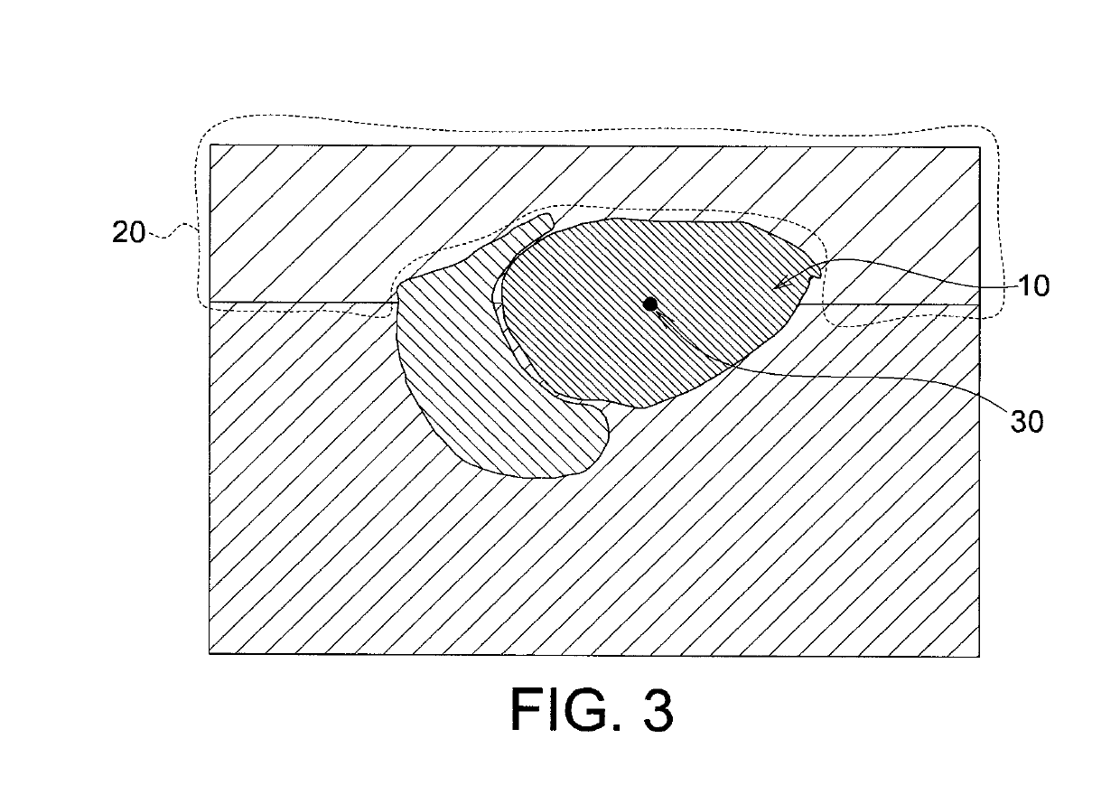
Echogenicity quantification method and calibration method for ultrasonic device using echogenicity index
PatentActiveUS10249037B2
Innovation
- An echogenicity quantification method that calculates an echogenicity index by averaging and normalizing grayscale values within a Region Of Interest (ROI) and a reference region, excluding outliers, to provide an objective and consistent measure across different ultrasonic devices and operators.

Imaging and Evaluating Embryos, Oocytes, and Stem Cells
PatentPendingUS20250020568A1
Innovation
- The use of time-lapse microscopy and image analysis to measure cellular parameters such as cytokinesis duration and gene expression levels, allowing for the determination of developmental potential by comparing these measurements to reference values, thereby guiding clinical decisions for embryo transfer.


Regulatory Framework for Prenatal Diagnostic Devices
The regulatory landscape governing prenatal diagnostic devices, particularly those utilizing ultrasound echogenicity for embryonic development assessment, encompasses multiple jurisdictional frameworks designed to ensure safety, efficacy, and clinical validity. In the United States, the Food and Drug Administration (FDA) classifies ultrasound systems as Class II medical devices under 21 CFR 892.1560, requiring premarket notification through the 510(k) pathway. Devices incorporating advanced algorithms for automated echogenicity analysis or artificial intelligence-enhanced anomaly detection may face more stringent scrutiny under Class III designation, necessitating premarket approval with comprehensive clinical validation data. The FDA's guidance documents specifically address software as a medical device (SaMD) considerations when computational methods are employed for diagnostic interpretation.
European Union regulations operate under the Medical Device Regulation (MDR 2017/745), which replaced the previous Medical Device Directive. Prenatal diagnostic ultrasound systems typically fall under Class IIb or Class III categories depending on their intended diagnostic claims. Manufacturers must demonstrate conformity through notified body assessment, providing clinical evidence that echogenicity-based diagnostic capabilities meet essential safety and performance requirements. The EU's emphasis on clinical evaluation reports demands robust documentation of diagnostic accuracy, particularly for detecting specific embryonic anomalies.
International harmonization efforts through the International Medical Device Regulators Forum (IMDRF) have established common principles for software validation and clinical performance assessment. The Global Harmonization Task Force guidelines provide frameworks for risk classification and quality management systems applicable across multiple jurisdictions. Countries including Japan, Canada, and Australia have adopted similar tiered classification systems, though specific requirements for echogenicity-based diagnostic claims vary.
Regulatory pathways increasingly emphasize real-world evidence and post-market surveillance, requiring manufacturers to maintain vigilance systems for adverse event reporting and performance monitoring. Special considerations apply to devices making claims about specific chromosomal abnormalities or structural malformations, often requiring prospective clinical studies with defined sensitivity and specificity thresholds. Compliance with standards such as IEC 60601 for electrical safety and IEC 62304 for medical device software lifecycle processes remains mandatory across most regulatory frameworks.
AI Integration in Embryonic Ultrasound Analysis
The integration of artificial intelligence into embryonic ultrasound analysis represents a transformative advancement in prenatal diagnostic capabilities, particularly for detecting developmental anomalies through echogenicity assessment. Machine learning algorithms, especially convolutional neural networks (CNNs) and deep learning architectures, have demonstrated remarkable proficiency in analyzing ultrasound image patterns that correlate with embryonic abnormalities. These AI systems can process vast quantities of echogenic data with consistency and speed unattainable through manual interpretation alone.
Current AI applications focus on automated feature extraction from ultrasound images, where algorithms identify subtle variations in tissue echogenicity that may indicate structural or chromosomal abnormalities. Deep learning models trained on extensive datasets of normal and abnormal embryonic development can recognize complex patterns in echo texture, intensity distribution, and spatial relationships. These systems achieve diagnostic accuracy rates exceeding 90% in controlled studies, significantly reducing inter-observer variability that traditionally challenges ultrasound interpretation.
Advanced AI frameworks now incorporate multi-modal data integration, combining echogenicity measurements with maternal biomarkers, genetic information, and temporal developmental tracking. This holistic approach enables more comprehensive risk stratification and early anomaly detection. Real-time AI assistance during ultrasound examinations provides clinicians with immediate decision support, highlighting regions of concern and suggesting optimal imaging angles for enhanced visualization.
The implementation of explainable AI (XAI) techniques addresses the critical need for transparency in clinical decision-making. These systems not only generate diagnostic predictions but also provide visual explanations indicating which echogenic features influenced their assessments, thereby building clinician trust and facilitating knowledge transfer. Cloud-based AI platforms enable continuous model improvement through federated learning, where algorithms refine their capabilities across diverse patient populations while maintaining data privacy.
Despite these advances, challenges remain in standardizing AI validation protocols, ensuring generalizability across different ultrasound equipment manufacturers, and establishing regulatory frameworks for clinical deployment. The ongoing development of AI-augmented ultrasound analysis promises to democratize access to expert-level prenatal diagnostics while maintaining the essential role of human clinical judgment in patient care.
Unlock deeper insights with PatSnap Eureka Quick Research — get a full tech report to explore trends and direct your research. Try now!
Generate Your Research Report Instantly with AI Agent
Supercharge your innovation with PatSnap Eureka AI Agent Platform!