

Dissecting Microscope vs Phase Contrast Microscope: Which for Transparent Tissue?
JUL 16, 20269 MIN READ
Generate Your Research Report Instantly with AI Agent
Patsnap Eureka helps you evaluate technical feasibility & market potential.
Microscopy Evolution for Transparent Tissue Observation
The observation of transparent tissue has long presented significant challenges in microscopy due to the inherent lack of contrast in such specimens. The evolution of microscopy techniques specifically addressing this challenge spans over a century, marked by pivotal innovations that fundamentally transformed biological research capabilities. Early microscopy relied primarily on bright-field illumination, which proved inadequate for visualizing unstained transparent specimens such as living cells, embryos, and tissue cultures.
The development trajectory began with the invention of the dissecting microscope in the late 19th century, which provided stereoscopic visualization at lower magnifications. This technology enabled researchers to observe three-dimensional structures of transparent specimens with enhanced depth perception, though it offered limited resolution and contrast enhancement. The dissecting microscope represented an important step forward by allowing manipulation and observation of living specimens without extensive preparation.
A revolutionary breakthrough occurred in the 1930s with Frits Zernike's invention of phase contrast microscopy, for which he received the Nobel Prize in Physics in 1953. This technique exploited the phase shifts of light passing through transparent specimens, converting invisible phase variations into visible amplitude differences. Phase contrast microscopy enabled unprecedented visualization of cellular structures, organelles, and dynamic processes in living cells without staining or fixation, fundamentally changing cell biology research.
The mid-20th century witnessed refinement and widespread adoption of phase contrast technology, with manufacturers developing standardized phase contrast objectives and condensers. Differential interference contrast (DIC) microscopy emerged in the 1950s as a complementary technique, offering enhanced resolution and optical sectioning capabilities. The late 20th century brought digital imaging integration, allowing real-time documentation and quantitative analysis of transparent tissue observations.
Contemporary developments include advanced computational imaging methods, super-resolution techniques adapted for transparent specimens, and hybrid systems combining multiple contrast-enhancement modalities. These innovations continue to expand the boundaries of what can be observed in living transparent tissues, supporting fields ranging from developmental biology to regenerative medicine.
The development trajectory began with the invention of the dissecting microscope in the late 19th century, which provided stereoscopic visualization at lower magnifications. This technology enabled researchers to observe three-dimensional structures of transparent specimens with enhanced depth perception, though it offered limited resolution and contrast enhancement. The dissecting microscope represented an important step forward by allowing manipulation and observation of living specimens without extensive preparation.
A revolutionary breakthrough occurred in the 1930s with Frits Zernike's invention of phase contrast microscopy, for which he received the Nobel Prize in Physics in 1953. This technique exploited the phase shifts of light passing through transparent specimens, converting invisible phase variations into visible amplitude differences. Phase contrast microscopy enabled unprecedented visualization of cellular structures, organelles, and dynamic processes in living cells without staining or fixation, fundamentally changing cell biology research.
The mid-20th century witnessed refinement and widespread adoption of phase contrast technology, with manufacturers developing standardized phase contrast objectives and condensers. Differential interference contrast (DIC) microscopy emerged in the 1950s as a complementary technique, offering enhanced resolution and optical sectioning capabilities. The late 20th century brought digital imaging integration, allowing real-time documentation and quantitative analysis of transparent tissue observations.
Contemporary developments include advanced computational imaging methods, super-resolution techniques adapted for transparent specimens, and hybrid systems combining multiple contrast-enhancement modalities. These innovations continue to expand the boundaries of what can be observed in living transparent tissues, supporting fields ranging from developmental biology to regenerative medicine.
Market Demand for Advanced Tissue Imaging Solutions
The global demand for advanced tissue imaging solutions has experienced substantial growth driven by expanding applications across biomedical research, clinical diagnostics, developmental biology, and pharmaceutical development. Transparent tissue observation represents a critical capability in these fields, where researchers require detailed visualization of cellular structures, tissue architecture, and dynamic biological processes without compromising sample integrity. The ability to observe living specimens in real-time has become increasingly essential for understanding disease mechanisms, evaluating therapeutic interventions, and advancing regenerative medicine applications.
Research institutions and academic laboratories constitute a primary demand segment, where transparent tissue imaging supports fundamental studies in embryology, neuroscience, and cellular biology. These facilities require cost-effective yet reliable imaging solutions capable of revealing structural details in unstained or minimally processed specimens. The growing emphasis on three-dimensional tissue culture systems and organoid research has further amplified the need for microscopy techniques that can penetrate thick specimens while maintaining optical clarity.
Clinical and diagnostic laboratories represent another significant market segment, particularly in pathology and surgical applications. The demand in this sector focuses on rapid, non-destructive tissue assessment methods that can guide intraoperative decisions or provide preliminary diagnostic information. Transparent tissue imaging capabilities enable pathologists to examine fresh tissue samples before fixation and staining, potentially accelerating diagnostic workflows and improving patient outcomes.
The pharmaceutical and biotechnology industries have emerged as major consumers of advanced tissue imaging technologies. Drug discovery processes increasingly rely on complex tissue models and organ-on-chip systems that require sophisticated imaging capabilities to assess compound efficacy and toxicity. The shift toward personalized medicine and biologics development has created demand for imaging solutions that can characterize tissue responses at cellular and subcellular levels with minimal sample preparation.
Market growth is further propelled by technological convergence trends, where traditional microscopy techniques are being integrated with digital imaging, artificial intelligence, and automated analysis platforms. End-users increasingly seek imaging solutions that not only provide superior optical performance but also offer compatibility with quantitative analysis workflows and data management systems. This evolution reflects a broader industry transition from purely observational microscopy toward comprehensive imaging informatics platforms that support data-driven decision-making in biological and medical research.
Research institutions and academic laboratories constitute a primary demand segment, where transparent tissue imaging supports fundamental studies in embryology, neuroscience, and cellular biology. These facilities require cost-effective yet reliable imaging solutions capable of revealing structural details in unstained or minimally processed specimens. The growing emphasis on three-dimensional tissue culture systems and organoid research has further amplified the need for microscopy techniques that can penetrate thick specimens while maintaining optical clarity.
Clinical and diagnostic laboratories represent another significant market segment, particularly in pathology and surgical applications. The demand in this sector focuses on rapid, non-destructive tissue assessment methods that can guide intraoperative decisions or provide preliminary diagnostic information. Transparent tissue imaging capabilities enable pathologists to examine fresh tissue samples before fixation and staining, potentially accelerating diagnostic workflows and improving patient outcomes.
The pharmaceutical and biotechnology industries have emerged as major consumers of advanced tissue imaging technologies. Drug discovery processes increasingly rely on complex tissue models and organ-on-chip systems that require sophisticated imaging capabilities to assess compound efficacy and toxicity. The shift toward personalized medicine and biologics development has created demand for imaging solutions that can characterize tissue responses at cellular and subcellular levels with minimal sample preparation.
Market growth is further propelled by technological convergence trends, where traditional microscopy techniques are being integrated with digital imaging, artificial intelligence, and automated analysis platforms. End-users increasingly seek imaging solutions that not only provide superior optical performance but also offer compatibility with quantitative analysis workflows and data management systems. This evolution reflects a broader industry transition from purely observational microscopy toward comprehensive imaging informatics platforms that support data-driven decision-making in biological and medical research.
Current Capabilities and Limitations of Both Microscopy Types
Dissecting microscopes, also known as stereo microscopes, excel in providing three-dimensional visualization of transparent tissues with working distances ranging from 20mm to 100mm. Their primary strength lies in offering magnifications between 6x and 50x, which proves ideal for macroscopic examination and manipulation of specimens. The dual optical paths create stereoscopic vision, enabling researchers to perceive depth and spatial relationships within transparent structures. These instruments maintain excellent field of view, typically spanning 10mm to 40mm in diameter, facilitating observation of larger tissue samples without frequent repositioning. The generous working distance accommodates dissection tools and micromanipulation instruments, making them indispensable for surgical procedures and specimen preparation.
However, dissecting microscopes face significant limitations when examining cellular-level details in transparent tissues. Their maximum useful magnification rarely exceeds 100x, restricting observation to gross anatomical features rather than subcellular structures. The optical design prioritizes depth perception over resolution, resulting in lower numerical apertures compared to compound microscopes. Consequently, fine structural details within transparent tissues remain poorly resolved, and contrast enhancement capabilities are minimal without additional illumination techniques.
Phase contrast microscopes demonstrate superior performance in revealing internal structures of transparent, unstained tissues through optical phase shift conversion. Operating at magnifications from 40x to 1000x, these instruments transform phase variations caused by refractive index differences into amplitude variations visible to the human eye. This capability enables visualization of cellular organelles, membrane boundaries, and intracellular components without chemical staining, preserving tissue viability for live-cell imaging applications. The technique proves particularly effective for observing thin tissue sections, typically 5-20 micrometers thick, where phase differences generate sufficient contrast.
The principal limitations of phase contrast microscopy include restricted working distance, usually under 10mm, which prevents manipulation during observation. The technique produces characteristic halo artifacts around specimen edges, potentially obscuring fine details at boundaries. Additionally, phase contrast systems require precise optical alignment and are optimized for specific specimen thicknesses, performing suboptimally with thick transparent tissues exceeding 50 micrometers. The narrow depth of field at higher magnifications necessitates frequent focal adjustments when examining three-dimensional structures, and the monocular optical path eliminates stereoscopic depth perception essential for spatial understanding.
However, dissecting microscopes face significant limitations when examining cellular-level details in transparent tissues. Their maximum useful magnification rarely exceeds 100x, restricting observation to gross anatomical features rather than subcellular structures. The optical design prioritizes depth perception over resolution, resulting in lower numerical apertures compared to compound microscopes. Consequently, fine structural details within transparent tissues remain poorly resolved, and contrast enhancement capabilities are minimal without additional illumination techniques.
Phase contrast microscopes demonstrate superior performance in revealing internal structures of transparent, unstained tissues through optical phase shift conversion. Operating at magnifications from 40x to 1000x, these instruments transform phase variations caused by refractive index differences into amplitude variations visible to the human eye. This capability enables visualization of cellular organelles, membrane boundaries, and intracellular components without chemical staining, preserving tissue viability for live-cell imaging applications. The technique proves particularly effective for observing thin tissue sections, typically 5-20 micrometers thick, where phase differences generate sufficient contrast.
The principal limitations of phase contrast microscopy include restricted working distance, usually under 10mm, which prevents manipulation during observation. The technique produces characteristic halo artifacts around specimen edges, potentially obscuring fine details at boundaries. Additionally, phase contrast systems require precise optical alignment and are optimized for specific specimen thicknesses, performing suboptimally with thick transparent tissues exceeding 50 micrometers. The narrow depth of field at higher magnifications necessitates frequent focal adjustments when examining three-dimensional structures, and the monocular optical path eliminates stereoscopic depth perception essential for spatial understanding.
Comparative Technical Solutions for Transparent Specimen Imaging
01 Phase contrast optical systems and components
Phase contrast microscopy utilizes specialized optical components including phase plates, annular apertures, and phase rings to convert phase shifts in transparent specimens into amplitude changes visible as contrast variations. These systems employ specific configurations of condensers and objectives with matched phase elements to enhance visualization of unstained biological samples and transparent materials without affecting specimen viability.- Phase contrast optical systems and components: Phase contrast microscopy utilizes specialized optical components including phase plates, annular apertures, and phase rings to convert phase differences in transparent specimens into amplitude differences visible to the human eye. These systems employ specific configurations of condensers and objective lenses with matched phase elements to enhance contrast in unstained biological samples. The optical design includes precise alignment mechanisms and adjustable phase shifting elements to optimize image quality.
- Digital image processing for contrast enhancement: Advanced digital imaging systems incorporate computational methods to enhance contrast in microscopy images. These techniques include algorithms for background correction, dynamic range adjustment, and real-time image processing. The systems utilize electronic sensors and processors to capture and manipulate image data, enabling improved visualization of low-contrast specimens through software-based enhancement methods.
- Illumination systems for improved transparency imaging: Specialized illumination configurations are designed to optimize the visualization of transparent specimens. These systems employ various light sources, including LED arrays and fiber optic delivery systems, combined with specific illumination geometries such as oblique, transmitted, or epi-illumination. The designs focus on controlling light intensity, wavelength, and directionality to maximize contrast while minimizing artifacts in transparent sample imaging.
- Stereoscopic and dissecting microscope optical designs: Dissecting microscopes feature dual optical paths providing stereoscopic viewing with enhanced depth perception and working distance. These designs incorporate zoom mechanisms, variable magnification systems, and ergonomic viewing angles optimized for specimen manipulation. The optical configurations balance resolution, field of view, and working distance to facilitate detailed examination and dissection of larger specimens.
- Modular and adaptive contrast enhancement mechanisms: Modern microscopy systems incorporate modular components allowing switching between multiple contrast modes including phase contrast, differential interference contrast, and brightfield illumination. These adaptive systems feature interchangeable optical elements, motorized turrets, and automated switching mechanisms. The designs enable users to optimize imaging parameters for different specimen types and transparency levels without changing the basic microscope configuration.
02 Digital image contrast enhancement methods
Digital processing techniques are applied to microscope images to improve contrast and visibility of transparent specimens. These methods include computational algorithms for contrast adjustment, background subtraction, edge enhancement, and dynamic range optimization. Software-based approaches enable real-time processing and post-acquisition enhancement of microscopic images to reveal fine structural details in low-contrast samples.Expand Specific Solutions03 Illumination systems for contrast optimization
Advanced illumination configurations are designed to maximize image contrast in dissecting and phase contrast microscopes. These systems incorporate adjustable light sources, oblique illumination arrangements, variable aperture diaphragms, and specialized filters to control light intensity and angle. Proper illumination geometry is critical for enhancing edge definition and three-dimensional perception of transparent specimens.Expand Specific Solutions04 Optical path design for stereoscopic and dissecting microscopes
Stereoscopic dissecting microscopes employ dual optical paths with specific angular separation to provide three-dimensional visualization of specimens. The optical design includes zoom mechanisms, variable magnification systems, and ergonomic viewing angles optimized for extended observation periods. These configurations balance working distance, field of view, and depth perception to facilitate manipulation and examination of larger transparent specimens.Expand Specific Solutions05 Hybrid and modular microscopy systems
Modern microscope designs integrate multiple contrast modes including phase contrast, brightfield, and darkfield capabilities within a single modular platform. These systems feature interchangeable optical components, motorized switching mechanisms, and adaptable configurations that allow users to select optimal visualization methods for different specimen types. Modular approaches provide flexibility for various applications while maintaining optical performance across different contrast techniques.Expand Specific Solutions
Leading Microscopy Manufacturers and Market Competition
The microscopy sector for transparent tissue observation represents a mature, competitive market dominated by established optical technology leaders. Major players including Evident Corp. (formerly Olympus), Nikon Corp., Leica Microsystems, Carl Zeiss Microscopy, and FUJIFILM Corp. command significant market share through comprehensive product portfolios spanning dissecting and phase contrast microscopy systems. These companies leverage decades of optical engineering expertise and continuous innovation in imaging software, automation, and AI-enhanced analysis capabilities. The market exhibits steady growth driven by expanding applications in life sciences, clinical diagnostics, and materials research. Technology maturity is high, with leading manufacturers like Evident, Leica, and Zeiss offering advanced modular platforms integrating confocal, super-resolution, and digital pathology workflows, while emerging players such as Ningbo Yong Xin Optics and Shanghai Ankon Medical Technology focus on cost-competitive solutions for regional markets.
Evident Corp.
Technical Solution: Evident (formerly Olympus Scientific Solutions) provides comprehensive solutions for both dissecting and phase contrast microscopy for transparent tissue observation. Their dissecting microscopes feature high numerical aperture objectives with working distances up to 115mm, enabling stereoscopic visualization of transparent specimens with excellent depth perception and minimal sample preparation. Their phase contrast microscopy systems utilize advanced optical designs with specialized phase rings and condenser configurations that convert phase shifts in transparent specimens into amplitude variations, making cellular structures and organelles visible without staining. The company integrates digital imaging capabilities with both modalities, offering real-time observation and documentation. Their systems are widely adopted in developmental biology, cell culture monitoring, and tissue engineering applications where non-invasive observation of living transparent tissues is critical.
Strengths: Market-leading optical quality, extensive product portfolio covering both modalities, strong integration with digital imaging systems, excellent technical support network. Weaknesses: Premium pricing positioning, complex system configurations may require specialized training, limited customization options for highly specialized research applications.
FUJIFILM Corp.
Technical Solution: FUJIFILM has developed microscopy solutions leveraging their optical and imaging expertise for transparent tissue observation applications. Their dissecting microscope systems incorporate high-performance zoom optics with extended depth of field capabilities, enabling three-dimensional visualization of transparent biological specimens with minimal chromatic aberration. For phase contrast microscopy, FUJIFILM integrates their advanced lens coating technologies and precision optical manufacturing to produce phase contrast systems that enhance contrast in transparent tissues while maintaining high light throughput. The company's solutions emphasize integration with digital imaging platforms, utilizing their expertise in image sensor technology and image processing algorithms to enhance visualization of subtle structural features in transparent specimens. Their systems find applications in pharmaceutical research, quality control in cell therapy manufacturing, and basic biological research requiring detailed observation of living transparent tissues without invasive staining procedures.
Strengths: Strong integration with digital imaging technologies, competitive pricing compared to traditional microscopy leaders, reliable optical performance, good technical support in Asian markets. Weaknesses: Smaller market share in microscopy compared to core business areas, limited product range compared to specialized microscopy manufacturers, less established brand recognition in Western research institutions.
Core Optical Principles in Contrast Enhancement Methods
Inverted microscope for high-contrast imaging
PatentActiveUS8228600B2
Innovation
- An inverted microscope design that allows multiple objectives with disposable modulators in the imaging beam path, using a lens system to generate intermediate images of objective pupils, enabling rapid selection and exchange of modulators at accessible locations for flexible implementation of various contrasting processes.
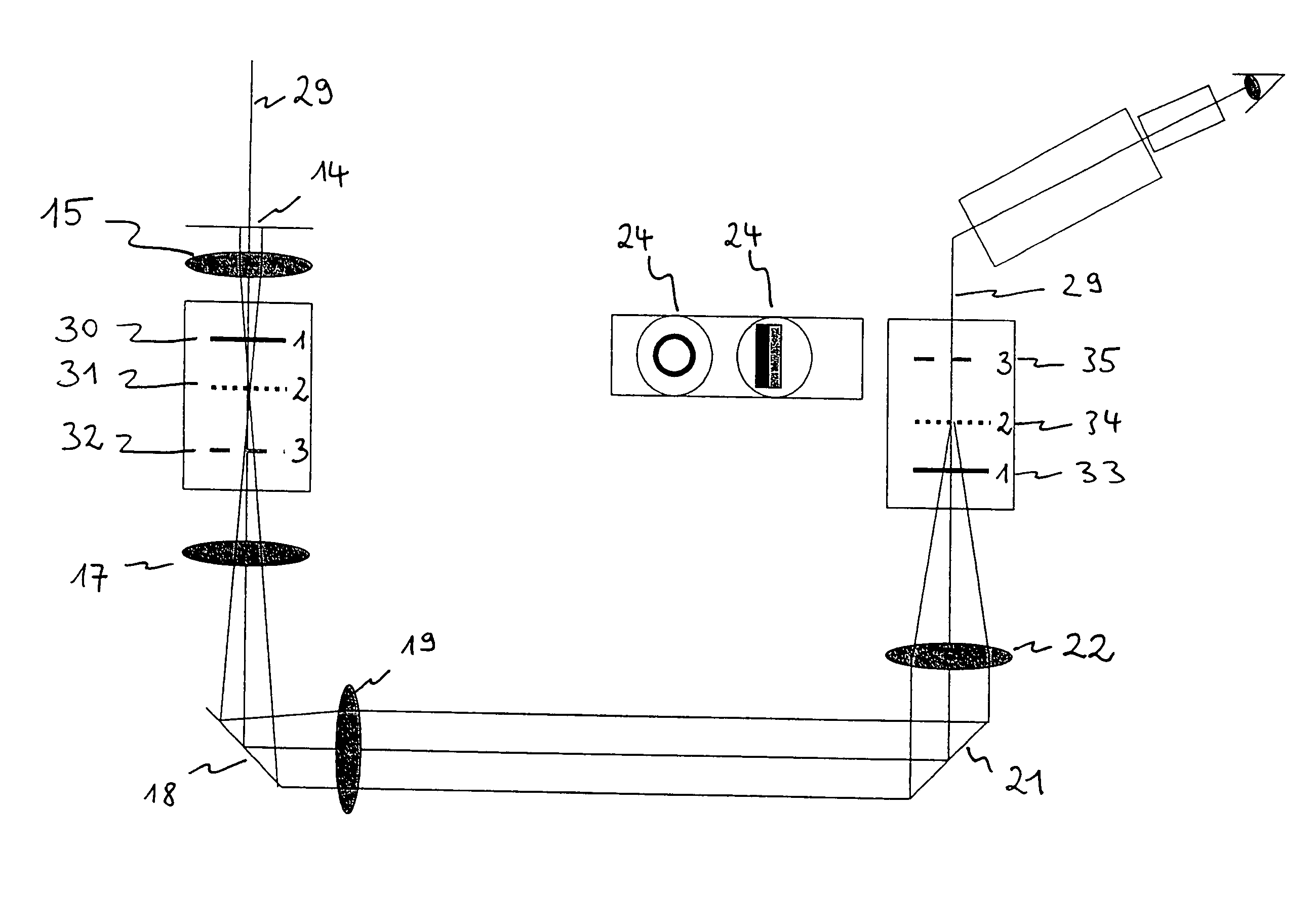
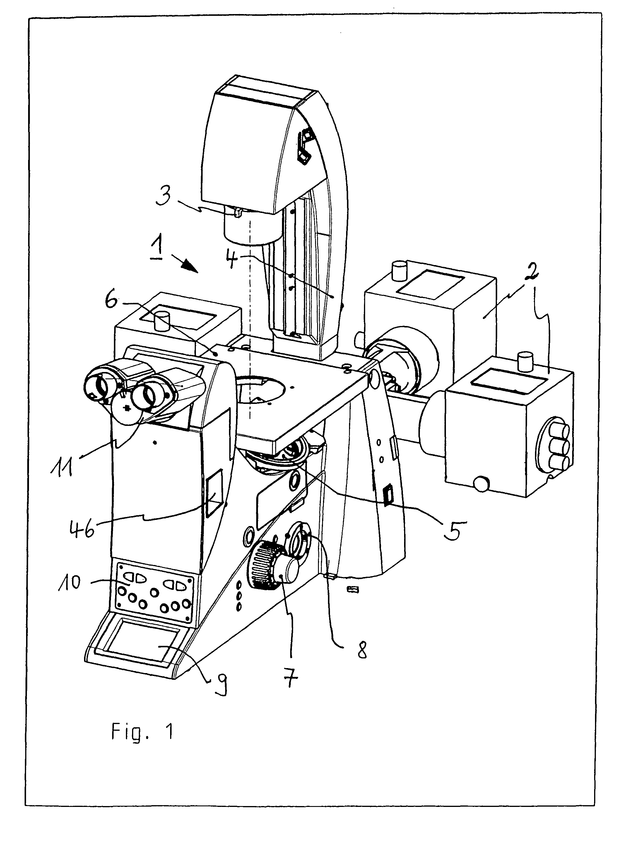
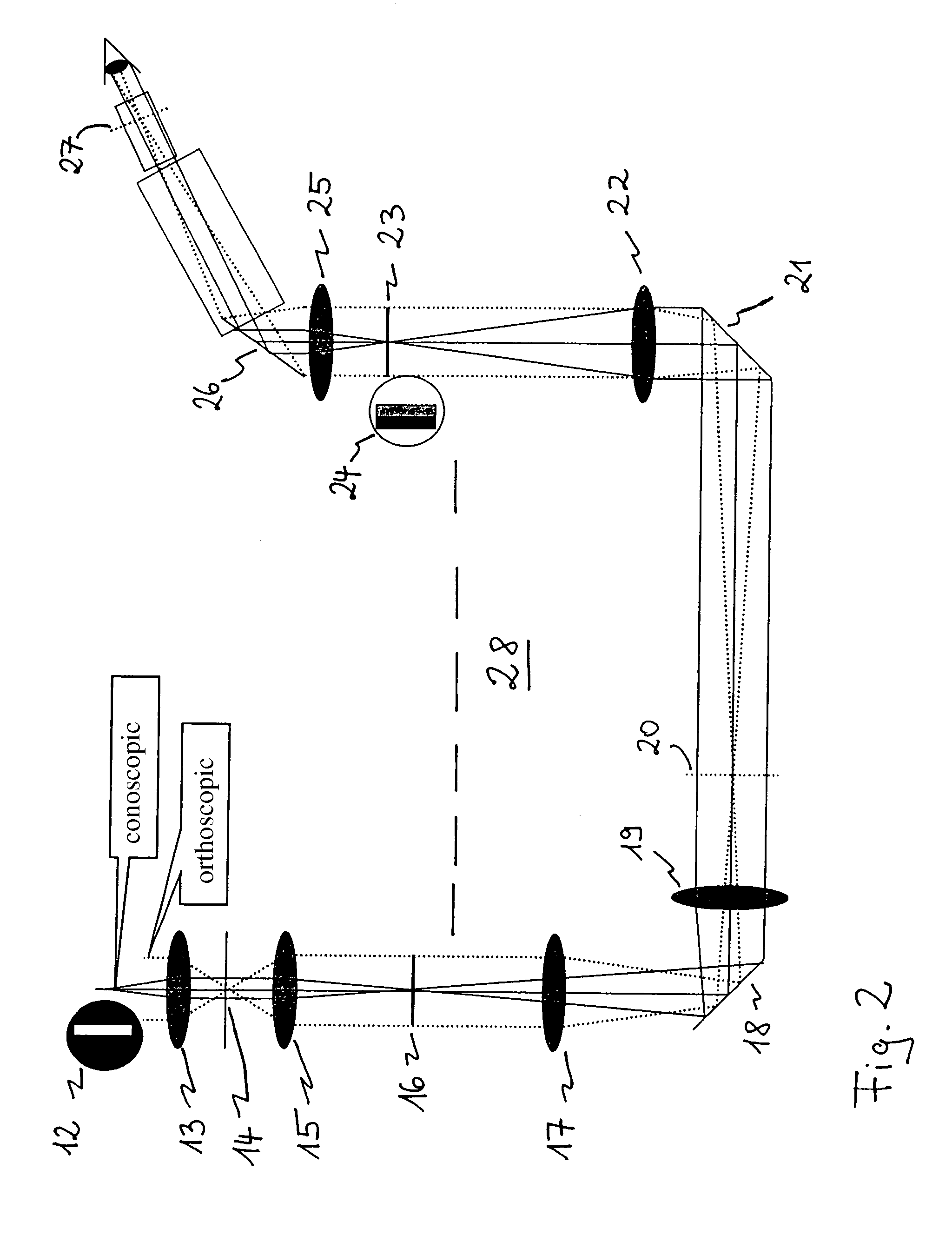

Observing device
PatentInactiveEP1956360A1
Innovation
- An observing apparatus with a surface illuminant having alternating bright and dark areas, an image-forming optical system, and an imaging unit, where the distance and pitch of the surface illuminant are optimized to satisfy specific numerical aperture and wavelength conditions, and optionally includes a liquid crystal spatial light modulator and image processing to enhance image quality.


Application-Specific Selection Criteria for Microscopy Techniques
When selecting between dissecting microscopes and phase contrast microscopes for transparent tissue observation, several application-specific criteria must be carefully evaluated to ensure optimal results. The decision-making process should be guided by the specific requirements of the research or clinical application, as each microscopy technique offers distinct advantages tailored to different observational needs.
The primary consideration involves the required magnification range and working distance. Dissecting microscopes typically provide lower magnification levels ranging from 5x to 50x with significantly larger working distances, making them ideal for manipulating specimens during observation. In contrast, phase contrast microscopes operate at higher magnifications, generally from 40x to 1000x, with shorter working distances, suited for detailed cellular and subcellular examination of transparent tissues.
Sample preparation requirements represent another critical selection factor. Applications requiring minimal sample preparation and the ability to observe living tissues in their natural state favor dissecting microscopes, as they accommodate thicker specimens and allow real-time manipulation. Phase contrast microscopy, while also enabling live tissue observation, demands more stringent sample preparation protocols, including specific thickness requirements and mounting procedures to achieve optimal contrast enhancement.
The depth of field and three-dimensional visualization needs significantly influence technique selection. Dissecting microscopes excel in applications requiring stereoscopic vision and depth perception, essential for surgical procedures, embryo manipulation, and gross anatomical studies. Phase contrast microscopes sacrifice depth of field for enhanced resolution of fine structural details within thin transparent specimens, making them preferable for cytological examinations and intracellular component visualization.
Budget constraints and operational complexity also factor into the selection process. Dissecting microscopes generally present lower initial investment costs and simpler operational requirements, suitable for routine screening and educational purposes. Phase contrast systems, despite higher costs and more complex alignment procedures, deliver superior performance for specialized research applications demanding high-resolution imaging of transparent structures. The intended throughput, user expertise level, and integration with documentation systems further refine the selection criteria, ensuring alignment between microscopy technique capabilities and specific application demands.
The primary consideration involves the required magnification range and working distance. Dissecting microscopes typically provide lower magnification levels ranging from 5x to 50x with significantly larger working distances, making them ideal for manipulating specimens during observation. In contrast, phase contrast microscopes operate at higher magnifications, generally from 40x to 1000x, with shorter working distances, suited for detailed cellular and subcellular examination of transparent tissues.
Sample preparation requirements represent another critical selection factor. Applications requiring minimal sample preparation and the ability to observe living tissues in their natural state favor dissecting microscopes, as they accommodate thicker specimens and allow real-time manipulation. Phase contrast microscopy, while also enabling live tissue observation, demands more stringent sample preparation protocols, including specific thickness requirements and mounting procedures to achieve optimal contrast enhancement.
The depth of field and three-dimensional visualization needs significantly influence technique selection. Dissecting microscopes excel in applications requiring stereoscopic vision and depth perception, essential for surgical procedures, embryo manipulation, and gross anatomical studies. Phase contrast microscopes sacrifice depth of field for enhanced resolution of fine structural details within thin transparent specimens, making them preferable for cytological examinations and intracellular component visualization.
Budget constraints and operational complexity also factor into the selection process. Dissecting microscopes generally present lower initial investment costs and simpler operational requirements, suitable for routine screening and educational purposes. Phase contrast systems, despite higher costs and more complex alignment procedures, deliver superior performance for specialized research applications demanding high-resolution imaging of transparent structures. The intended throughput, user expertise level, and integration with documentation systems further refine the selection criteria, ensuring alignment between microscopy technique capabilities and specific application demands.
Cost-Benefit Analysis of Microscopy Investment Strategies
When evaluating microscopy investment strategies for transparent tissue observation, organizations must weigh the financial implications against operational benefits of dissecting microscopes versus phase contrast microscopes. The initial capital expenditure represents a critical decision point, as dissecting microscopes typically range from $500 to $5,000 for quality instruments, while phase contrast microscopes command higher prices between $3,000 and $15,000 due to their specialized optical components and precision engineering requirements. This substantial price differential necessitates careful consideration of institutional budgets and research priorities.
Beyond acquisition costs, operational expenses significantly impact long-term financial planning. Phase contrast systems generally incur higher maintenance costs due to their complex optical configurations, requiring specialized alignment procedures and periodic calibration by trained technicians. Replacement parts, particularly phase rings and condenser assemblies, represent ongoing investments. Conversely, dissecting microscopes offer simpler maintenance protocols with lower associated costs, though they may require more frequent bulb replacements depending on usage intensity.
The return on investment calculation must incorporate productivity metrics and research output quality. Phase contrast microscopy enables cellular-level observation and detailed structural analysis of transparent specimens, potentially accelerating research timelines and enhancing publication quality. This capability justifies higher costs for institutions focused on cellular biology or developmental studies. However, for applications requiring rapid specimen screening, gross morphological assessment, or educational purposes, dissecting microscopes deliver adequate performance at substantially lower total cost of ownership.
Institutional scale and utilization patterns further influence cost-effectiveness. High-throughput laboratories with multiple users benefit from investing in several dissecting microscopes to maximize accessibility, while centralizing phase contrast capabilities in core facilities optimizes expensive equipment utilization. Training costs also merit consideration, as phase contrast techniques require more extensive user education compared to the intuitive operation of dissecting microscopes. Strategic procurement decisions should align equipment capabilities with specific research objectives, user expertise levels, and anticipated specimen types to maximize value realization across the investment lifecycle.
Beyond acquisition costs, operational expenses significantly impact long-term financial planning. Phase contrast systems generally incur higher maintenance costs due to their complex optical configurations, requiring specialized alignment procedures and periodic calibration by trained technicians. Replacement parts, particularly phase rings and condenser assemblies, represent ongoing investments. Conversely, dissecting microscopes offer simpler maintenance protocols with lower associated costs, though they may require more frequent bulb replacements depending on usage intensity.
The return on investment calculation must incorporate productivity metrics and research output quality. Phase contrast microscopy enables cellular-level observation and detailed structural analysis of transparent specimens, potentially accelerating research timelines and enhancing publication quality. This capability justifies higher costs for institutions focused on cellular biology or developmental studies. However, for applications requiring rapid specimen screening, gross morphological assessment, or educational purposes, dissecting microscopes deliver adequate performance at substantially lower total cost of ownership.
Institutional scale and utilization patterns further influence cost-effectiveness. High-throughput laboratories with multiple users benefit from investing in several dissecting microscopes to maximize accessibility, while centralizing phase contrast capabilities in core facilities optimizes expensive equipment utilization. Training costs also merit consideration, as phase contrast techniques require more extensive user education compared to the intuitive operation of dissecting microscopes. Strategic procurement decisions should align equipment capabilities with specific research objectives, user expertise levels, and anticipated specimen types to maximize value realization across the investment lifecycle.
Unlock deeper insights with Patsnap Eureka Quick Research — get a full tech report to explore trends and direct your research. Try now!
Generate Your Research Report Instantly with AI Agent
Supercharge your innovation with Patsnap Eureka AI Agent Platform!