

Dissecting Microscope vs Stereo Microscope: Depth Perception Compared
JUL 16, 20269 MIN READ
Generate Your Research Report Instantly with AI Agent
Patsnap Eureka helps you evaluate technical feasibility & market potential.
Depth Perception in Microscopy: Background and Objectives
Depth perception represents a fundamental capability in microscopy that enables researchers and practitioners to accurately interpret three-dimensional structures of specimens. This perceptual ability has evolved significantly alongside microscope technology development, transitioning from monocular systems with limited depth cues to sophisticated stereoscopic configurations that provide enhanced spatial understanding. The historical progression of microscopy reveals that early instruments offered minimal depth information, compelling users to rely heavily on focus adjustments and specimen manipulation to construct mental three-dimensional models.
The emergence of stereoscopic microscopy marked a pivotal advancement in addressing depth perception limitations. Two primary stereoscopic approaches have dominated the field: dissecting microscopes and stereo microscopes. While these terms are sometimes used interchangeably in casual contexts, they represent distinct optical architectures with fundamentally different mechanisms for generating depth perception. Dissecting microscopes typically employ a common main objective design, whereas true stereo microscopes utilize separate optical paths for each eye, creating genuine parallax effects that more closely mimic natural human binocular vision.
Contemporary applications across biological research, materials science, quality control, and surgical procedures demand increasingly precise depth discrimination capabilities. The ability to accurately perceive depth influences critical tasks including specimen dissection, micromanipulation, defect inspection, and microsurgical interventions. As specimen complexity increases and working distances become more constrained, the limitations of different stereoscopic approaches become more pronounced, directly impacting operational efficiency and outcome quality.
The primary objective of investigating depth perception differences between dissecting and stereo microscopes centers on establishing quantitative and qualitative performance benchmarks. This research aims to identify specific operational scenarios where each technology demonstrates superior depth discrimination, evaluate the physiological and cognitive factors affecting user perception under different optical configurations, and determine how optical design parameters influence three-dimensional spatial accuracy. Understanding these distinctions enables informed equipment selection decisions, guides future optical system optimization, and establishes foundations for developing enhanced depth perception technologies that address current limitations in microscopic observation and manipulation tasks.
The emergence of stereoscopic microscopy marked a pivotal advancement in addressing depth perception limitations. Two primary stereoscopic approaches have dominated the field: dissecting microscopes and stereo microscopes. While these terms are sometimes used interchangeably in casual contexts, they represent distinct optical architectures with fundamentally different mechanisms for generating depth perception. Dissecting microscopes typically employ a common main objective design, whereas true stereo microscopes utilize separate optical paths for each eye, creating genuine parallax effects that more closely mimic natural human binocular vision.
Contemporary applications across biological research, materials science, quality control, and surgical procedures demand increasingly precise depth discrimination capabilities. The ability to accurately perceive depth influences critical tasks including specimen dissection, micromanipulation, defect inspection, and microsurgical interventions. As specimen complexity increases and working distances become more constrained, the limitations of different stereoscopic approaches become more pronounced, directly impacting operational efficiency and outcome quality.
The primary objective of investigating depth perception differences between dissecting and stereo microscopes centers on establishing quantitative and qualitative performance benchmarks. This research aims to identify specific operational scenarios where each technology demonstrates superior depth discrimination, evaluate the physiological and cognitive factors affecting user perception under different optical configurations, and determine how optical design parameters influence three-dimensional spatial accuracy. Understanding these distinctions enables informed equipment selection decisions, guides future optical system optimization, and establishes foundations for developing enhanced depth perception technologies that address current limitations in microscopic observation and manipulation tasks.
Market Demand for Enhanced Microscopic Depth Visualization
The demand for enhanced microscopic depth visualization has intensified across multiple professional sectors, driven by the increasing complexity of precision tasks and the need for improved operational accuracy. In biological research laboratories, scientists conducting dissection procedures and specimen manipulation require superior three-dimensional perception to navigate delicate tissue structures without causing damage. The ability to accurately judge depth relationships between cellular layers and anatomical features directly impacts research outcomes and specimen preservation quality.
Manufacturing industries, particularly electronics assembly and quality control operations, represent another significant market segment. As electronic components continue to miniaturize, assembly technicians and quality inspectors face mounting challenges in evaluating solder joint quality, detecting surface defects, and positioning micro-components. Enhanced depth perception capabilities enable faster defect identification and reduce assembly errors, translating directly into improved production efficiency and reduced waste rates.
The medical device sector demonstrates growing requirements for advanced depth visualization during microsurgical procedures and device assembly. Ophthalmic surgeons, neurosurgeons, and reconstructive specialists depend on accurate depth perception when operating on microscopic anatomical structures. Similarly, dental professionals performing endodontic treatments and implant placements require precise spatial awareness to avoid complications and achieve optimal clinical outcomes.
Educational institutions and training facilities constitute an emerging market segment, where enhanced depth visualization supports skill development in microscopy techniques. Students in biological sciences, materials engineering, and medical programs benefit from improved three-dimensional understanding when learning specimen examination and manipulation procedures. Training programs increasingly recognize that superior depth perception accelerates skill acquisition and reduces learning curves.
The jewelry and watchmaking industries also exhibit sustained demand for enhanced microscopic depth visualization. Craftsmen performing intricate engraving, stone setting, and component assembly operations require exceptional spatial judgment to maintain quality standards and prevent costly errors. As luxury goods markets expand globally, the need for precision visualization tools continues to grow, supporting both traditional craftsmanship and modern manufacturing approaches.
Manufacturing industries, particularly electronics assembly and quality control operations, represent another significant market segment. As electronic components continue to miniaturize, assembly technicians and quality inspectors face mounting challenges in evaluating solder joint quality, detecting surface defects, and positioning micro-components. Enhanced depth perception capabilities enable faster defect identification and reduce assembly errors, translating directly into improved production efficiency and reduced waste rates.
The medical device sector demonstrates growing requirements for advanced depth visualization during microsurgical procedures and device assembly. Ophthalmic surgeons, neurosurgeons, and reconstructive specialists depend on accurate depth perception when operating on microscopic anatomical structures. Similarly, dental professionals performing endodontic treatments and implant placements require precise spatial awareness to avoid complications and achieve optimal clinical outcomes.
Educational institutions and training facilities constitute an emerging market segment, where enhanced depth visualization supports skill development in microscopy techniques. Students in biological sciences, materials engineering, and medical programs benefit from improved three-dimensional understanding when learning specimen examination and manipulation procedures. Training programs increasingly recognize that superior depth perception accelerates skill acquisition and reduces learning curves.
The jewelry and watchmaking industries also exhibit sustained demand for enhanced microscopic depth visualization. Craftsmen performing intricate engraving, stone setting, and component assembly operations require exceptional spatial judgment to maintain quality standards and prevent costly errors. As luxury goods markets expand globally, the need for precision visualization tools continues to grow, supporting both traditional craftsmanship and modern manufacturing approaches.
Current Depth Perception Capabilities and Limitations
Dissecting microscopes and stereo microscopes represent two distinct optical configurations that fundamentally differ in their depth perception mechanisms. Dissecting microscopes, also known as stereomicroscopes in some contexts, typically employ dual optical paths with convergent angles ranging from 10 to 15 degrees, creating genuine stereoscopic vision. This binocular arrangement enables operators to perceive three-dimensional structures naturally, with depth discrimination capabilities extending from approximately 0.5mm to 5mm depending on magnification levels and working distances.
Current stereo microscopes achieve depth perception through either Greenough or common main objective (CMO) designs. Greenough systems utilize two independent optical paths inclined at fixed angles, delivering superior depth perception with working distances between 60mm and 120mm. The parallax effect generated by these separated optical axes provides robust three-dimensional visualization, particularly effective for specimens requiring manipulation under magnification. However, this configuration introduces optical aberrations at higher magnifications, limiting depth accuracy beyond 50x magnification.
CMO stereo microscopes employ a single objective lens with beam-splitting prisms to create stereoscopic images. While offering superior optical correction and extended zoom ranges, these systems demonstrate reduced depth perception compared to Greenough designs due to smaller effective interpupillary distances. The depth discrimination threshold in CMO systems typically ranges from 1mm to 3mm, constraining precision in applications requiring fine depth judgment.
Both microscope types face inherent limitations in quantitative depth measurement. The subjective nature of human stereoscopic vision introduces variability between operators, with depth perception accuracy declining significantly beyond 100x magnification. Working distance constraints further limit depth perception capabilities, as reduced working distances compress the effective depth of field. Additionally, illumination quality critically impacts depth perception, with inadequate contrast or shadowing effects diminishing the ability to distinguish subtle surface variations.
Contemporary systems struggle with digital depth representation, as traditional stereoscopic viewing cannot be fully replicated through standard display technologies. This limitation becomes particularly evident in telemicroscopy applications where remote operators rely on two-dimensional screen representations, fundamentally compromising depth perception capabilities compared to direct binocular observation.
Current stereo microscopes achieve depth perception through either Greenough or common main objective (CMO) designs. Greenough systems utilize two independent optical paths inclined at fixed angles, delivering superior depth perception with working distances between 60mm and 120mm. The parallax effect generated by these separated optical axes provides robust three-dimensional visualization, particularly effective for specimens requiring manipulation under magnification. However, this configuration introduces optical aberrations at higher magnifications, limiting depth accuracy beyond 50x magnification.
CMO stereo microscopes employ a single objective lens with beam-splitting prisms to create stereoscopic images. While offering superior optical correction and extended zoom ranges, these systems demonstrate reduced depth perception compared to Greenough designs due to smaller effective interpupillary distances. The depth discrimination threshold in CMO systems typically ranges from 1mm to 3mm, constraining precision in applications requiring fine depth judgment.
Both microscope types face inherent limitations in quantitative depth measurement. The subjective nature of human stereoscopic vision introduces variability between operators, with depth perception accuracy declining significantly beyond 100x magnification. Working distance constraints further limit depth perception capabilities, as reduced working distances compress the effective depth of field. Additionally, illumination quality critically impacts depth perception, with inadequate contrast or shadowing effects diminishing the ability to distinguish subtle surface variations.
Contemporary systems struggle with digital depth representation, as traditional stereoscopic viewing cannot be fully replicated through standard display technologies. This limitation becomes particularly evident in telemicroscopy applications where remote operators rely on two-dimensional screen representations, fundamentally compromising depth perception capabilities compared to direct binocular observation.
Existing Depth Perception Solutions Comparison
01 Dual optical path stereo microscope systems
Stereo microscopes utilize dual optical paths to provide separate images to each eye, creating depth perception through binocular vision. This fundamental design includes two separate objective lenses or beam splitters that capture the specimen from slightly different angles, mimicking natural human vision. The angular separation between the optical axes enables three-dimensional visualization of specimens with enhanced depth cues.- Dual optical path design for stereoscopic vision: Stereo microscopes utilize two separate optical paths to provide distinct images to each eye, creating depth perception through binocular vision. This design incorporates dual objective lenses and eyepieces positioned at specific angles to simulate natural human vision. The convergence angle and interpupillary distance adjustments enable users to perceive three-dimensional structures of specimens with enhanced spatial awareness.
- Variable magnification zoom systems: Advanced zoom mechanisms allow continuous magnification adjustment while maintaining stereoscopic depth perception throughout the magnification range. These systems employ complex lens arrangements that preserve the parallax angle necessary for three-dimensional viewing. The zoom functionality enables users to examine specimens at different scales without losing depth information or requiring objective lens changes.
- Digital imaging integration for depth visualization: Modern stereo microscopes incorporate digital cameras and image processing systems to capture and enhance depth information. These systems may include dual-sensor configurations that replicate the stereoscopic viewing experience in digital format. Advanced algorithms process the parallax information to generate three-dimensional digital representations, enabling measurement and analysis of specimen depth characteristics.
- Ergonomic viewing angle and optical axis configuration: The optical design incorporates specific angles between viewing axes and adjustable eyepiece positions to optimize depth perception while reducing user fatigue. These configurations account for natural eye positioning and allow customization for different users. The optical axis arrangement ensures that both eyes receive properly aligned images with appropriate convergence for comfortable stereoscopic observation over extended periods.
- Illumination systems for enhanced depth contrast: Specialized lighting arrangements including oblique, coaxial, and transmitted illumination enhance the perception of depth by creating shadows and highlights that emphasize surface topology. These illumination systems can be adjusted in intensity, angle, and color temperature to optimize contrast for different specimen types. The lighting design works synergistically with the stereoscopic optical system to maximize three-dimensional visualization capabilities.
02 Variable magnification and zoom mechanisms
Advanced stereo microscopes incorporate zoom mechanisms that allow continuous magnification adjustment while maintaining depth perception and focus. These systems use complex lens arrangements and optical components that preserve the stereoscopic effect across different magnification levels. The zoom functionality enables users to examine specimens at various scales without losing the three-dimensional viewing capability.Expand Specific Solutions03 Digital imaging and electronic depth enhancement
Modern stereo microscopes integrate digital imaging systems that capture stereoscopic images and can electronically process depth information. These systems may include dual cameras, image sensors, or computational methods to enhance or reconstruct depth perception. Digital processing allows for measurement, analysis, and documentation of three-dimensional structures while maintaining or improving depth visualization.Expand Specific Solutions04 Illumination systems for depth visualization
Specialized illumination techniques enhance depth perception in stereo microscopes by providing optimal lighting conditions that emphasize surface topology and three-dimensional features. These systems include oblique lighting, coaxial illumination, and adjustable light sources positioned to create shadows and highlights that accentuate depth. Proper illumination is critical for maximizing the stereoscopic effect and revealing fine structural details.Expand Specific Solutions05 Ergonomic viewing and adjustable interpupillary distance
Stereo microscopes feature adjustable eyepiece configurations that accommodate different users' interpupillary distances to optimize depth perception and viewing comfort. These adjustments ensure that each user can properly align their eyes with the optical paths for maximum stereoscopic effect. Ergonomic designs include adjustable viewing angles, diopter corrections, and eyepiece spacing mechanisms that maintain proper binocular fusion for accurate depth perception.Expand Specific Solutions
Leading Microscope Manufacturers and Market Position
The depth perception comparison between dissecting and stereo microscopes represents a mature technology domain within the broader optical microscopy market, currently experiencing steady growth driven by surgical precision demands and industrial inspection applications. The market is dominated by established players including Leica Instruments, Carl Zeiss Meditec AG, and Olympus Corp., who leverage decades of optical engineering expertise. These companies, alongside Siemens Healthineers AG and Koninklijke Philips NV in medical applications, demonstrate high technological maturity through advanced stereoscopic imaging systems and integrated digital solutions. The competitive landscape shows consolidation around premium optical precision, with emerging differentiation through AI-enhanced depth perception and augmented reality integration, as evidenced by specialized entrants like Photonic Sensors & Algorithms SL and SurgAR SAS targeting next-generation 3D visualization capabilities in surgical and industrial contexts.
Leica Microsystems (Schweiz) AG
Technical Solution: Leica Microsystems specializes in high-performance stereo microscopes with advanced depth perception capabilities through their proprietary FusionOptics technology. This technology combines high resolution in the dominant eye with increased depth of field in the secondary eye, creating enhanced three-dimensional perception compared to conventional stereo microscopes. Their M series stereo microscopes feature Greenough optical design with two separate optical paths angled at 12-15 degrees, providing natural stereoscopic depth perception. The systems incorporate encoded magnification carriers that automatically adjust illumination and working distance parameters to maintain optimal depth visualization. Compared to dissecting microscopes which typically offer basic stereo vision with limited working distances, Leica's stereo microscopes provide working distances up to 300mm with maintained depth accuracy, making them suitable for precision assembly and quality inspection tasks requiring superior depth discrimination.
Strengths: FusionOptics technology provides unique depth perception advantages, excellent ergonomics with flexible working distances, robust build quality for industrial applications. Weaknesses: Premium pricing structure, complex optical system requiring specialized maintenance, learning curve for FusionOptics adaptation.
Carl Zeiss Meditec AG
Technical Solution: Carl Zeiss Meditec develops advanced stereo microscope systems with integrated depth perception technologies for surgical and diagnostic applications. Their stereo microscopes utilize dual optical paths with precise angular separation to create stereoscopic vision, enabling accurate depth discrimination through binocular parallax. The company's OPMI series surgical microscopes incorporate apochromatic optics and variable magnification systems that maintain consistent depth perception across different zoom levels. These systems feature optimized interpupillary distance adjustment and convergence angles to enhance three-dimensional visualization. Compared to traditional dissecting microscopes with fixed magnification, Zeiss stereo microscopes provide superior depth resolution through continuously variable magnification ranging from 6x to 40x, allowing surgeons to perceive depth cues more naturally during microsurgical procedures.
Strengths: Industry-leading optical quality with apochromatic correction, excellent depth resolution through optimized stereoscopic angles, wide magnification range maintaining depth perception. Weaknesses: Higher cost compared to basic dissecting microscopes, requires precise alignment and calibration, larger footprint limiting portability.
Core Optical Technologies for Depth Imaging
Personalized hand-eye coordinated digital stereo microscopic systems and methods
PatentActiveUS20180344413A1
Innovation
- A personalized method for digital stereo microscope systems that includes a camera system producing stereo pairs of images, with a test procedure to determine and adjust parameters like resolution, magnification, and disparity, and store these settings as default values for individual surgeons, optimizing the system for their specific needs.
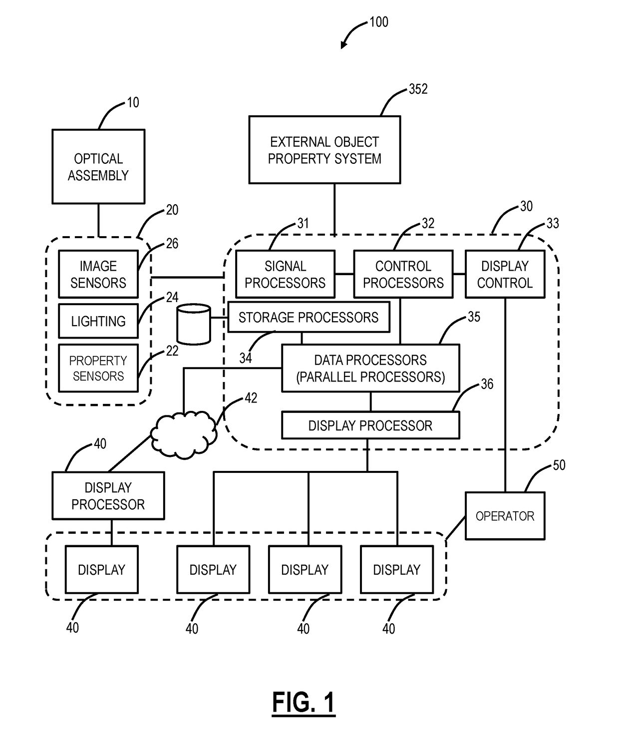
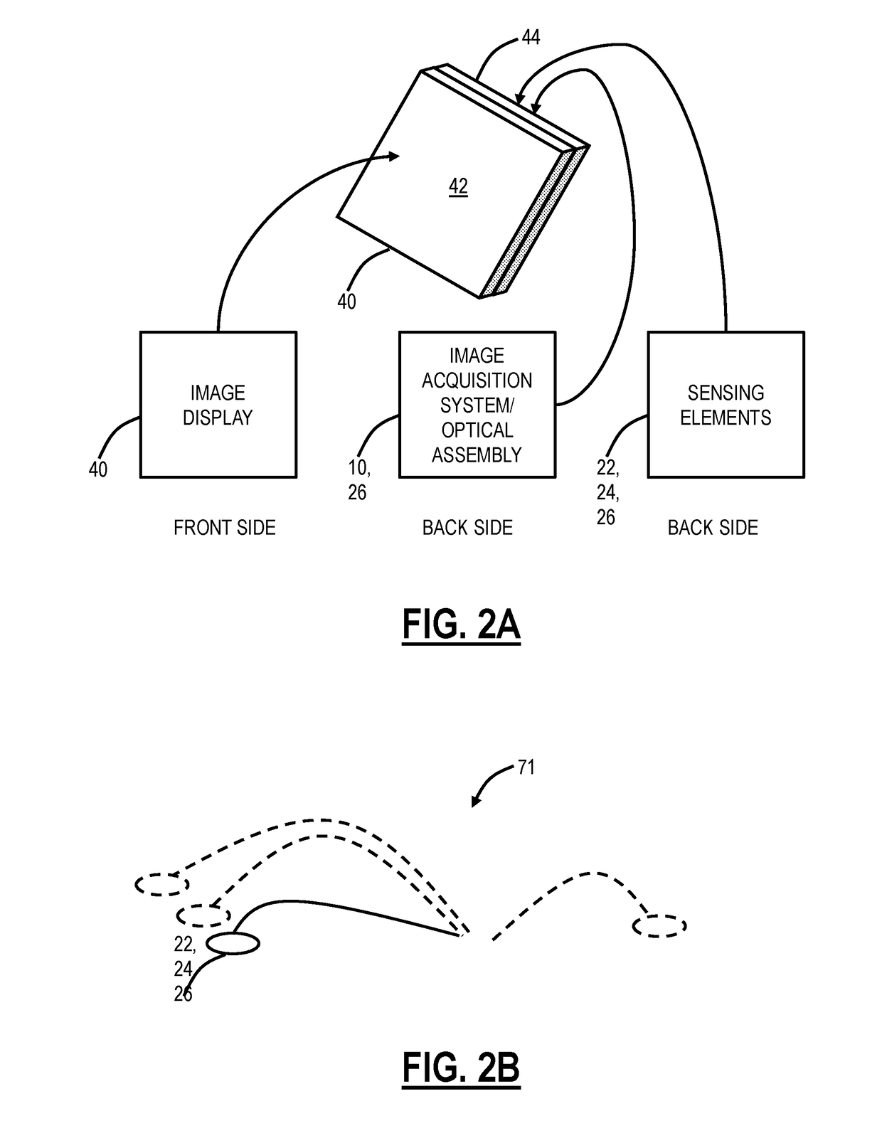
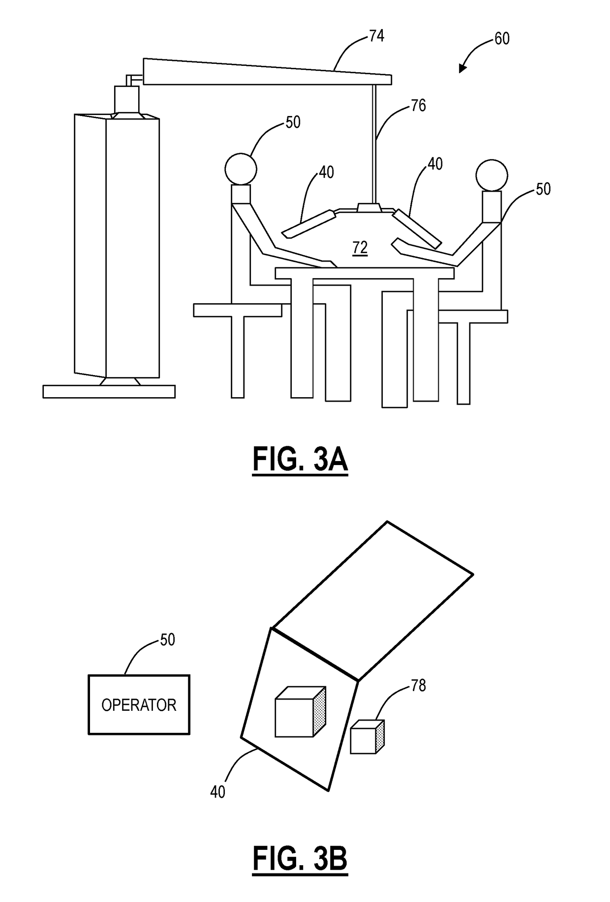

Patent
Innovation
- Enhanced optical path design that improves depth perception accuracy by optimizing the convergence angle between left and right optical channels in stereo microscope systems.
- Implementation of digital image processing algorithms to quantify and enhance depth cues, providing measurable depth perception metrics for comparison between dissecting and stereo microscopes.
- Novel ergonomic eyepiece configuration that reduces eye strain while maintaining superior depth perception through optimized interpupillary distance adjustment mechanisms.
Optical Design Standards for Stereo Microscopes
Optical design standards for stereo microscopes establish fundamental principles that directly influence depth perception capabilities, distinguishing them from conventional dissecting microscopes. The International Organization for Standardization (ISO) 19012 series provides comprehensive guidelines for microscope optical systems, emphasizing stereoscopic angle requirements, optical axis convergence, and magnification consistency across both optical paths. These standards mandate minimum stereoscopic angles typically ranging from 10 to 16 degrees, which creates the parallax necessary for three-dimensional visualization. The convergence point of dual optical axes must align precisely with the working distance to ensure optimal depth discrimination.
Modern stereo microscope designs adhere to the Greenough or Common Main Objective (CMO) configurations, each governed by distinct optical principles. The Greenough system employs two independent optical paths inclined at specific angles, typically 12-15 degrees, creating natural stereoscopic viewing. Standards require precise alignment tolerances within 0.1 degrees to prevent image distortion or viewer fatigue. The CMO design utilizes a shared objective lens with beam-splitting prisms, demanding stringent specifications for prism angle accuracy and coating uniformity to maintain equal light transmission and image quality between channels.
Numerical aperture (NA) specifications significantly impact depth of field and resolution characteristics. Standards typically specify NA ranges from 0.05 to 0.3 for stereo microscopes, balancing resolution requirements against the extended working distances necessary for manipulation tasks. The relationship between NA, working distance, and depth perception follows established optical formulas, with standards requiring manufacturers to provide detailed specifications enabling users to calculate expected depth discrimination capabilities.
Chromatic aberration correction standards have evolved substantially, with modern achromatic and apochromatic designs minimizing color fringing that can compromise depth judgment. Standards mandate specific correction levels across the visible spectrum, typically requiring residual chromatic aberration below 2 micrometers across the field of view. Additionally, field flatness specifications ensure consistent focus and depth perception across the entire viewing area, with maximum deviation standards typically set at less than 5% of the depth of field at the field periphery.
Modern stereo microscope designs adhere to the Greenough or Common Main Objective (CMO) configurations, each governed by distinct optical principles. The Greenough system employs two independent optical paths inclined at specific angles, typically 12-15 degrees, creating natural stereoscopic viewing. Standards require precise alignment tolerances within 0.1 degrees to prevent image distortion or viewer fatigue. The CMO design utilizes a shared objective lens with beam-splitting prisms, demanding stringent specifications for prism angle accuracy and coating uniformity to maintain equal light transmission and image quality between channels.
Numerical aperture (NA) specifications significantly impact depth of field and resolution characteristics. Standards typically specify NA ranges from 0.05 to 0.3 for stereo microscopes, balancing resolution requirements against the extended working distances necessary for manipulation tasks. The relationship between NA, working distance, and depth perception follows established optical formulas, with standards requiring manufacturers to provide detailed specifications enabling users to calculate expected depth discrimination capabilities.
Chromatic aberration correction standards have evolved substantially, with modern achromatic and apochromatic designs minimizing color fringing that can compromise depth judgment. Standards mandate specific correction levels across the visible spectrum, typically requiring residual chromatic aberration below 2 micrometers across the field of view. Additionally, field flatness specifications ensure consistent focus and depth perception across the entire viewing area, with maximum deviation standards typically set at less than 5% of the depth of field at the field periphery.
Ergonomic Factors in Microscope Depth Perception
Ergonomic considerations play a critical role in determining the effectiveness of depth perception when operating dissecting and stereo microscopes. The physical design and operational characteristics of each microscope type impose distinct postural and visual demands on users, which directly influence their ability to accurately perceive three-dimensional structures. Prolonged microscope use often leads to musculoskeletal strain, visual fatigue, and reduced perceptual accuracy, making ergonomic optimization essential for maintaining both operator comfort and diagnostic precision.
The working distance between the objective lens and specimen differs significantly between dissecting and stereo microscopes, affecting user posture and head positioning. Dissecting microscopes typically offer longer working distances, allowing operators to maintain a more natural neck angle and reducing cervical strain during extended observation periods. This extended working space also facilitates manual manipulation of specimens without compromising visual access. Conversely, stereo microscopes with shorter working distances may require users to adopt forward-leaning postures, increasing the risk of neck and shoulder discomfort that can indirectly impair depth judgment through fatigue-induced attention deficits.
Interpupillary distance adjustment mechanisms represent another crucial ergonomic factor influencing depth perception quality. Both microscope types require proper alignment with the user's interpupillary distance to achieve optimal stereoscopic fusion. However, the ease and precision of these adjustments vary across designs, with some systems offering continuous adjustment while others provide stepped increments. Misalignment forces the visual system to compensate through vergence adjustments, causing eye strain and reducing the reliability of depth cues over time.
Eyepiece height and angle adjustability further impact the ergonomic profile of depth perception tasks. Microscopes with flexible eyepiece positioning enable users to maintain neutral spine alignment while viewing, reducing cumulative fatigue during lengthy procedures. Fixed eyepiece configurations may force operators into compromised postures that accelerate the onset of discomfort and diminish sustained attention to subtle depth variations. The integration of adjustable diopter settings in individual eyepieces also accommodates users with refractive differences between eyes, ensuring balanced binocular input essential for accurate stereopsis.
Illumination ergonomics constitute an often-overlooked factor affecting depth perception performance. Glare, uneven lighting, and excessive brightness levels can cause visual discomfort that disrupts the delicate balance of binocular vision required for depth discrimination. Microscope designs incorporating adjustable, diffused lighting systems with appropriate color temperature support prolonged observation without inducing photostress, thereby maintaining consistent depth perception capabilities throughout extended work sessions.
The working distance between the objective lens and specimen differs significantly between dissecting and stereo microscopes, affecting user posture and head positioning. Dissecting microscopes typically offer longer working distances, allowing operators to maintain a more natural neck angle and reducing cervical strain during extended observation periods. This extended working space also facilitates manual manipulation of specimens without compromising visual access. Conversely, stereo microscopes with shorter working distances may require users to adopt forward-leaning postures, increasing the risk of neck and shoulder discomfort that can indirectly impair depth judgment through fatigue-induced attention deficits.
Interpupillary distance adjustment mechanisms represent another crucial ergonomic factor influencing depth perception quality. Both microscope types require proper alignment with the user's interpupillary distance to achieve optimal stereoscopic fusion. However, the ease and precision of these adjustments vary across designs, with some systems offering continuous adjustment while others provide stepped increments. Misalignment forces the visual system to compensate through vergence adjustments, causing eye strain and reducing the reliability of depth cues over time.
Eyepiece height and angle adjustability further impact the ergonomic profile of depth perception tasks. Microscopes with flexible eyepiece positioning enable users to maintain neutral spine alignment while viewing, reducing cumulative fatigue during lengthy procedures. Fixed eyepiece configurations may force operators into compromised postures that accelerate the onset of discomfort and diminish sustained attention to subtle depth variations. The integration of adjustable diopter settings in individual eyepieces also accommodates users with refractive differences between eyes, ensuring balanced binocular input essential for accurate stereopsis.
Illumination ergonomics constitute an often-overlooked factor affecting depth perception performance. Glare, uneven lighting, and excessive brightness levels can cause visual discomfort that disrupts the delicate balance of binocular vision required for depth discrimination. Microscope designs incorporating adjustable, diffused lighting systems with appropriate color temperature support prolonged observation without inducing photostress, thereby maintaining consistent depth perception capabilities throughout extended work sessions.
Unlock deeper insights with Patsnap Eureka Quick Research — get a full tech report to explore trends and direct your research. Try now!
Generate Your Research Report Instantly with AI Agent
Supercharge your innovation with Patsnap Eureka AI Agent Platform!