X-ray diagnostic device

a diagnostic device and x-ray technology, applied in the field of x-ray diagnostic devices, can solve the problems of large space occupation, inability to provide a good display of soft tissue, and relatively expensive ct devices

Inactive Publication Date: 2007-11-22
SIEMENS HEALTHCARE GMBH
View PDF16 Cites 66 Cited by
  • Summary
  • Abstract
  • Description
  • Claims
  • Application Information

AI Technical Summary

Benefits of technology

[0020]The device for correcting can advantageously be embodied in such a way as to effect calibration of the image recording system, for example geometry, distortion correction, intensity and/or

Problems solved by technology

What, though, is disadvantageous is that CT examinations can be carried out only on a patient who is in a lying position; moreover, a CT device is a relativ

Method used


Image

  • X-ray diagnostic device
  • X-ray diagnostic device
  • X-ray diagnostic device

Examples


Embodiment Construction

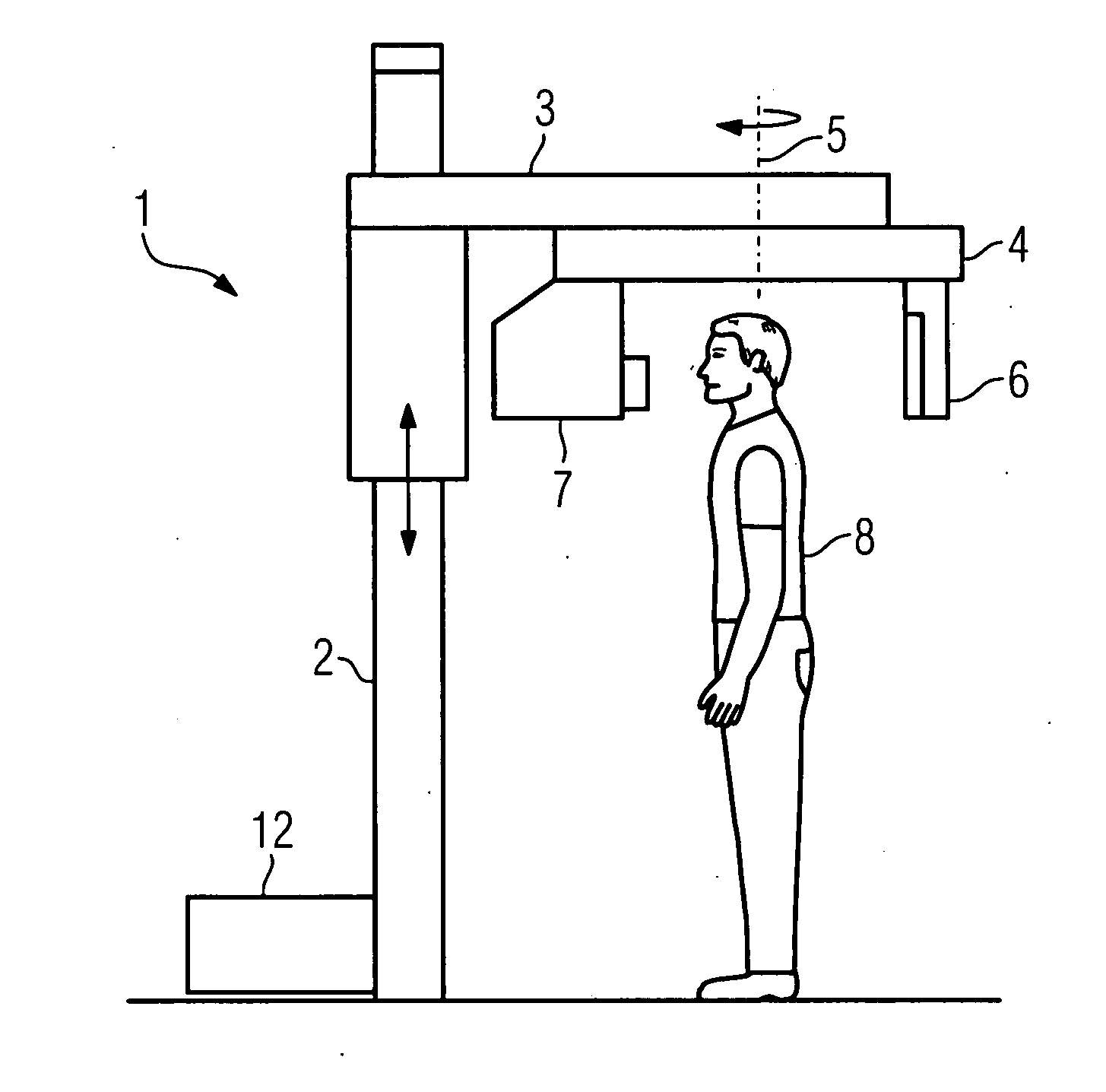
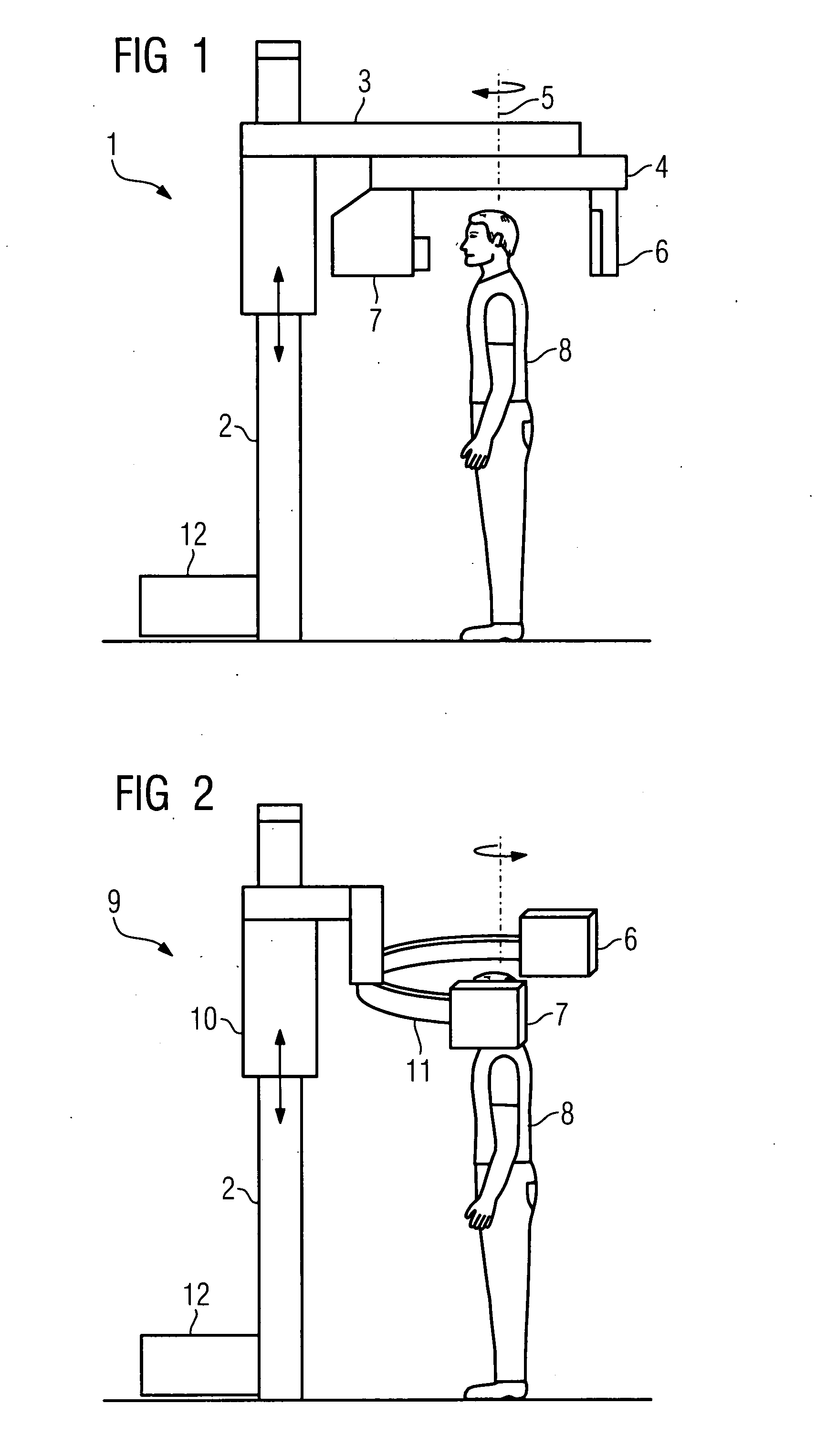
[0029]The X-ray diagnostic device 1 shown in FIG. 1 includes a stand 2 that is embodied as a floor stand and to which is attached a height-adjustable support arm 3. The support arm 3 is embodied as a boom; a second support arm 4 is attached thereto. The second support arm 4 is rotatable around a vertical axis 5. Attached to one end of the support arm 4 is an image detector embodied as a flat-panel detector 6. Attached to the other end of the support arm 4 is an X-ray emitter 7. The X-ray emitter 7 or, as the case may be, emitter unit includes an X-ray tube, a diaphragm, and a filter. As is shown in FIG. 1, the cranium of a patient 8 can be examined by means of the X-ray diagnostic device 1; dental or orthopedic examinations can also be performed on a patient who is in a seated or lying position. The support arm 4 and hence the flat-panel detector 6 and X-ray emitter 7 rotate during the examination so that projection images are recorded in rapid succession from different projections....


PUM


Abstract

There is described an X-ray diagnostic device for performing cephalometric, dental or orthopedic examinations on a patient who is seated or standing. The X-ray diagnostic device comprises an X-ray emitter and an image detector embodied as a flat-panel detector that are arranged situated opposite each other on an orbitally moveable mount. The X-ray diagnostic device further comprises means for adjusting the height of the X-ray emitter and the image detector, a digital image system for recording a projection image using rotation angiography, a device for image processing for reconstructing the projection image into a 3D volume image; and a device for correcting physical effects or artifacts for representing soft tissue in the projection image and in the 3D volume image reconstructed therefrom.

Description

CROSS REFERENCE TO RELATED APPLICATIONS[0001]This application claims priority of German application No. 10 2006 021 373.4 DE filed May 8, 2006, which is incorporated by reference herein in its entirety.FIELD OF INVENTION[0002]The invention relates to an X-ray diagnostic device for performing cephalometric, dental or orthopedic examinations on a patient who is seated or standing.BACKGROUND OF INVENTION[0003]Dental and orthopedic disorders occur very frequently, so in the event of said disorders fast and reliable diagnosis and directly initiated therapy are of particular importance for the healing process.[0004]The diagnosing of disorders of said type is supported by imaging methods, with an X-ray or CT examination frequently being performed for the relevant purpose. What, though, is disadvantageous is that CT examinations can be carried out only on a patient who is in a lying position; moreover, a CT device is a relatively expensive examination modality requiring a large amount of sp...

Claims


Application Information

Patent Timeline
IPC IPC(8): A61B6/14
CPCA61B6/4441A61B6/14
Inventor MASCHKE, MICHAEL
Owner SIEMENS HEALTHCARE GMBH
Why Eureka
Try Eureka