
X-ray photography apparatus
a technology of x-ray and photography equipment, which is applied in the direction of radiation diagnostic diaphragms, patient positioning for diagnostics, applications, etc., can solve the problems of large burden on the patient, inability to take x-ray images in image diagnosis, and shortening the time of tooth taking
Patent Information
- Authority / Receiving Office
- US · United States
- Patent Type
- Patents(United States)
- Current Assignee / Owner
- Publication Date
- 2021-02-02
Smart Images
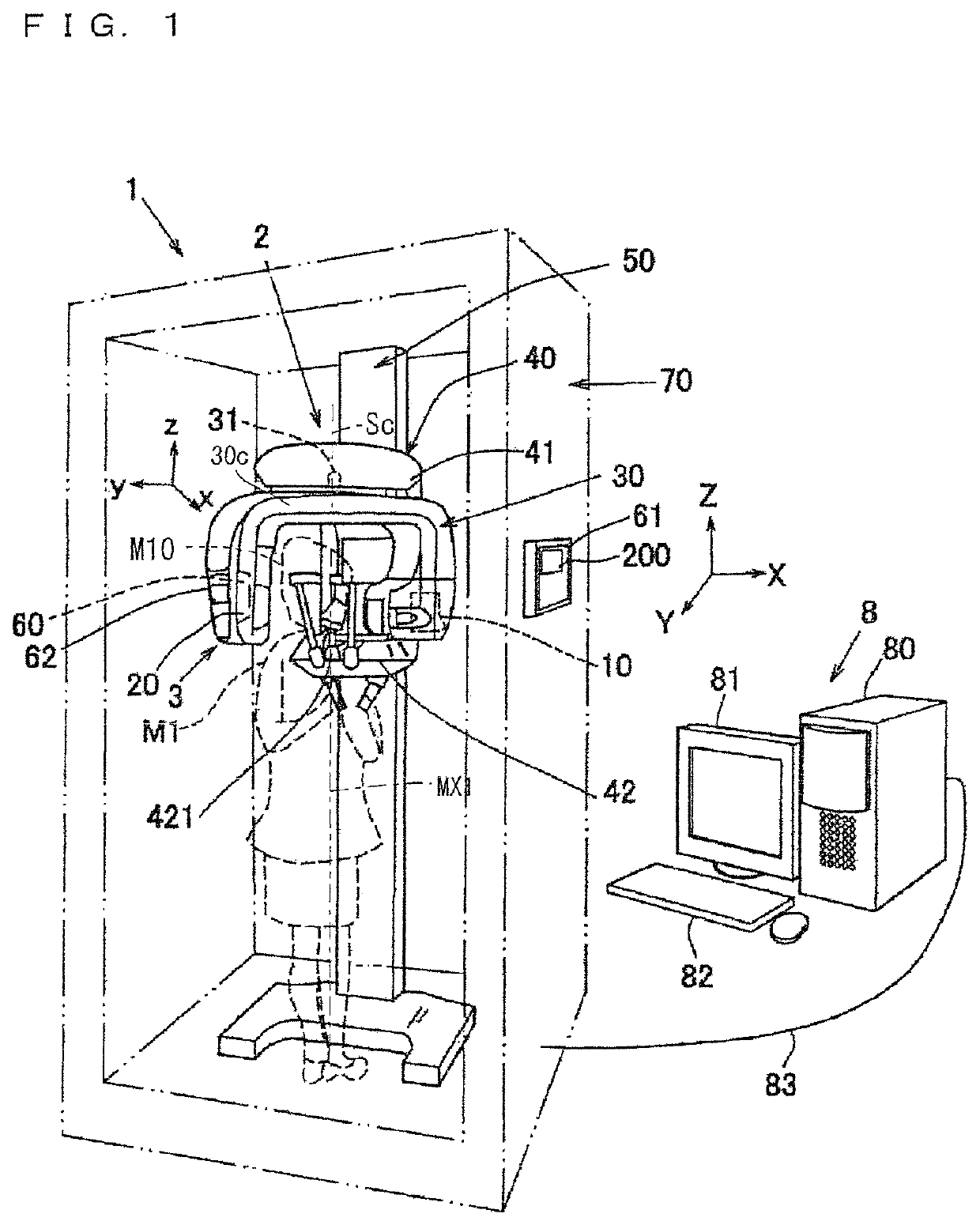
Figure 1 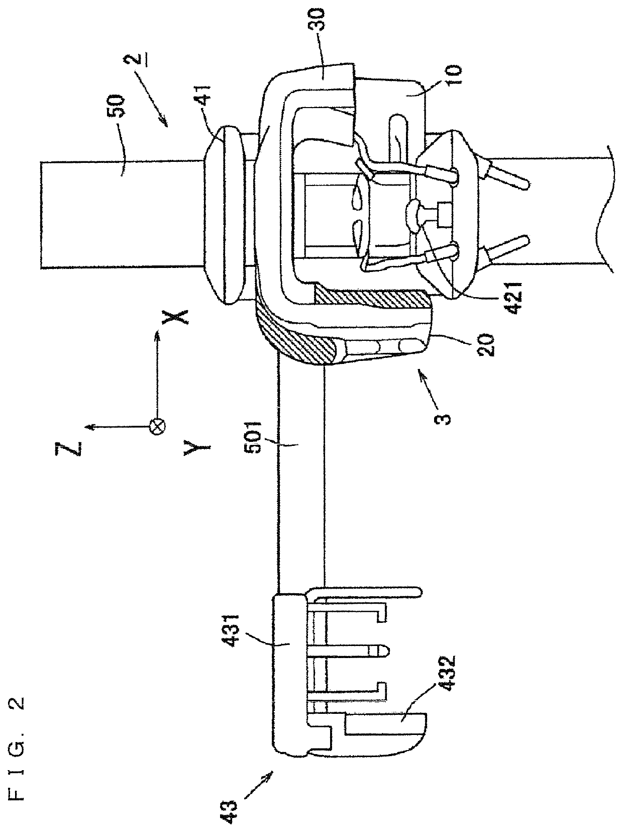
Figure 2 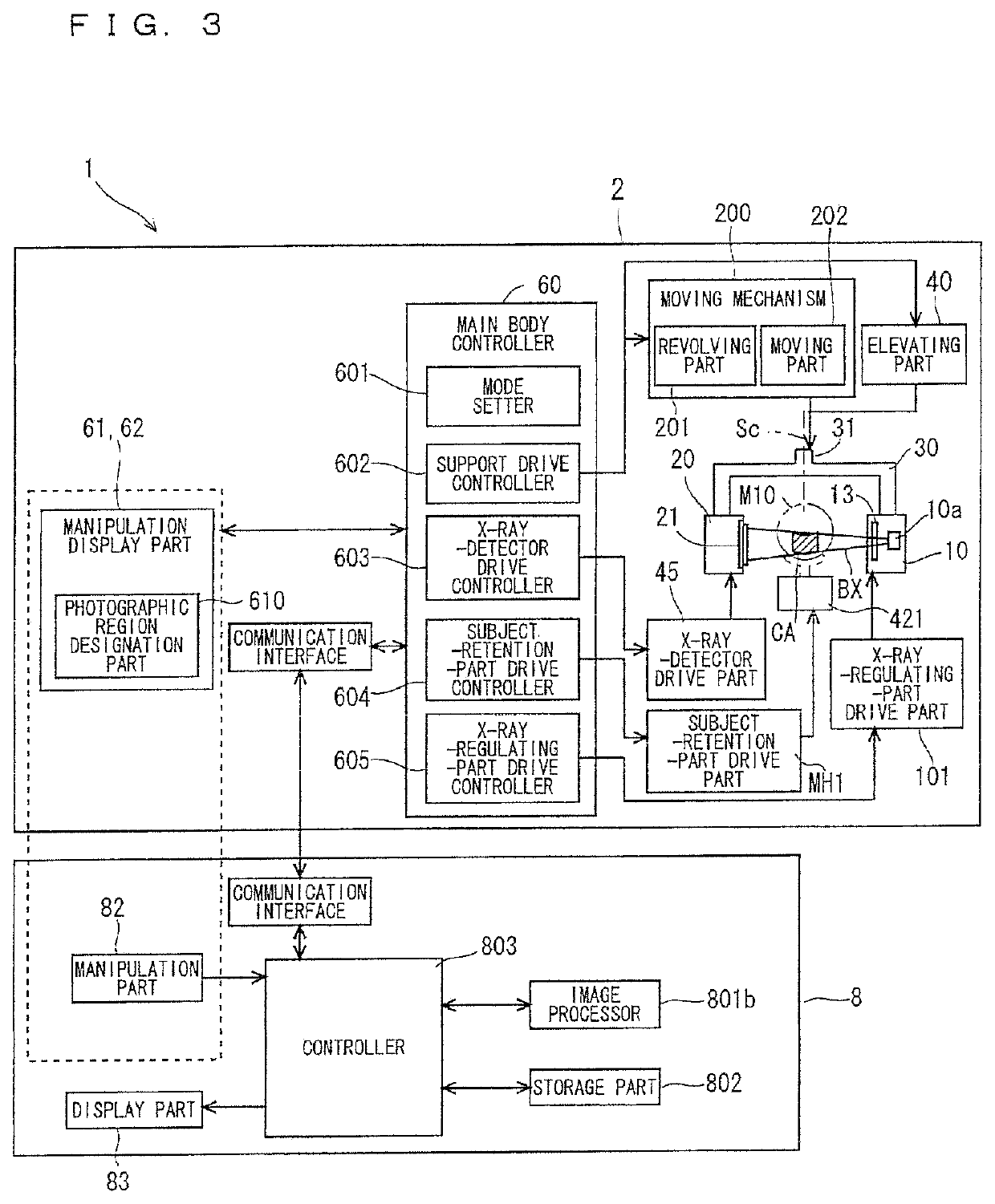
Figure 3
Abstract
Description
BACKGROUND OF THE INVENTION
[0001] 1. Field of the Invention
[0002] The present invention relates to an X-ray photography apparatus.
[0003] 2. Description of the Background Art
[0004] Conventionally what is called intraoral radiography (dental radiography), in which an X-ray detection means (such as an X-ray film and an X-ray sensor panel) is set in the oral cavity of a patient to photograph part of a row of teeth or gums, is performed in X-ray photography of the dental field. Because only a local region is irradiated with an X-ray in the intraoral radiography, advantageously the photographing is simply performed and X-ray exposure is reduced. However, it is necessary to dispose in advance the X-ray detection means in the oral cavity of the patient, and thus a large burden is placed on the patient.
[0005] Therefore, Japanese Patent Application Laid-Open No. 2007-136163 has made a proposal that panoramic photography is performed using a panoramic image photographing device and a tomographic im...
Examples
Embodiment Construction
[0073]Hereinafter, preferred embodiments of the present invention will be described with reference to the accompanying drawings. In the drawings, for the sake of convenience, occasionally, the size or the number of pieces of each component is indicated while magnified or simplified as occasion demands.
[0074]1. Preferred Embodiment
[0075]FIG. 1 is a schematic perspective view of an X-ray photography apparatus 1 according to a preferred embodiment of the present invention. FIG. 2 is a partial front view of the X-ray photography apparatus 1 on which a cephalostat 43 is mounted. FIG. 3 is a block diagram of the configuration of the X-ray photography apparatus 1.
[0076]The X-ray photography apparatus 1 is substantially comprised of manipulation display parts 61 and 62, a main body 2, and an image processing device 8. The manipulation display parts 61 and 62 act as display element while setting a photographic region CA. The main body 2 collects X-ray projection data (frame data) by performi...