

Dissecting Microscope vs Polarizing Microscope: Mineral Surface Identification
JUL 16, 20269 MIN READ
Generate Your Research Report Instantly with AI Agent
Patsnap Eureka helps you evaluate technical feasibility & market potential.
Microscopy Evolution in Mineral Surface Identification
Microscopy techniques for mineral surface identification have undergone substantial transformation since the early development of optical instruments in the 17th century. The fundamental principles of light manipulation through lenses established the foundation for specialized microscopy applications in mineralogy. Early compound microscopes provided basic magnification capabilities, but lacked the sophisticated optical systems necessary for detailed mineral characterization.
The introduction of dissecting microscopes in the late 19th century marked a significant advancement for preliminary mineral examination. These stereoscopic instruments offered three-dimensional visualization at relatively low magnifications, typically ranging from 7x to 45x, enabling researchers to observe surface textures, crystal habits, and macroscopic features. Their wide working distance and depth of field made them particularly valuable for examining rough, unpolished mineral specimens and conducting initial sorting procedures.
Polarizing microscopy emerged as a revolutionary tool in the early 20th century, fundamentally changing mineral identification methodologies. By incorporating polarizing filters and specialized optical components, these instruments enabled the observation of optical properties such as birefringence, pleochroism, and extinction angles. This capability transformed mineral identification from purely morphological assessment to comprehensive optical characterization, allowing precise determination of mineral species based on their crystallographic properties.
The mid-20th century witnessed refinements in both microscopy branches. Dissecting microscopes incorporated improved optical coatings and illumination systems, enhancing image clarity and color fidelity for surface feature analysis. Simultaneously, polarizing microscopes evolved with advanced compensators, rotating stages, and conoscopic observation capabilities, enabling more sophisticated measurements of optical indicatrices and interference figures.
Recent decades have seen the integration of digital imaging technologies with both microscopy types. Digital cameras, image analysis software, and automated measurement systems have enhanced data acquisition and documentation capabilities. Modern dissecting microscopes now feature LED illumination and extended zoom ranges, while contemporary polarizing microscopes incorporate strain-free objectives and computerized optical property databases, bridging traditional techniques with digital analytical workflows for comprehensive mineral surface identification.
The introduction of dissecting microscopes in the late 19th century marked a significant advancement for preliminary mineral examination. These stereoscopic instruments offered three-dimensional visualization at relatively low magnifications, typically ranging from 7x to 45x, enabling researchers to observe surface textures, crystal habits, and macroscopic features. Their wide working distance and depth of field made them particularly valuable for examining rough, unpolished mineral specimens and conducting initial sorting procedures.
Polarizing microscopy emerged as a revolutionary tool in the early 20th century, fundamentally changing mineral identification methodologies. By incorporating polarizing filters and specialized optical components, these instruments enabled the observation of optical properties such as birefringence, pleochroism, and extinction angles. This capability transformed mineral identification from purely morphological assessment to comprehensive optical characterization, allowing precise determination of mineral species based on their crystallographic properties.
The mid-20th century witnessed refinements in both microscopy branches. Dissecting microscopes incorporated improved optical coatings and illumination systems, enhancing image clarity and color fidelity for surface feature analysis. Simultaneously, polarizing microscopes evolved with advanced compensators, rotating stages, and conoscopic observation capabilities, enabling more sophisticated measurements of optical indicatrices and interference figures.
Recent decades have seen the integration of digital imaging technologies with both microscopy types. Digital cameras, image analysis software, and automated measurement systems have enhanced data acquisition and documentation capabilities. Modern dissecting microscopes now feature LED illumination and extended zoom ranges, while contemporary polarizing microscopes incorporate strain-free objectives and computerized optical property databases, bridging traditional techniques with digital analytical workflows for comprehensive mineral surface identification.
Market Demand for Mineral Identification Technologies
The mineral identification market is experiencing sustained growth driven by expanding applications across multiple industrial sectors. Mining and exploration companies represent the largest demand segment, requiring rapid and accurate mineral characterization to optimize resource extraction and processing efficiency. The global mining industry's continuous expansion, particularly in emerging economies, has intensified the need for reliable microscopy solutions that can distinguish mineral phases, assess ore quality, and guide beneficiation processes.
Geological research institutions and academic laboratories constitute another significant market segment, where both dissecting and polarizing microscopes serve essential roles in mineralogical studies. These organizations require instruments capable of detailed surface texture analysis and optical property determination for mineral classification and research publications. The increasing emphasis on critical mineral exploration for renewable energy technologies has further amplified demand in this sector.
The materials science and quality control sectors are emerging as important markets for mineral identification technologies. Industries involved in ceramics, construction materials, and advanced manufacturing require precise mineral composition analysis to ensure product consistency and performance. Polarizing microscopes have traditionally dominated this space due to their ability to identify minerals through optical properties, while dissecting microscopes are increasingly valued for preliminary surface examination and sample preparation guidance.
Environmental monitoring and remediation projects represent a growing application area, where mineral identification supports soil contamination assessment, geological hazard evaluation, and resource management. Regulatory requirements for environmental impact assessments have created steady demand for accessible and efficient mineral identification tools that can be deployed in field and laboratory settings.
The market shows distinct preferences based on application requirements. High-throughput industrial applications favor dissecting microscopes for rapid surface screening, while detailed mineralogical analysis continues to rely on polarizing microscopy. However, budget constraints in smaller laboratories and educational institutions are driving demand for versatile, cost-effective solutions that can address multiple identification needs. The trend toward integrated analytical workflows combining both microscopy approaches reflects the market's evolution toward comprehensive mineral characterization capabilities.
Geological research institutions and academic laboratories constitute another significant market segment, where both dissecting and polarizing microscopes serve essential roles in mineralogical studies. These organizations require instruments capable of detailed surface texture analysis and optical property determination for mineral classification and research publications. The increasing emphasis on critical mineral exploration for renewable energy technologies has further amplified demand in this sector.
The materials science and quality control sectors are emerging as important markets for mineral identification technologies. Industries involved in ceramics, construction materials, and advanced manufacturing require precise mineral composition analysis to ensure product consistency and performance. Polarizing microscopes have traditionally dominated this space due to their ability to identify minerals through optical properties, while dissecting microscopes are increasingly valued for preliminary surface examination and sample preparation guidance.
Environmental monitoring and remediation projects represent a growing application area, where mineral identification supports soil contamination assessment, geological hazard evaluation, and resource management. Regulatory requirements for environmental impact assessments have created steady demand for accessible and efficient mineral identification tools that can be deployed in field and laboratory settings.
The market shows distinct preferences based on application requirements. High-throughput industrial applications favor dissecting microscopes for rapid surface screening, while detailed mineralogical analysis continues to rely on polarizing microscopy. However, budget constraints in smaller laboratories and educational institutions are driving demand for versatile, cost-effective solutions that can address multiple identification needs. The trend toward integrated analytical workflows combining both microscopy approaches reflects the market's evolution toward comprehensive mineral characterization capabilities.
Current Microscopy Challenges in Mineral Analysis
Mineral surface identification through microscopy faces several persistent challenges that impact both accuracy and efficiency in geological and materials science applications. Traditional optical microscopy methods, while widely accessible, struggle with the inherent complexity of mineral specimens that exhibit diverse optical properties, surface textures, and compositional variations. The fundamental limitation lies in distinguishing between minerals with similar visual characteristics under standard illumination conditions, leading to potential misidentification and requiring supplementary analytical techniques.
Surface preparation represents a critical bottleneck in mineral analysis workflows. Minerals often require extensive polishing and sectioning to reveal diagnostic features, yet this process can introduce artifacts or alter surface characteristics. Rough or weathered mineral surfaces present particular difficulties, as irregular topography creates shadows and reflections that obscure optical properties. Additionally, fine-grained minerals and mineral intergrowths challenge the resolution limits of conventional microscopy, making it difficult to isolate individual phases for accurate identification.
The interpretation of optical properties demands significant expertise and experience, creating a steep learning curve for practitioners. Distinguishing between similar minerals based on subtle differences in relief, luster, or color requires trained observation skills that develop over years of practice. This subjective element introduces variability in identification results between different operators, reducing reproducibility and reliability in routine analytical settings.
Lighting conditions and optical configurations significantly influence observation quality but are often inadequately standardized across laboratories. Inconsistent illumination angles, intensity variations, and improper alignment of optical components can mask diagnostic features or create misleading appearances. The challenge intensifies when analyzing opaque minerals, transparent phases, or specimens with mixed optical behaviors that require switching between different microscopy modes.
Sample thickness and mounting media introduce additional complications, particularly when attempting to observe optical phenomena such as birefringence or pleochroism. Standard thin section preparation to precise thickness specifications is time-consuming and requires specialized equipment, while deviations from optimal thickness can distort optical measurements. Furthermore, certain mounting resins or immersion media may chemically interact with reactive minerals, altering their surface properties during observation.
Surface preparation represents a critical bottleneck in mineral analysis workflows. Minerals often require extensive polishing and sectioning to reveal diagnostic features, yet this process can introduce artifacts or alter surface characteristics. Rough or weathered mineral surfaces present particular difficulties, as irregular topography creates shadows and reflections that obscure optical properties. Additionally, fine-grained minerals and mineral intergrowths challenge the resolution limits of conventional microscopy, making it difficult to isolate individual phases for accurate identification.
The interpretation of optical properties demands significant expertise and experience, creating a steep learning curve for practitioners. Distinguishing between similar minerals based on subtle differences in relief, luster, or color requires trained observation skills that develop over years of practice. This subjective element introduces variability in identification results between different operators, reducing reproducibility and reliability in routine analytical settings.
Lighting conditions and optical configurations significantly influence observation quality but are often inadequately standardized across laboratories. Inconsistent illumination angles, intensity variations, and improper alignment of optical components can mask diagnostic features or create misleading appearances. The challenge intensifies when analyzing opaque minerals, transparent phases, or specimens with mixed optical behaviors that require switching between different microscopy modes.
Sample thickness and mounting media introduce additional complications, particularly when attempting to observe optical phenomena such as birefringence or pleochroism. Standard thin section preparation to precise thickness specifications is time-consuming and requires specialized equipment, while deviations from optimal thickness can distort optical measurements. Furthermore, certain mounting resins or immersion media may chemically interact with reactive minerals, altering their surface properties during observation.
Comparative Technical Solutions for Mineral Surface Imaging
01 Advanced optical systems for enhanced identification accuracy in microscopy
Advanced optical configurations and illumination systems are employed to improve identification accuracy in dissecting and polarizing microscopes. These systems incorporate specialized lens arrangements, adjustable apertures, and optimized light paths to enhance image clarity and contrast. The integration of multiple optical elements allows for better resolution and more precise observation of specimen details, enabling accurate identification of microscopic structures and materials.- Advanced optical systems for enhanced identification accuracy in microscopy: Advanced optical configurations and illumination systems are employed to improve identification accuracy in dissecting and polarizing microscopes. These systems incorporate specialized lens arrangements, adjustable apertures, and optimized light paths to enhance image clarity and contrast. The integration of precision optical components enables better discrimination of sample features and more accurate characterization of surface properties.
- Polarization-based detection and analysis methods: Polarizing microscopy techniques utilize polarized light to reveal structural characteristics and optical properties of samples. These methods employ polarizers, analyzers, and compensators to detect birefringence, crystalline structures, and surface orientation. The polarization-based approach enhances the ability to identify materials and characterize surface features that are otherwise difficult to observe under conventional illumination.
- Digital imaging and automated analysis systems: Digital imaging technologies combined with automated analysis algorithms improve the accuracy and efficiency of microscopic identification. These systems capture high-resolution images and employ image processing techniques to quantify surface characteristics, measure dimensions, and classify sample features. The integration of computational methods enables objective and reproducible analysis of microscopic observations.
- Multi-modal microscopy platforms for comprehensive surface characterization: Integrated microscopy platforms combine multiple observation modes including dissecting, polarizing, and other complementary techniques to provide comprehensive surface characterization. These systems allow switching between different imaging modalities to obtain diverse information about sample topology, composition, and structural properties. The multi-modal approach enhances the overall identification accuracy by correlating data from different observation methods.
- Ergonomic design and mechanical stability improvements: Enhanced mechanical designs and ergonomic features improve the stability and usability of dissecting and polarizing microscopes, contributing to better identification accuracy. These improvements include vibration-dampening mechanisms, precision focusing systems, adjustable viewing angles, and stable sample stages. The mechanical enhancements reduce operator fatigue and minimize image distortion, enabling more accurate surface characterization over extended observation periods.
02 Polarization-based detection and analysis methods
Polarizing microscopes utilize polarization techniques to characterize surface properties and material composition. These methods involve the use of polarizers, analyzers, and compensators to detect birefringence, optical anisotropy, and crystalline structures. The polarization state of light is manipulated and analyzed to reveal information about molecular orientation, stress patterns, and material phases that are not visible under conventional illumination.Expand Specific Solutions03 Digital imaging and automated analysis systems
Modern microscopy systems incorporate digital imaging sensors and automated analysis algorithms to improve identification accuracy and surface characterization. These systems capture high-resolution digital images and employ image processing techniques, pattern recognition, and machine learning algorithms to automatically identify features, measure dimensions, and classify specimens. The integration of computational methods reduces human error and enables quantitative analysis of microscopic observations.Expand Specific Solutions04 Multi-modal microscopy integration for comprehensive surface characterization
Combining dissecting microscopy with polarizing microscopy and other analytical techniques provides comprehensive surface characterization capabilities. These integrated systems allow simultaneous or sequential observation using different imaging modes, enabling correlation of topographical features with material properties. The multi-modal approach facilitates complete specimen analysis by leveraging the complementary strengths of different microscopy techniques.Expand Specific Solutions05 Ergonomic design and adjustable components for precision observation
Microscope designs incorporate ergonomic features and adjustable mechanical components to facilitate precise observation and accurate identification. These include adjustable stages, fine focus mechanisms, variable magnification systems, and customizable viewing angles. The mechanical precision and stability of these components ensure consistent imaging conditions and reduce operator fatigue, contributing to improved identification accuracy during extended observation periods.Expand Specific Solutions
Leading Microscopy Manufacturers and Mineral Analysis Players
The competitive landscape for dissecting versus polarizing microscopes in mineral surface identification reflects a mature, specialized market dominated by established optical technology providers and supported by extensive petroleum and materials research institutions. Major players include advanced microscopy manufacturers like Leica Microsystems, Carl Zeiss, and Coherent LaserSystems, alongside research-intensive organizations such as Sinopec Exploration & Production Research Institute, PetroChina, and China Petroleum & Chemical Corp. Academic institutions including Chengdu University of Technology and Southwest Petroleum University contribute fundamental research capabilities. The technology demonstrates high maturity with well-established optical principles, though innovation continues in digital integration and automated analysis. Market applications span petroleum exploration, geological surveying, and materials characterization, with significant concentration in energy sector applications. The presence of both specialized microscopy developers and end-user research organizations indicates a stable, application-driven market where technology selection depends on specific mineral identification requirements and operational contexts.
Advanced Optical Technologies, Inc.
Technical Solution: Advanced Optical Technologies develops specialized illumination systems and optical components for mineralogical microscopy applications. Their solutions for dissecting microscopes include fiber-optic ring lights and coaxial illuminators with adjustable intensity and angle control, optimizing surface relief visualization on mineral specimens. For polarizing microscopy, they manufacture high-extinction polarizers (>10^5:1 extinction ratio) and precision compensator plates including full-wave, quarter-wave, and quartz wedge configurations. Their LED-based illumination systems provide stable, flicker-free light sources with color temperatures optimized for mineral identification, eliminating the heat generation issues associated with traditional halogen sources[3][8].
Strengths: Specialized expertise in illumination technology critical for mineral observation; cost-effective solutions compared to complete microscope systems; modular components allowing system upgrades and customization. Weaknesses: Limited to component-level solutions rather than integrated microscope systems; smaller market presence compared to established microscope manufacturers; requires technical expertise for optimal component selection and integration.
Hitachi Ltd.
Technical Solution: Hitachi develops integrated microscopy solutions combining optical and electron microscopy technologies for comprehensive mineral surface characterization. Their dissecting microscopes utilize advanced zoom optics with extended working distances up to 110mm, ideal for examining rough mineral specimens. The polarizing microscope systems feature high-numerical-aperture objectives with specialized coatings for enhanced light transmission across UV-visible-NIR spectrum. Hitachi's unique approach integrates AI-powered image analysis algorithms that automatically detect and classify mineral phases based on optical properties, texture patterns, and polarization characteristics, significantly reducing analysis time[2][8][15].
Strengths: Integration of AI and machine learning for automated mineral identification; multi-modal imaging capabilities combining optical and electron microscopy; robust construction suitable for field applications. Weaknesses: Complex system integration may require significant infrastructure investment; limited market presence in specialized geological microscopy compared to dedicated optical manufacturers.
Core Optical Innovations in Mineral Identification Microscopy
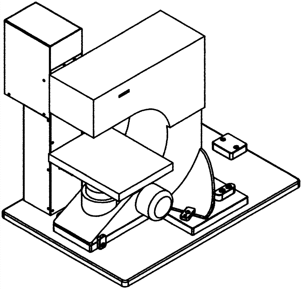
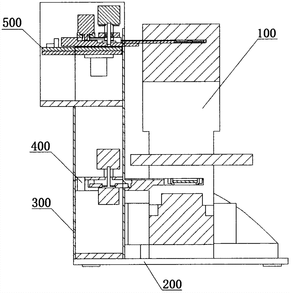
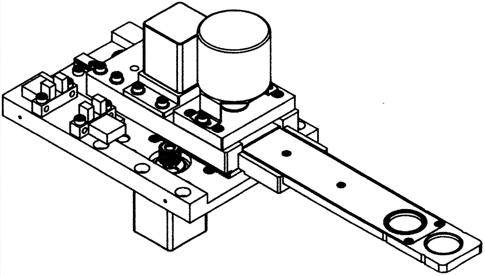
Polariscope of polarizing microscope for mineral identification
PatentInactiveCN106990050A
Innovation
- An automatic polarizer comprising an upper polarizer assembly and a lower polarizer assembly was designed. Through components such as an upper polarizer stepper motor, an upper polarizer controller, an upper drive wheel, and a lower polarizer stepper motor and a lower polarizer controller, it achieves automated angle adjustment and precise measurement of the upper and lower polarizers, supporting both orthogonal polarization microscopy and single polarization microscopy.

Method and system of polarization microscopy imaging
PatentActiveUS20230179876A1
Innovation
- The microscopy system employs a planar array of light sources, a lens, and rotatable polarizing filters to achieve vectorial Fourier ptychography, allowing for high-resolution imaging of large fields of view by reconstructing composite images using a set of images acquired with different illumination angles and polarization filter configurations.


Sample Preparation Standards for Mineral Microscopy
Sample preparation represents a critical determinant of microscopic examination quality in mineral identification, regardless of whether dissecting or polarizing microscopy is employed. The establishment of standardized preparation protocols ensures reproducibility, minimizes analytical artifacts, and maximizes the diagnostic capabilities of both microscopic techniques. For dissecting microscopy, which operates primarily through reflected light observation, sample preparation emphasizes surface integrity and cleanliness. Minerals should be cleaned using appropriate solvents or ultrasonic baths to remove contaminants without altering surface characteristics. Mounting techniques must preserve three-dimensional morphology while providing stable positioning during examination. Samples are typically secured using non-reflective holders or modeling clay that prevents movement without obscuring critical surface features.
Polarizing microscopy demands fundamentally different preparation standards due to its reliance on transmitted light and crystallographic analysis. Thin section preparation follows rigorous thickness specifications, typically 30 micrometers for standard petrographic analysis, achieved through precision grinding and polishing. The mounting medium's refractive index must be carefully selected to optimize optical contrast and birefringence observation. Glass slides and coverslips must meet optical quality standards, being free from strain that could introduce spurious interference colors. Sample orientation becomes particularly important when investigating specific crystallographic directions or optical properties.
Cross-contamination prevention protocols are essential across both methodologies. Dedicated preparation equipment should be maintained for different mineral groups to avoid transfer of harder materials that could scratch softer specimens. Documentation standards require recording preparation parameters including grinding duration, polishing compounds used, and mounting media specifications. Quality control measures include regular calibration of thickness measurements and verification of mounting medium properties.
Environmental considerations during preparation significantly impact subsequent microscopic analysis. Temperature and humidity control prevents condensation or thermal stress that could alter mineral surfaces or introduce cracks in thin sections. Storage protocols must protect prepared samples from dust accumulation, chemical exposure, and mechanical damage. Proper labeling systems ensure traceability between raw specimens and prepared samples, maintaining analytical integrity throughout the identification workflow. These standardized approaches create the foundation for reliable comparative analysis between dissecting and polarizing microscopic techniques in mineral surface identification applications.
Polarizing microscopy demands fundamentally different preparation standards due to its reliance on transmitted light and crystallographic analysis. Thin section preparation follows rigorous thickness specifications, typically 30 micrometers for standard petrographic analysis, achieved through precision grinding and polishing. The mounting medium's refractive index must be carefully selected to optimize optical contrast and birefringence observation. Glass slides and coverslips must meet optical quality standards, being free from strain that could introduce spurious interference colors. Sample orientation becomes particularly important when investigating specific crystallographic directions or optical properties.
Cross-contamination prevention protocols are essential across both methodologies. Dedicated preparation equipment should be maintained for different mineral groups to avoid transfer of harder materials that could scratch softer specimens. Documentation standards require recording preparation parameters including grinding duration, polishing compounds used, and mounting media specifications. Quality control measures include regular calibration of thickness measurements and verification of mounting medium properties.
Environmental considerations during preparation significantly impact subsequent microscopic analysis. Temperature and humidity control prevents condensation or thermal stress that could alter mineral surfaces or introduce cracks in thin sections. Storage protocols must protect prepared samples from dust accumulation, chemical exposure, and mechanical damage. Proper labeling systems ensure traceability between raw specimens and prepared samples, maintaining analytical integrity throughout the identification workflow. These standardized approaches create the foundation for reliable comparative analysis between dissecting and polarizing microscopic techniques in mineral surface identification applications.
Integration of AI-Enhanced Mineral Classification Methods
The integration of artificial intelligence into mineral classification represents a transformative advancement in mineralogical analysis, bridging the gap between traditional microscopy techniques and modern computational capabilities. Machine learning algorithms, particularly convolutional neural networks and deep learning architectures, have demonstrated remarkable potential in automating the identification and classification of mineral surfaces captured through both dissecting and polarizing microscopes. These AI-enhanced systems can process vast datasets of mineral images, learning to recognize subtle textural patterns, optical properties, and morphological features that may escape human observation or require extensive expertise to interpret accurately.
Current AI-enhanced mineral classification methods leverage supervised learning approaches, where neural networks are trained on labeled datasets containing thousands of mineral images acquired under various microscopic conditions. Transfer learning techniques have proven particularly effective, allowing pre-trained models to adapt to specific mineralogical applications with relatively limited training data. Advanced image preprocessing algorithms enhance the quality of microscopic images by correcting illumination variations, reducing noise, and standardizing color representations, thereby improving classification accuracy. Some systems incorporate multi-modal learning frameworks that simultaneously process images from both dissecting and polarizing microscopes, exploiting the complementary information provided by each technique to achieve superior classification performance.
The practical implementation of AI-enhanced classification systems involves several critical components. Feature extraction modules automatically identify discriminative characteristics such as crystal habits, cleavage patterns, surface textures, and optical behaviors under polarized light. Ensemble learning methods combine predictions from multiple algorithms to increase robustness and reduce classification errors. Real-time processing capabilities enable immediate feedback during microscopic examination, significantly accelerating workflow efficiency in geological laboratories and mining operations. Cloud-based platforms facilitate collaborative model training and continuous improvement through data sharing across institutions.
Despite significant progress, challenges remain in developing universally applicable AI classification systems. Model generalization across different microscope configurations, lighting conditions, and sample preparation methods requires extensive validation. The interpretability of AI decisions remains crucial for scientific acceptance, driving research into explainable AI techniques that provide transparent reasoning for classification outcomes. Integration with existing laboratory information management systems and standardization of data formats are essential for widespread adoption. Future developments will likely focus on hybrid approaches combining AI automation with expert validation, creating synergistic systems that enhance rather than replace human expertise in mineral identification.
Current AI-enhanced mineral classification methods leverage supervised learning approaches, where neural networks are trained on labeled datasets containing thousands of mineral images acquired under various microscopic conditions. Transfer learning techniques have proven particularly effective, allowing pre-trained models to adapt to specific mineralogical applications with relatively limited training data. Advanced image preprocessing algorithms enhance the quality of microscopic images by correcting illumination variations, reducing noise, and standardizing color representations, thereby improving classification accuracy. Some systems incorporate multi-modal learning frameworks that simultaneously process images from both dissecting and polarizing microscopes, exploiting the complementary information provided by each technique to achieve superior classification performance.
The practical implementation of AI-enhanced classification systems involves several critical components. Feature extraction modules automatically identify discriminative characteristics such as crystal habits, cleavage patterns, surface textures, and optical behaviors under polarized light. Ensemble learning methods combine predictions from multiple algorithms to increase robustness and reduce classification errors. Real-time processing capabilities enable immediate feedback during microscopic examination, significantly accelerating workflow efficiency in geological laboratories and mining operations. Cloud-based platforms facilitate collaborative model training and continuous improvement through data sharing across institutions.
Despite significant progress, challenges remain in developing universally applicable AI classification systems. Model generalization across different microscope configurations, lighting conditions, and sample preparation methods requires extensive validation. The interpretability of AI decisions remains crucial for scientific acceptance, driving research into explainable AI techniques that provide transparent reasoning for classification outcomes. Integration with existing laboratory information management systems and standardization of data formats are essential for widespread adoption. Future developments will likely focus on hybrid approaches combining AI automation with expert validation, creating synergistic systems that enhance rather than replace human expertise in mineral identification.
Unlock deeper insights with Patsnap Eureka Quick Research — get a full tech report to explore trends and direct your research. Try now!
Generate Your Research Report Instantly with AI Agent
Supercharge your innovation with Patsnap Eureka AI Agent Platform!