

Introducing Novel Algorithms for Enhanced Echogenicity Analysis
JAN 20, 20269 MIN READ
Generate Your Research Report Instantly with AI Agent
Patsnap Eureka helps you evaluate technical feasibility & market potential.
Echogenicity Analysis Background and Objectives
Echogenicity analysis represents a fundamental component of ultrasound imaging interpretation, serving as a critical diagnostic tool across multiple medical disciplines. The technique measures and characterizes the intensity of reflected ultrasound waves from tissue structures, providing essential information about tissue composition, pathological changes, and anatomical variations. Since the advent of clinical ultrasound in the 1950s, echogenicity assessment has evolved from subjective visual interpretation to increasingly sophisticated quantitative methodologies, yet significant challenges remain in achieving consistent, reproducible, and clinically actionable results.
Traditional echogenicity analysis relies heavily on operator expertise and subjective visual assessment, leading to considerable inter-observer variability and limited reproducibility. The grayscale representation of ultrasound images, while intuitive, often fails to capture subtle tissue characteristics that may indicate early pathological changes. Furthermore, conventional analysis methods struggle with artifacts, noise interference, and the inherent complexity of heterogeneous tissue structures, limiting diagnostic accuracy and clinical utility.
The primary objective of introducing novel algorithms for enhanced echogenicity analysis is to transform subjective interpretation into objective, quantifiable measurements that can support more accurate diagnosis and treatment planning. These advanced computational approaches aim to extract deeper tissue characterization information beyond what human visual perception can discern, enabling early disease detection and precise monitoring of treatment responses.
Key technical objectives include developing robust algorithms capable of automated tissue classification, reducing operator dependency, and establishing standardized quantitative metrics for echogenicity assessment. The algorithms must effectively address challenges such as speckle noise reduction, boundary detection enhancement, and real-time processing capabilities while maintaining clinical workflow integration. Additionally, these innovations should facilitate the creation of comprehensive tissue signature databases that can support machine learning applications and artificial intelligence-driven diagnostic systems.
Ultimately, enhanced echogenicity analysis algorithms seek to bridge the gap between raw ultrasound data and clinically meaningful insights, transforming ultrasound imaging into a more powerful, reliable, and accessible diagnostic modality across diverse medical applications including oncology, cardiology, obstetrics, and musculoskeletal imaging.
Traditional echogenicity analysis relies heavily on operator expertise and subjective visual assessment, leading to considerable inter-observer variability and limited reproducibility. The grayscale representation of ultrasound images, while intuitive, often fails to capture subtle tissue characteristics that may indicate early pathological changes. Furthermore, conventional analysis methods struggle with artifacts, noise interference, and the inherent complexity of heterogeneous tissue structures, limiting diagnostic accuracy and clinical utility.
The primary objective of introducing novel algorithms for enhanced echogenicity analysis is to transform subjective interpretation into objective, quantifiable measurements that can support more accurate diagnosis and treatment planning. These advanced computational approaches aim to extract deeper tissue characterization information beyond what human visual perception can discern, enabling early disease detection and precise monitoring of treatment responses.
Key technical objectives include developing robust algorithms capable of automated tissue classification, reducing operator dependency, and establishing standardized quantitative metrics for echogenicity assessment. The algorithms must effectively address challenges such as speckle noise reduction, boundary detection enhancement, and real-time processing capabilities while maintaining clinical workflow integration. Additionally, these innovations should facilitate the creation of comprehensive tissue signature databases that can support machine learning applications and artificial intelligence-driven diagnostic systems.
Ultimately, enhanced echogenicity analysis algorithms seek to bridge the gap between raw ultrasound data and clinically meaningful insights, transforming ultrasound imaging into a more powerful, reliable, and accessible diagnostic modality across diverse medical applications including oncology, cardiology, obstetrics, and musculoskeletal imaging.
Market Demand for Advanced Ultrasound Imaging
The global ultrasound imaging market is experiencing robust growth driven by increasing demand for non-invasive diagnostic procedures, rising prevalence of chronic diseases, and expanding applications across multiple medical specialties. Healthcare providers worldwide are seeking advanced imaging solutions that deliver superior diagnostic accuracy while maintaining cost-effectiveness and patient safety. The shift toward point-of-care diagnostics and portable ultrasound devices has further accelerated market expansion, particularly in emerging economies where access to traditional imaging infrastructure remains limited.
Enhanced echogenicity analysis represents a critical advancement area within this expanding market. Clinical practitioners consistently express the need for improved tissue characterization capabilities, particularly in complex diagnostic scenarios involving liver disease staging, cardiac tissue assessment, and oncological applications. Current limitations in distinguishing subtle variations in tissue echogenicity often necessitate additional imaging modalities or invasive procedures, increasing both healthcare costs and patient burden. The demand for algorithmic innovations that can extract more meaningful information from ultrasound data continues to intensify as healthcare systems prioritize early disease detection and precision medicine approaches.
The market opportunity extends across multiple healthcare segments. Radiology departments seek solutions that enhance workflow efficiency and diagnostic confidence, while specialized fields including cardiology, obstetrics, and emergency medicine require tailored echogenicity analysis capabilities. The growing adoption of artificial intelligence in medical imaging has created heightened expectations for intelligent analysis tools that can assist clinicians in real-time decision-making. Healthcare institutions are increasingly willing to invest in advanced ultrasound technologies that demonstrate measurable improvements in diagnostic outcomes and operational efficiency.
Regulatory trends and reimbursement policies are also shaping market demand. Healthcare payers are showing greater willingness to support advanced imaging technologies that demonstrate clear clinical value and potential for reducing downstream healthcare costs. This creates favorable conditions for innovative echogenicity analysis solutions that can substantiate their clinical utility through rigorous validation studies. The convergence of technological capability, clinical need, and economic incentive establishes a compelling market environment for novel algorithmic approaches to ultrasound image analysis.
Enhanced echogenicity analysis represents a critical advancement area within this expanding market. Clinical practitioners consistently express the need for improved tissue characterization capabilities, particularly in complex diagnostic scenarios involving liver disease staging, cardiac tissue assessment, and oncological applications. Current limitations in distinguishing subtle variations in tissue echogenicity often necessitate additional imaging modalities or invasive procedures, increasing both healthcare costs and patient burden. The demand for algorithmic innovations that can extract more meaningful information from ultrasound data continues to intensify as healthcare systems prioritize early disease detection and precision medicine approaches.
The market opportunity extends across multiple healthcare segments. Radiology departments seek solutions that enhance workflow efficiency and diagnostic confidence, while specialized fields including cardiology, obstetrics, and emergency medicine require tailored echogenicity analysis capabilities. The growing adoption of artificial intelligence in medical imaging has created heightened expectations for intelligent analysis tools that can assist clinicians in real-time decision-making. Healthcare institutions are increasingly willing to invest in advanced ultrasound technologies that demonstrate measurable improvements in diagnostic outcomes and operational efficiency.
Regulatory trends and reimbursement policies are also shaping market demand. Healthcare payers are showing greater willingness to support advanced imaging technologies that demonstrate clear clinical value and potential for reducing downstream healthcare costs. This creates favorable conditions for innovative echogenicity analysis solutions that can substantiate their clinical utility through rigorous validation studies. The convergence of technological capability, clinical need, and economic incentive establishes a compelling market environment for novel algorithmic approaches to ultrasound image analysis.
Current Echogenicity Analysis Limitations and Challenges
Echogenicity analysis remains a cornerstone of ultrasound imaging diagnostics, yet current methodologies face substantial technical constraints that limit their clinical effectiveness. Traditional approaches predominantly rely on subjective visual assessment by sonographers, introducing significant inter-observer variability that can reach up to 30% in diagnostic concordance rates. This subjectivity stems from the inherent complexity of interpreting grayscale intensity patterns, where tissue characterization depends heavily on operator experience and training levels.
Quantitative analysis methods, while offering improved objectivity, encounter fundamental challenges in standardization across different ultrasound equipment manufacturers and imaging protocols. Variations in transducer frequencies, gain settings, and time-gain compensation parameters create inconsistencies in echogenicity measurements, making cross-institutional comparisons problematic. The absence of universally accepted reference standards further complicates the establishment of reliable diagnostic thresholds for various pathological conditions.
Current computational algorithms struggle with several technical bottlenecks. Speckle noise inherent to ultrasound imaging significantly degrades image quality and obscures subtle echogenicity variations critical for early disease detection. Existing noise reduction techniques often compromise spatial resolution, creating a trade-off between image clarity and diagnostic precision. Additionally, conventional histogram-based analysis methods fail to capture complex spatial relationships and textural patterns that characterize different tissue types.
The dynamic nature of ultrasound imaging presents another layer of complexity. Real-time acquisition introduces motion artifacts and temporal variations that current algorithms inadequately address. Tissue heterogeneity within regions of interest further challenges automated segmentation and classification systems, particularly in organs with naturally variable echogenic properties such as the liver or thyroid gland.
Deep learning approaches, despite showing promise, face limitations in medical ultrasound applications. The scarcity of large, well-annotated datasets specific to echogenicity analysis restricts model training effectiveness. Furthermore, the black-box nature of many neural network architectures raises concerns regarding clinical interpretability and regulatory approval pathways. Current models also demonstrate limited generalization capabilities when applied to imaging data from different equipment or patient populations, necessitating extensive retraining and validation efforts.
Quantitative analysis methods, while offering improved objectivity, encounter fundamental challenges in standardization across different ultrasound equipment manufacturers and imaging protocols. Variations in transducer frequencies, gain settings, and time-gain compensation parameters create inconsistencies in echogenicity measurements, making cross-institutional comparisons problematic. The absence of universally accepted reference standards further complicates the establishment of reliable diagnostic thresholds for various pathological conditions.
Current computational algorithms struggle with several technical bottlenecks. Speckle noise inherent to ultrasound imaging significantly degrades image quality and obscures subtle echogenicity variations critical for early disease detection. Existing noise reduction techniques often compromise spatial resolution, creating a trade-off between image clarity and diagnostic precision. Additionally, conventional histogram-based analysis methods fail to capture complex spatial relationships and textural patterns that characterize different tissue types.
The dynamic nature of ultrasound imaging presents another layer of complexity. Real-time acquisition introduces motion artifacts and temporal variations that current algorithms inadequately address. Tissue heterogeneity within regions of interest further challenges automated segmentation and classification systems, particularly in organs with naturally variable echogenic properties such as the liver or thyroid gland.
Deep learning approaches, despite showing promise, face limitations in medical ultrasound applications. The scarcity of large, well-annotated datasets specific to echogenicity analysis restricts model training effectiveness. Furthermore, the black-box nature of many neural network architectures raises concerns regarding clinical interpretability and regulatory approval pathways. Current models also demonstrate limited generalization capabilities when applied to imaging data from different equipment or patient populations, necessitating extensive retraining and validation efforts.
Existing Echogenicity Analysis Solutions
01 Machine learning algorithms for echogenicity analysis
Novel machine learning and artificial intelligence algorithms are developed to analyze echogenicity patterns in medical imaging. These algorithms utilize deep learning networks, neural networks, and pattern recognition techniques to automatically classify and quantify tissue echogenicity levels. The algorithms can process ultrasound images to identify abnormal echogenic regions, distinguish between different tissue types based on their acoustic properties, and provide diagnostic assistance to medical professionals.- Machine learning algorithms for echogenicity analysis: Novel machine learning and artificial intelligence algorithms are developed to analyze echogenicity patterns in medical imaging. These algorithms utilize deep learning networks, neural networks, and pattern recognition techniques to automatically classify and quantify tissue echogenicity. The algorithms can process ultrasound images to identify abnormal echogenic regions, distinguish between different tissue types based on their acoustic properties, and provide diagnostic support for medical professionals.
- Image processing techniques for echogenicity enhancement: Advanced image processing methods are employed to enhance and optimize echogenicity visualization in ultrasound imaging. These techniques include noise reduction algorithms, contrast enhancement methods, and signal processing approaches that improve the clarity and interpretability of echogenic structures. The methods enable better differentiation of tissue boundaries and improved detection of subtle echogenicity variations that may indicate pathological conditions.
- Quantitative echogenicity measurement systems: Automated systems for quantitative assessment of echogenicity are developed to provide objective measurements of tissue acoustic properties. These systems utilize computational algorithms to calculate echogenicity indices, texture parameters, and statistical features from ultrasound data. The quantitative approach reduces subjective interpretation variability and enables standardized comparison of echogenicity across different examinations and patients.
- Real-time echogenicity detection and classification: Real-time processing algorithms are implemented for immediate echogenicity detection and classification during ultrasound examinations. These algorithms perform instantaneous analysis of incoming ultrasound data streams, enabling dynamic assessment of tissue echogenicity during scanning procedures. The real-time capability facilitates immediate clinical decision-making and allows operators to adjust scanning parameters based on echogenicity feedback.
- Multi-modal echogenicity analysis frameworks: Comprehensive frameworks integrating multiple imaging modalities and data sources are developed for enhanced echogenicity analysis. These frameworks combine ultrasound echogenicity data with other diagnostic information, utilize cross-modal correlation algorithms, and implement fusion techniques to provide comprehensive tissue characterization. The multi-modal approach improves diagnostic accuracy by leveraging complementary information from different sources.
02 Image processing techniques for echogenicity enhancement
Advanced image processing methods are employed to enhance and optimize echogenicity visualization in ultrasound imaging. These techniques include noise reduction algorithms, contrast enhancement methods, and signal processing approaches that improve the clarity and interpretability of echogenic structures. The methods enable better differentiation of tissue boundaries and improved detection of subtle echogenicity variations that may indicate pathological conditions.Expand Specific Solutions03 Quantitative echogenicity measurement systems
Automated systems for quantitative assessment of echogenicity are developed using novel computational algorithms. These systems provide objective measurements of tissue echogenicity through statistical analysis, histogram evaluation, and standardized scoring methods. The algorithms calculate echogenicity indices, perform comparative analysis across different regions, and generate reproducible quantitative metrics that reduce subjective interpretation variability.Expand Specific Solutions04 Real-time echogenicity detection and classification
Real-time processing algorithms enable immediate detection and classification of echogenicity during ultrasound examinations. These algorithms implement fast computational methods, parallel processing techniques, and optimized classification models that operate with minimal latency. The systems can provide instant feedback to operators, facilitate guided procedures, and support dynamic monitoring of echogenic changes during interventional procedures.Expand Specific Solutions05 Multi-modal echogenicity analysis frameworks
Comprehensive frameworks integrate multiple analytical approaches for echogenicity assessment across different imaging modalities and clinical applications. These frameworks combine various algorithmic techniques including texture analysis, feature extraction, and multi-parametric evaluation methods. The integrated systems support cross-modal comparison, longitudinal tracking of echogenicity changes, and correlation with other diagnostic parameters to provide holistic diagnostic insights.Expand Specific Solutions
Key Players in Medical Imaging Algorithm Development
The echogenicity analysis field represents a maturing technology sector experiencing steady growth, driven by increasing demand for advanced medical imaging diagnostics. The competitive landscape spans established diagnostic giants like Illumina, Agilent Technologies, and Roche Diagnostics Operations alongside specialized players such as AmCad BioMed Corp., which pioneered FDA-cleared ultrasound CAD devices. Technology maturity varies significantly across players: while companies like Promega Corp. and ProteinSimple offer established genomic and protein analysis platforms, emerging firms like Congenica Ltd. and GRAIL Inc. are advancing AI-driven diagnostic algorithms. Academic institutions including Cornell University, University of Birmingham, and South China University of Technology contribute foundational research, while research organizations like Fraunhofer-Gesellschaft bridge innovation gaps. The market demonstrates consolidation trends, evidenced by Thermo Fisher's acquisition of Affymetrix, indicating movement toward integrated diagnostic solutions combining novel algorithmic approaches with established imaging technologies.
Agilent Technologies, Inc.
Technical Solution: Agilent Technologies has developed advanced signal processing algorithms for echogenicity analysis primarily targeting preclinical and research ultrasound applications. Their technology emphasizes high-frequency ultrasound imaging with specialized algorithms for small animal and cellular-level tissue characterization. The system employs advanced beamforming techniques including coherent plane-wave compounding and synthetic aperture focusing to enhance spatial resolution and contrast. Their algorithms incorporate quantitative acoustic microscopy methods that analyze frequency-dependent backscatter for microstructural tissue assessment. Agilent's solution features sophisticated motion compensation algorithms that correct for physiological movements during image acquisition, ensuring accurate echogenicity measurements in dynamic tissues. The platform includes parametric imaging capabilities that generate color-coded maps of acoustic parameters including attenuation coefficients, sound speed variations, and scatterer size distributions. Their algorithms support multi-dimensional data analysis, enabling correlation of echogenicity patterns with molecular and histological findings in translational research settings.
Strengths: Exceptional high-resolution imaging capabilities, strong research and development focus, comprehensive parametric analysis tools. Weaknesses: Primarily research-oriented with limited clinical deployment, higher cost structure, requires specialized expertise for operation.
Roche Diagnostics Operations, Inc.
Technical Solution: Roche has developed sophisticated echogenicity analysis algorithms focusing on quantitative ultrasound (QUS) parameters for tissue characterization. Their approach combines traditional statistical methods with machine learning classifiers to extract acoustic features from raw radiofrequency (RF) ultrasound data. The technology employs spectral analysis techniques including power spectrum estimation and backscatter coefficient calculations to assess tissue microstructure. Their algorithms incorporate envelope statistics modeling using Nakagami and Rayleigh distributions to characterize scatterer properties in biological tissues. The system features automated region-of-interest (ROI) selection using edge detection and segmentation algorithms, reducing operator dependency. Roche's solution integrates with their diagnostic platforms, providing standardized echogenicity metrics for longitudinal patient monitoring. The algorithms include compensation mechanisms for depth-dependent attenuation and beam focusing effects, ensuring measurement consistency across different imaging depths and transducer configurations.
Strengths: Strong clinical validation, seamless integration with diagnostic ecosystems, robust quality control mechanisms. Weaknesses: Limited to specific ultrasound modalities, requires specialized hardware, slower adoption of cutting-edge AI techniques.
Core Innovations in Novel Echogenicity Algorithms
Echogenicity quantification method and calibration method for ultrasonic device using echogenicity index
PatentActiveUS10249037B2
Innovation
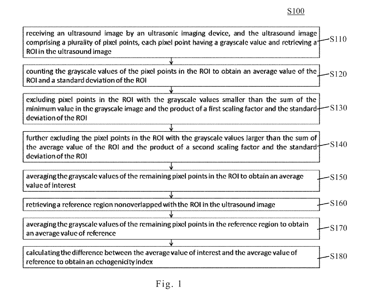
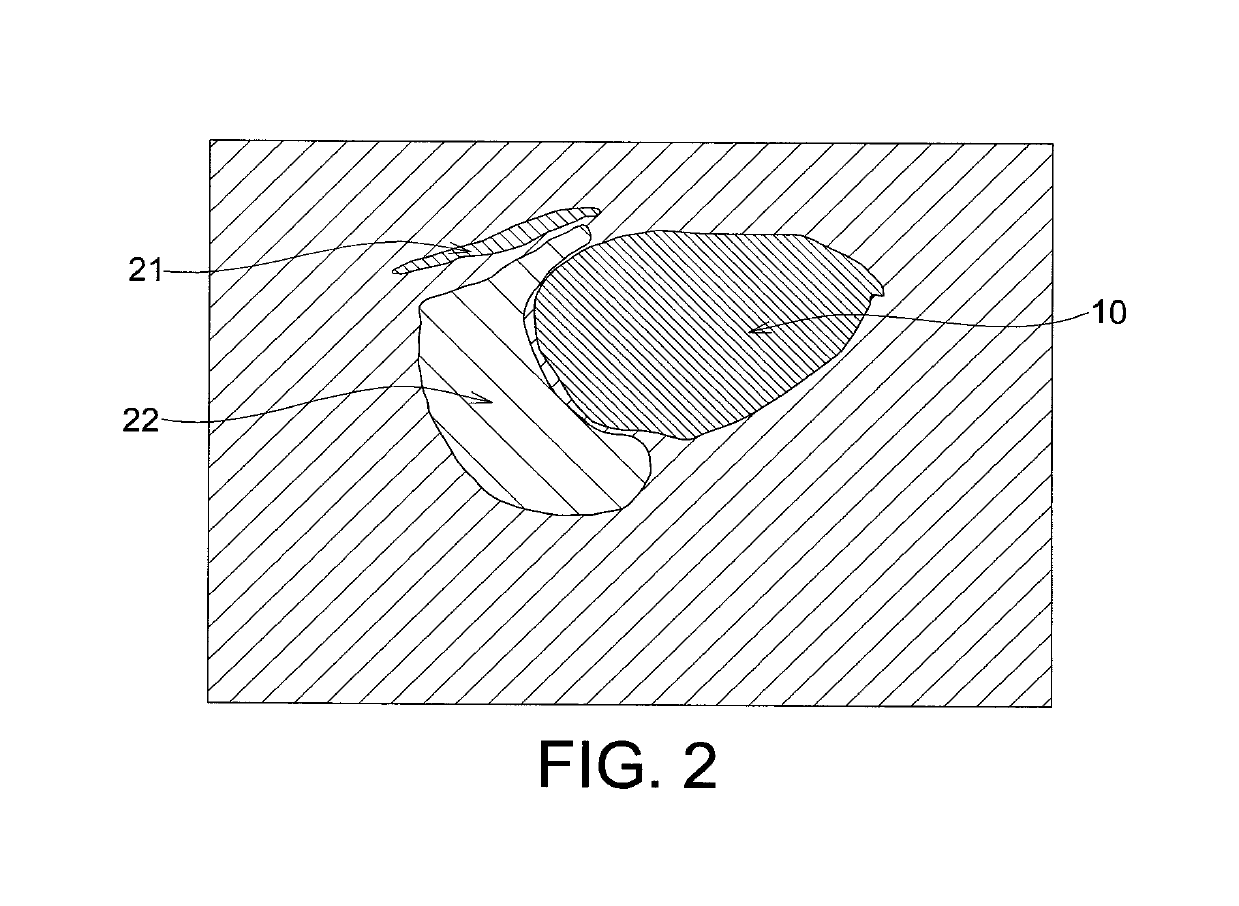
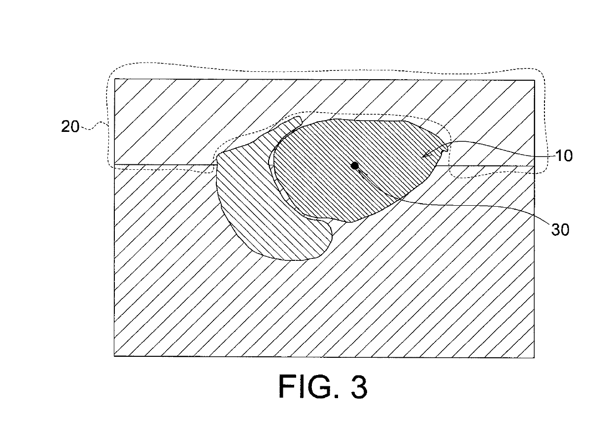
- An echogenicity quantification method that calculates an echogenicity index by averaging and normalizing grayscale values within a Region Of Interest (ROI) and a reference region, excluding outliers, to provide an objective and consistent measure across different ultrasonic devices and operators.

Medical devices with enhanced echogenicity
PatentPendingUS20250366825A1
Innovation
- Incorporation of hyperechoic particles, such as microspheres or air pockets, into medical devices like catheters and access cannulas to enhance their echogenicity, allowing for improved ultrasound visualization.


Medical Device Regulatory Requirements
The development and deployment of novel algorithms for enhanced echogenicity analysis must navigate a complex landscape of medical device regulatory requirements across different jurisdictions. In the United States, the Food and Drug Administration (FDA) classifies ultrasound imaging software and analytical algorithms as medical devices, typically falling under Class II or Class III depending on their intended use and risk profile. Enhanced echogenicity analysis algorithms that provide diagnostic information or influence clinical decision-making require premarket notification through the 510(k) pathway or premarket approval (PMA), necessitating comprehensive documentation of algorithm validation, clinical performance data, and risk management protocols.
The European Union's Medical Device Regulation (MDR 2017/745) imposes stringent requirements for software as a medical device (SaMD), mandating conformity assessment procedures and CE marking before market entry. Novel algorithms must demonstrate compliance with essential safety and performance requirements, including clinical evaluation reports that substantiate clinical benefits and acceptable risk-benefit ratios. The classification under MDR rules depends on the algorithm's purpose, with diagnostic algorithms generally classified as Class IIa or IIb, requiring involvement of notified bodies for conformity assessment.
International harmonization efforts through the International Medical Device Regulators Forum (IMDRF) have established guidelines specifically for SaMD, providing a risk-based framework that categorizes software based on the significance of information provided and the healthcare situation or condition. Enhanced echogenicity analysis algorithms must be evaluated against these criteria, considering factors such as whether they inform clinical management, drive clinical action, or diagnose disease states.
Quality management system compliance according to ISO 13485 standards is mandatory across most regulatory jurisdictions, requiring documented processes for software development lifecycle, verification and validation activities, and post-market surveillance. Additionally, algorithms incorporating artificial intelligence or machine learning components face emerging regulatory scrutiny, with agencies like the FDA developing specific frameworks for adaptive algorithms that may change over time. Cybersecurity considerations under standards such as IEC 62304 and FDA guidance on premarket cybersecurity are increasingly critical for software-based medical devices.
Regulatory pathways also demand comprehensive technical documentation including software design specifications, algorithm training and testing datasets, performance metrics, and intended use statements that clearly define the clinical context and user population for enhanced echogenicity analysis applications.
The European Union's Medical Device Regulation (MDR 2017/745) imposes stringent requirements for software as a medical device (SaMD), mandating conformity assessment procedures and CE marking before market entry. Novel algorithms must demonstrate compliance with essential safety and performance requirements, including clinical evaluation reports that substantiate clinical benefits and acceptable risk-benefit ratios. The classification under MDR rules depends on the algorithm's purpose, with diagnostic algorithms generally classified as Class IIa or IIb, requiring involvement of notified bodies for conformity assessment.
International harmonization efforts through the International Medical Device Regulators Forum (IMDRF) have established guidelines specifically for SaMD, providing a risk-based framework that categorizes software based on the significance of information provided and the healthcare situation or condition. Enhanced echogenicity analysis algorithms must be evaluated against these criteria, considering factors such as whether they inform clinical management, drive clinical action, or diagnose disease states.
Quality management system compliance according to ISO 13485 standards is mandatory across most regulatory jurisdictions, requiring documented processes for software development lifecycle, verification and validation activities, and post-market surveillance. Additionally, algorithms incorporating artificial intelligence or machine learning components face emerging regulatory scrutiny, with agencies like the FDA developing specific frameworks for adaptive algorithms that may change over time. Cybersecurity considerations under standards such as IEC 62304 and FDA guidance on premarket cybersecurity are increasingly critical for software-based medical devices.
Regulatory pathways also demand comprehensive technical documentation including software design specifications, algorithm training and testing datasets, performance metrics, and intended use statements that clearly define the clinical context and user population for enhanced echogenicity analysis applications.
Clinical Validation and Performance Metrics
Clinical validation of novel algorithms for enhanced echogenicity analysis requires rigorous evaluation frameworks that demonstrate both technical accuracy and clinical utility. The validation process must encompass diverse patient populations, multiple anatomical regions, and various pathological conditions to ensure generalizability. Prospective multicenter studies serve as the gold standard, comparing algorithmic outputs against established reference standards including histopathological findings, expert radiologist consensus, and longitudinal clinical outcomes. These studies should incorporate blinded assessments to minimize bias and establish inter-observer reliability metrics that quantify consistency across different operators and imaging systems.
Performance metrics for echogenicity analysis algorithms extend beyond traditional accuracy measures to include sensitivity, specificity, positive predictive value, and negative predictive value across different tissue types and pathological states. Receiver operating characteristic curves and area under the curve calculations provide comprehensive assessment of diagnostic performance at various threshold settings. Additionally, quantitative metrics such as mean absolute error, root mean square error, and correlation coefficients evaluate the precision of echogenicity measurements against reference standards. Computational efficiency metrics including processing time per image and memory requirements are critical for real-time clinical implementation.
The clinical validation framework must address algorithm robustness under varying imaging conditions, including different ultrasound frequencies, gain settings, and tissue depths. Subgroup analyses stratified by patient demographics, body mass index, and disease severity provide insights into algorithm performance across heterogeneous populations. Failure case analysis identifies specific scenarios where algorithmic performance degrades, guiding iterative refinement and establishing appropriate use limitations.
Regulatory compliance necessitates documentation of validation methodologies aligned with medical device standards and guidelines from regulatory bodies. Post-market surveillance mechanisms track real-world performance, capturing adverse events and long-term outcome data that inform continuous algorithm optimization. Comparative effectiveness studies against existing diagnostic modalities establish the incremental clinical value and potential cost-effectiveness of enhanced echogenicity analysis in routine practice.
Performance metrics for echogenicity analysis algorithms extend beyond traditional accuracy measures to include sensitivity, specificity, positive predictive value, and negative predictive value across different tissue types and pathological states. Receiver operating characteristic curves and area under the curve calculations provide comprehensive assessment of diagnostic performance at various threshold settings. Additionally, quantitative metrics such as mean absolute error, root mean square error, and correlation coefficients evaluate the precision of echogenicity measurements against reference standards. Computational efficiency metrics including processing time per image and memory requirements are critical for real-time clinical implementation.
The clinical validation framework must address algorithm robustness under varying imaging conditions, including different ultrasound frequencies, gain settings, and tissue depths. Subgroup analyses stratified by patient demographics, body mass index, and disease severity provide insights into algorithm performance across heterogeneous populations. Failure case analysis identifies specific scenarios where algorithmic performance degrades, guiding iterative refinement and establishing appropriate use limitations.
Regulatory compliance necessitates documentation of validation methodologies aligned with medical device standards and guidelines from regulatory bodies. Post-market surveillance mechanisms track real-world performance, capturing adverse events and long-term outcome data that inform continuous algorithm optimization. Comparative effectiveness studies against existing diagnostic modalities establish the incremental clinical value and potential cost-effectiveness of enhanced echogenicity analysis in routine practice.
Unlock deeper insights with Patsnap Eureka Quick Research — get a full tech report to explore trends and direct your research. Try now!
Generate Your Research Report Instantly with AI Agent
Supercharge your innovation with Patsnap Eureka AI Agent Platform!