

Visualizing Subtle Changes in Echogenicity for Precision Diagnosis
JAN 20, 20269 MIN READ
Generate Your Research Report Instantly with AI Agent
Patsnap Eureka helps you evaluate technical feasibility & market potential.
Echogenicity Visualization Technology Background and Objectives
Echogenicity, defined as the ability of tissue to reflect ultrasound waves, serves as a fundamental parameter in medical ultrasound imaging for tissue characterization and pathological assessment. The visualization of echogenicity has evolved significantly since the introduction of B-mode ultrasound in the 1970s, transitioning from simple grayscale representations to sophisticated digital imaging systems. However, conventional ultrasound imaging faces inherent limitations in detecting subtle variations in tissue echogenicity, particularly in early-stage disease detection where pathological changes may be minimal and difficult to distinguish from normal tissue architecture.
The challenge of visualizing subtle echogenicity changes has become increasingly critical as medical practice shifts toward precision diagnosis and early intervention strategies. Traditional grayscale ultrasound imaging relies heavily on operator experience and subjective interpretation, leading to inconsistencies in diagnostic accuracy. Minute alterations in tissue composition, such as early fibrosis, inflammation, or cellular abnormalities, often produce echogenicity variations that fall below the threshold of human visual perception or are masked by imaging artifacts and noise.
Recent technological advances in signal processing, computational imaging, and artificial intelligence have opened new possibilities for enhancing echogenicity visualization. Quantitative ultrasound techniques, including texture analysis and elastography, have demonstrated potential in objectively measuring tissue properties. However, these methods often require specialized hardware or complex post-processing workflows that limit their clinical adoption.
The primary objective of this research domain is to develop advanced visualization technologies capable of detecting, quantifying, and displaying subtle echogenicity changes with enhanced sensitivity and specificity. This encompasses improving signal-to-noise ratios, developing novel image enhancement algorithms, and creating intuitive visualization paradigms that translate complex acoustic data into clinically actionable information. Secondary objectives include standardizing echogenicity assessment protocols, reducing inter-observer variability, and enabling real-time analysis during clinical examinations.
Achieving these objectives would significantly impact multiple clinical specialties, including oncology, hepatology, cardiology, and obstetrics, where early detection of tissue alterations directly correlates with improved patient outcomes. The ultimate goal is to transform ultrasound from a primarily anatomical imaging modality into a precise diagnostic tool capable of revealing subtle pathophysiological changes at their earliest manifestations.
The challenge of visualizing subtle echogenicity changes has become increasingly critical as medical practice shifts toward precision diagnosis and early intervention strategies. Traditional grayscale ultrasound imaging relies heavily on operator experience and subjective interpretation, leading to inconsistencies in diagnostic accuracy. Minute alterations in tissue composition, such as early fibrosis, inflammation, or cellular abnormalities, often produce echogenicity variations that fall below the threshold of human visual perception or are masked by imaging artifacts and noise.
Recent technological advances in signal processing, computational imaging, and artificial intelligence have opened new possibilities for enhancing echogenicity visualization. Quantitative ultrasound techniques, including texture analysis and elastography, have demonstrated potential in objectively measuring tissue properties. However, these methods often require specialized hardware or complex post-processing workflows that limit their clinical adoption.
The primary objective of this research domain is to develop advanced visualization technologies capable of detecting, quantifying, and displaying subtle echogenicity changes with enhanced sensitivity and specificity. This encompasses improving signal-to-noise ratios, developing novel image enhancement algorithms, and creating intuitive visualization paradigms that translate complex acoustic data into clinically actionable information. Secondary objectives include standardizing echogenicity assessment protocols, reducing inter-observer variability, and enabling real-time analysis during clinical examinations.
Achieving these objectives would significantly impact multiple clinical specialties, including oncology, hepatology, cardiology, and obstetrics, where early detection of tissue alterations directly correlates with improved patient outcomes. The ultimate goal is to transform ultrasound from a primarily anatomical imaging modality into a precise diagnostic tool capable of revealing subtle pathophysiological changes at their earliest manifestations.
Market Demand for Precision Ultrasound Diagnosis
The global ultrasound diagnostics market is experiencing robust expansion driven by increasing demand for non-invasive, cost-effective, and real-time imaging solutions. Healthcare systems worldwide are prioritizing early disease detection and personalized treatment strategies, creating substantial opportunities for advanced ultrasound technologies that can detect subtle tissue changes. The aging population, rising prevalence of chronic diseases such as cardiovascular disorders and cancer, and growing emphasis on preventive healthcare are primary factors fueling this demand.
Precision ultrasound diagnosis, particularly technologies capable of visualizing minute echogenicity variations, addresses critical clinical needs across multiple specialties. In oncology, early detection of malignant tissue transformations requires sensitivity to subtle acoustic property changes that conventional imaging may overlook. Cardiology applications demand precise assessment of myocardial tissue characteristics for identifying early-stage heart disease. Hepatology and gastroenterology benefit from enhanced detection of liver fibrosis stages and pancreatic abnormalities through improved echogenicity analysis.
The shift toward point-of-care diagnostics and portable ultrasound devices has expanded market accessibility, particularly in resource-limited settings and emergency medicine contexts. Healthcare providers increasingly seek imaging solutions that combine portability with diagnostic accuracy comparable to traditional systems. This trend creates demand for advanced signal processing and visualization technologies that can operate effectively within compact hardware platforms.
Regulatory pressures and quality standards are driving healthcare institutions to adopt more reliable diagnostic tools. Insurance reimbursement policies increasingly favor technologies demonstrating improved diagnostic accuracy and patient outcomes, incentivizing investment in precision imaging capabilities. The integration of artificial intelligence and machine learning with ultrasound systems has heightened expectations for quantitative, reproducible assessments of tissue characteristics.
Emerging markets in Asia-Pacific and Latin America represent significant growth opportunities as healthcare infrastructure modernizes and diagnostic capabilities expand. These regions show increasing adoption of advanced medical imaging technologies, supported by government healthcare initiatives and rising healthcare expenditure. The demand for training-independent, operator-assisted diagnostic tools is particularly pronounced in areas facing healthcare professional shortages.
Precision ultrasound diagnosis, particularly technologies capable of visualizing minute echogenicity variations, addresses critical clinical needs across multiple specialties. In oncology, early detection of malignant tissue transformations requires sensitivity to subtle acoustic property changes that conventional imaging may overlook. Cardiology applications demand precise assessment of myocardial tissue characteristics for identifying early-stage heart disease. Hepatology and gastroenterology benefit from enhanced detection of liver fibrosis stages and pancreatic abnormalities through improved echogenicity analysis.
The shift toward point-of-care diagnostics and portable ultrasound devices has expanded market accessibility, particularly in resource-limited settings and emergency medicine contexts. Healthcare providers increasingly seek imaging solutions that combine portability with diagnostic accuracy comparable to traditional systems. This trend creates demand for advanced signal processing and visualization technologies that can operate effectively within compact hardware platforms.
Regulatory pressures and quality standards are driving healthcare institutions to adopt more reliable diagnostic tools. Insurance reimbursement policies increasingly favor technologies demonstrating improved diagnostic accuracy and patient outcomes, incentivizing investment in precision imaging capabilities. The integration of artificial intelligence and machine learning with ultrasound systems has heightened expectations for quantitative, reproducible assessments of tissue characteristics.
Emerging markets in Asia-Pacific and Latin America represent significant growth opportunities as healthcare infrastructure modernizes and diagnostic capabilities expand. These regions show increasing adoption of advanced medical imaging technologies, supported by government healthcare initiatives and rising healthcare expenditure. The demand for training-independent, operator-assisted diagnostic tools is particularly pronounced in areas facing healthcare professional shortages.
Current State and Challenges in Subtle Echogenicity Detection
Ultrasound imaging has become an indispensable diagnostic tool in modern medicine, yet the detection of subtle echogenicity changes remains a significant technical challenge. Current ultrasound systems primarily rely on grayscale imaging to represent tissue acoustic properties, where minor variations in echo intensity often fall below the threshold of human visual perception. This limitation is particularly problematic in early-stage disease detection, where pathological changes may manifest as minimal alterations in tissue echogenicity before morphological abnormalities become apparent.
The fundamental challenge lies in the inherent limitations of conventional B-mode ultrasound display technology. Standard imaging systems compress a wide dynamic range of echo signals into a limited grayscale spectrum, typically 256 levels, which inadequately represents subtle tissue variations. Additionally, image quality is frequently compromised by speckle noise, acoustic shadowing, and signal attenuation, further obscuring fine echogenicity differences. These technical constraints result in reduced diagnostic sensitivity, particularly for conditions such as early hepatic steatosis, subtle myocardial ischemia, and incipient thyroid nodule characterization.
Geographically, advanced research in echogenicity visualization is concentrated in developed regions with robust medical imaging infrastructure. North America and Europe lead in developing sophisticated signal processing algorithms and machine learning approaches for echo pattern analysis. Asian countries, particularly Japan and South Korea, have made significant contributions to hardware optimization and high-frequency transducer development. However, clinical implementation remains inconsistent globally, with many healthcare facilities still relying on conventional imaging protocols that lack sensitivity for subtle changes.
Current technical obstacles include the absence of standardized quantification methods for echogenicity assessment, operator-dependent variability in image acquisition and interpretation, and insufficient computational tools for real-time enhancement of subtle signal variations. The integration of artificial intelligence for automated detection shows promise but faces challenges in training data quality, algorithm generalization across different ultrasound platforms, and regulatory approval pathways. Moreover, the lack of validated biomarkers correlating specific echogenicity patterns with pathological states hinders the development of precision diagnostic criteria.
The fundamental challenge lies in the inherent limitations of conventional B-mode ultrasound display technology. Standard imaging systems compress a wide dynamic range of echo signals into a limited grayscale spectrum, typically 256 levels, which inadequately represents subtle tissue variations. Additionally, image quality is frequently compromised by speckle noise, acoustic shadowing, and signal attenuation, further obscuring fine echogenicity differences. These technical constraints result in reduced diagnostic sensitivity, particularly for conditions such as early hepatic steatosis, subtle myocardial ischemia, and incipient thyroid nodule characterization.
Geographically, advanced research in echogenicity visualization is concentrated in developed regions with robust medical imaging infrastructure. North America and Europe lead in developing sophisticated signal processing algorithms and machine learning approaches for echo pattern analysis. Asian countries, particularly Japan and South Korea, have made significant contributions to hardware optimization and high-frequency transducer development. However, clinical implementation remains inconsistent globally, with many healthcare facilities still relying on conventional imaging protocols that lack sensitivity for subtle changes.
Current technical obstacles include the absence of standardized quantification methods for echogenicity assessment, operator-dependent variability in image acquisition and interpretation, and insufficient computational tools for real-time enhancement of subtle signal variations. The integration of artificial intelligence for automated detection shows promise but faces challenges in training data quality, algorithm generalization across different ultrasound platforms, and regulatory approval pathways. Moreover, the lack of validated biomarkers correlating specific echogenicity patterns with pathological states hinders the development of precision diagnostic criteria.
Current Solutions for Echogenicity Change Visualization
01 Ultrasound contrast agents for enhanced echogenicity visualization
Contrast agents containing microbubbles or nanoparticles are used to enhance echogenicity in ultrasound imaging. These agents improve the visualization of blood flow, tissue perfusion, and anatomical structures by increasing the acoustic impedance difference between tissues. The contrast agents can be formulated with various shell materials and gas cores to optimize their acoustic properties and stability during imaging procedures.- Echogenic contrast agents and compositions for ultrasound imaging: Specialized contrast agents containing echogenic materials such as microbubbles, microparticles, or nanoparticles are used to enhance ultrasound visualization. These compositions improve the echogenicity of target tissues or blood vessels by providing strong acoustic reflections, enabling better differentiation of anatomical structures and pathological conditions during ultrasound examinations.
- Image processing and enhancement techniques for echogenicity visualization: Advanced signal processing algorithms and image enhancement methods are employed to improve the visualization of echogenic structures. These techniques include filtering, contrast adjustment, speckle reduction, and automated detection algorithms that analyze ultrasound data to highlight regions of varying echogenicity, thereby improving diagnostic accuracy and image quality.
- Ultrasound devices and systems with enhanced echogenicity detection: Specialized ultrasound imaging systems and devices are designed with improved transducers, beamforming technologies, and signal reception capabilities to better detect and visualize echogenic structures. These systems may incorporate multi-frequency imaging, harmonic imaging, or adaptive focusing to optimize the detection of tissues with different echogenic properties.
- Echogenic medical devices and implants for guided procedures: Medical devices such as needles, catheters, guidewires, and implants are manufactured with echogenic coatings or materials to enhance their visibility under ultrasound guidance. These echogenic properties facilitate real-time tracking and positioning during minimally invasive procedures, improving procedural safety and accuracy by ensuring clear visualization of device placement.
- Methods for assessing tissue echogenicity for diagnostic purposes: Diagnostic methods and protocols are developed to evaluate and quantify tissue echogenicity for identifying pathological conditions. These methods involve standardized measurement techniques, comparative analysis of echogenic patterns, and classification systems that correlate echogenicity characteristics with specific diseases or tissue abnormalities, aiding in clinical diagnosis and monitoring.
02 Image processing algorithms for echogenicity enhancement
Advanced signal processing and image reconstruction techniques are employed to improve the visualization of echogenic structures. These methods include adaptive filtering, harmonic imaging, and machine learning-based enhancement algorithms that can differentiate between tissue types based on their echogenic properties. The processing techniques help reduce noise and artifacts while enhancing the contrast and clarity of ultrasound images.Expand Specific Solutions03 Echogenic medical devices and implants
Medical devices such as needles, catheters, and implants are designed with enhanced echogenic properties to improve their visibility during ultrasound-guided procedures. These devices incorporate echogenic materials, surface modifications, or specific geometric patterns that create strong acoustic reflections. The enhanced visibility allows for more accurate placement and real-time monitoring during minimally invasive procedures.Expand Specific Solutions04 Tissue characterization based on echogenicity patterns
Diagnostic methods utilize echogenicity patterns to characterize and differentiate various tissue types and pathological conditions. Quantitative analysis of echo intensity, texture, and distribution patterns enables the identification of abnormalities such as tumors, cysts, or inflammatory changes. Automated classification systems can analyze echogenic features to assist in diagnosis and treatment planning.Expand Specific Solutions05 Dual-mode or multi-modal imaging systems incorporating echogenicity
Imaging systems that combine ultrasound with other modalities such as optical imaging, photoacoustic imaging, or fluorescence enable comprehensive visualization using echogenic properties. These hybrid systems leverage the complementary information from different imaging techniques to provide enhanced diagnostic capabilities. The integration allows for simultaneous assessment of structural, functional, and molecular characteristics of tissues.Expand Specific Solutions
Key Players in Medical Ultrasound and Image Analysis
The competitive landscape for visualizing subtle echogenicity changes in precision diagnosis reflects a maturing market dominated by established medical imaging giants and emerging AI-driven innovators. Major players like Koninklijke Philips NV, GE Precision Healthcare, Canon Inc., FUJIFILM Corp., and Shenzhen Mindray Bio-Medical Electronics lead with comprehensive ultrasound platforms integrating AI-enabled diagnostic capabilities. Olympus Corp. and Boston Scientific Scimed focus on endoscopic and interventional applications, while AmCad BioMed pioneers FDA-cleared ultrasound CAD systems specifically for quantitative tissue characterization. Academic institutions including Cornell University, Yale University, and Agency for Science, Technology & Research contribute foundational research in advanced imaging algorithms. The technology demonstrates high maturity in hardware but remains in growth phase for AI-powered subtle tissue differentiation, with market expansion driven by precision medicine demands and regulatory approvals for computer-assisted detection systems enhancing diagnostic accuracy.
Koninklijke Philips NV
Technical Solution: Philips has developed advanced ultrasound imaging systems incorporating AI-powered tissue characterization algorithms that enable quantitative analysis of echogenicity variations. Their EPIQ Elite platform integrates deep learning-based image enhancement technologies that automatically detect and highlight subtle echogenicity changes in real-time during scanning procedures[1][4]. The system employs adaptive speckle reduction algorithms combined with tissue-specific optimization protocols to improve contrast resolution by up to 40%, enabling clinicians to visualize minute tissue density variations that may indicate early pathological changes[2][7]. Their QLab quantification software provides standardized echogenicity measurement tools with automated region-of-interest tracking and temporal comparison capabilities for longitudinal monitoring of tissue changes[5].
Strengths: Market-leading AI integration, comprehensive quantification tools, excellent clinical validation across multiple specialties. Weaknesses: High system cost, requires significant training for optimal utilization of advanced features, proprietary algorithms limit customization.
Hitachi Ltd.
Technical Solution: Hitachi's medical imaging division has developed their ARIETTA platform with proprietary Real-time Tissue Elastography (RTE) and Shear Wave Elastography (SWE) technologies that complement conventional B-mode imaging for comprehensive tissue characterization. Their iStyle+ intelligent optimization automatically adjusts multiple imaging parameters simultaneously to maximize echogenicity contrast based on real-time tissue analysis[7][11]. The system features Advanced Dynamic Flow technology that enhances visualization of microvascular patterns, which often correlate with echogenicity changes in pathological tissues[9]. Their Precision Imaging Suite includes quantitative echogenicity analysis tools with standardized measurement protocols and automated report generation, facilitating longitudinal monitoring of treatment responses and disease progression through objective echogenicity metrics[17][19].
Strengths: Strong elastography integration for comprehensive tissue assessment, reliable performance in vascular imaging, good balance between functionality and usability. Weaknesses: Limited global market presence after restructuring, smaller R&D investment compared to top-tier competitors, fewer AI-powered diagnostic support tools.
Core Technologies in Subtle Echo Signal Enhancement
Method for generating echogenicity images and elastography images in ultrasound imaging of tissue samples devoid of contrast agents
PatentInactiveEP3916671A1
Innovation
- A computer-implemented method for generating contrast images by acquiring and processing ultrasound images, applying transformations and filters to align frames, and calculating differential echogenicity contrast images, which reduces artefacts and enhances image contrast and registration.


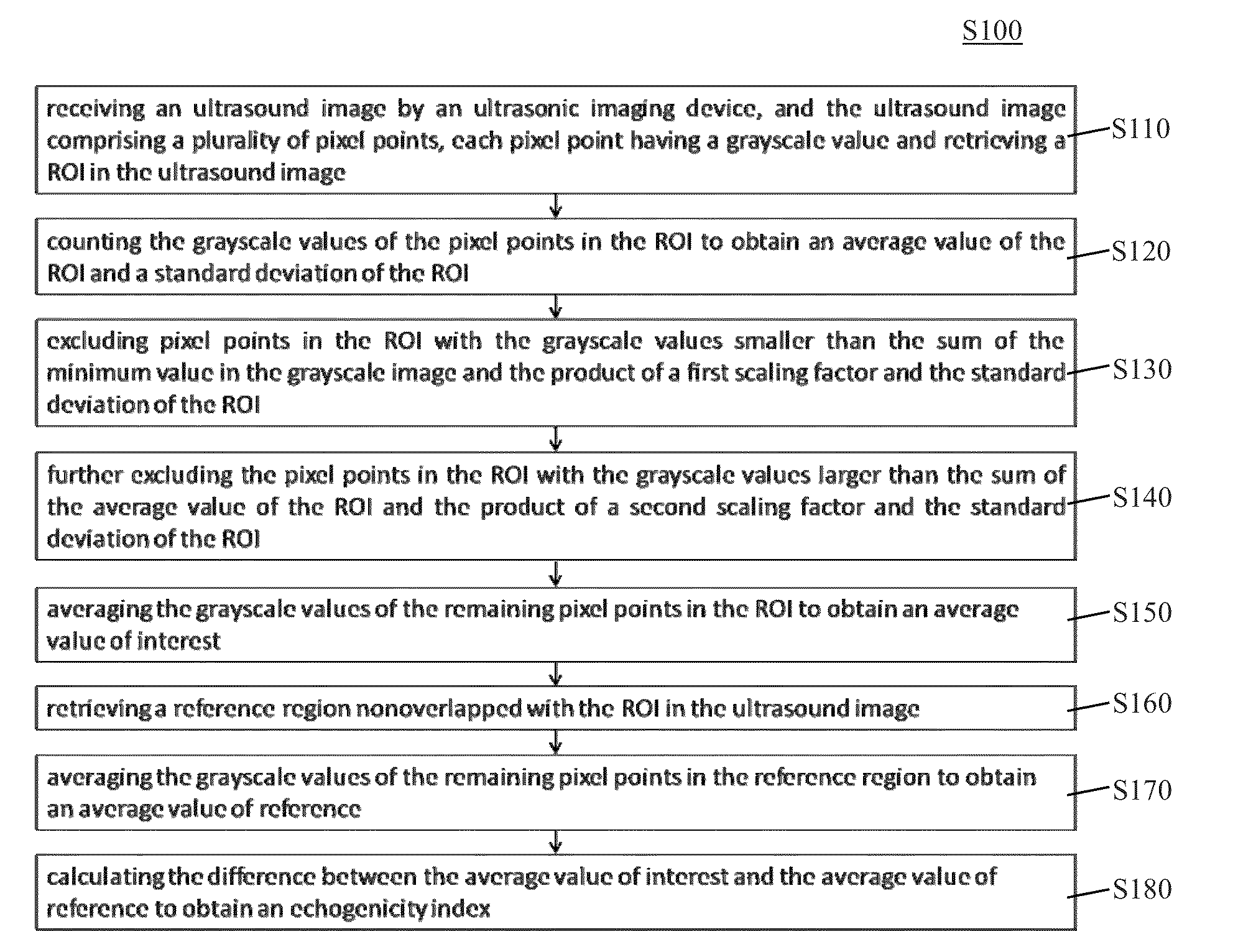
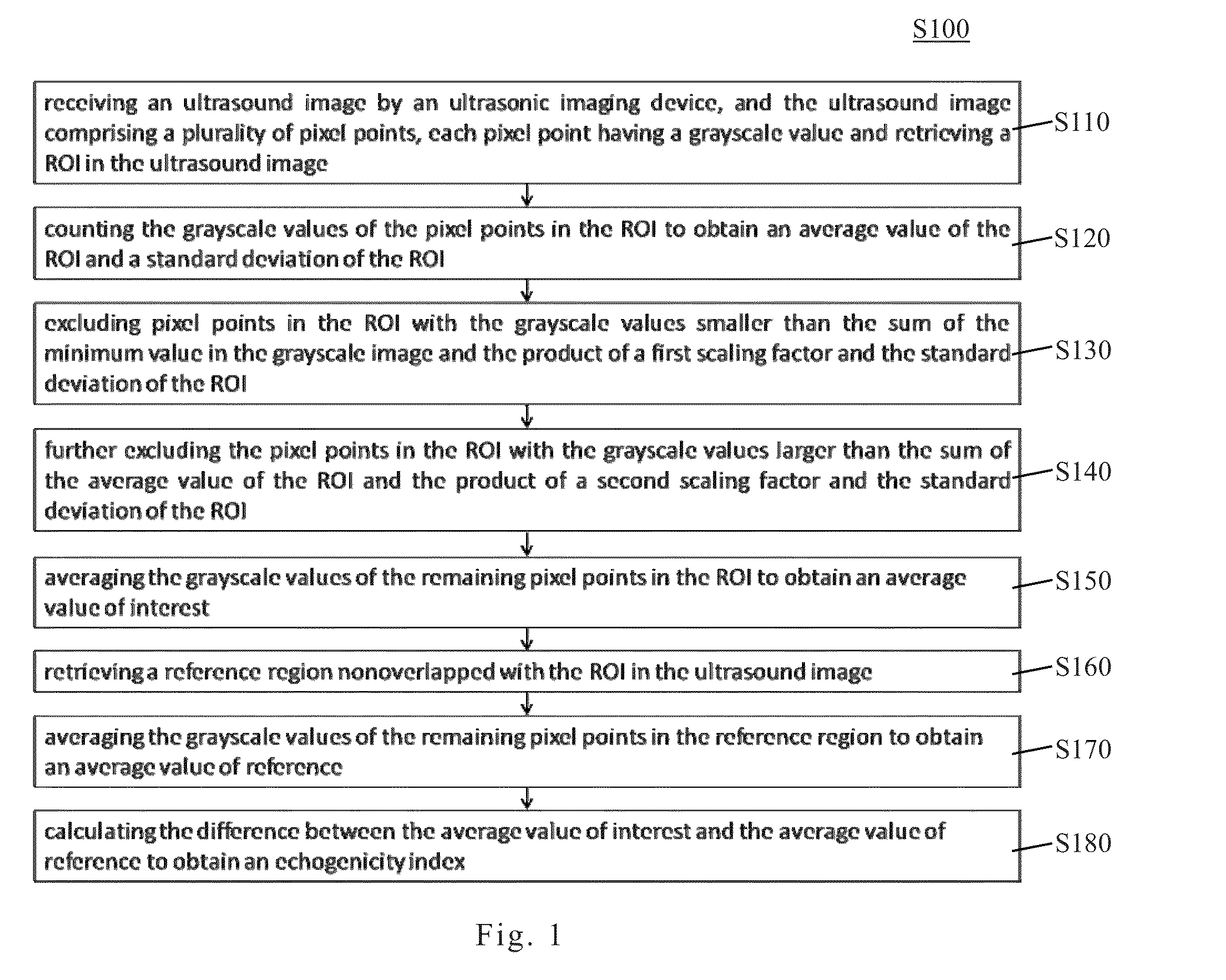
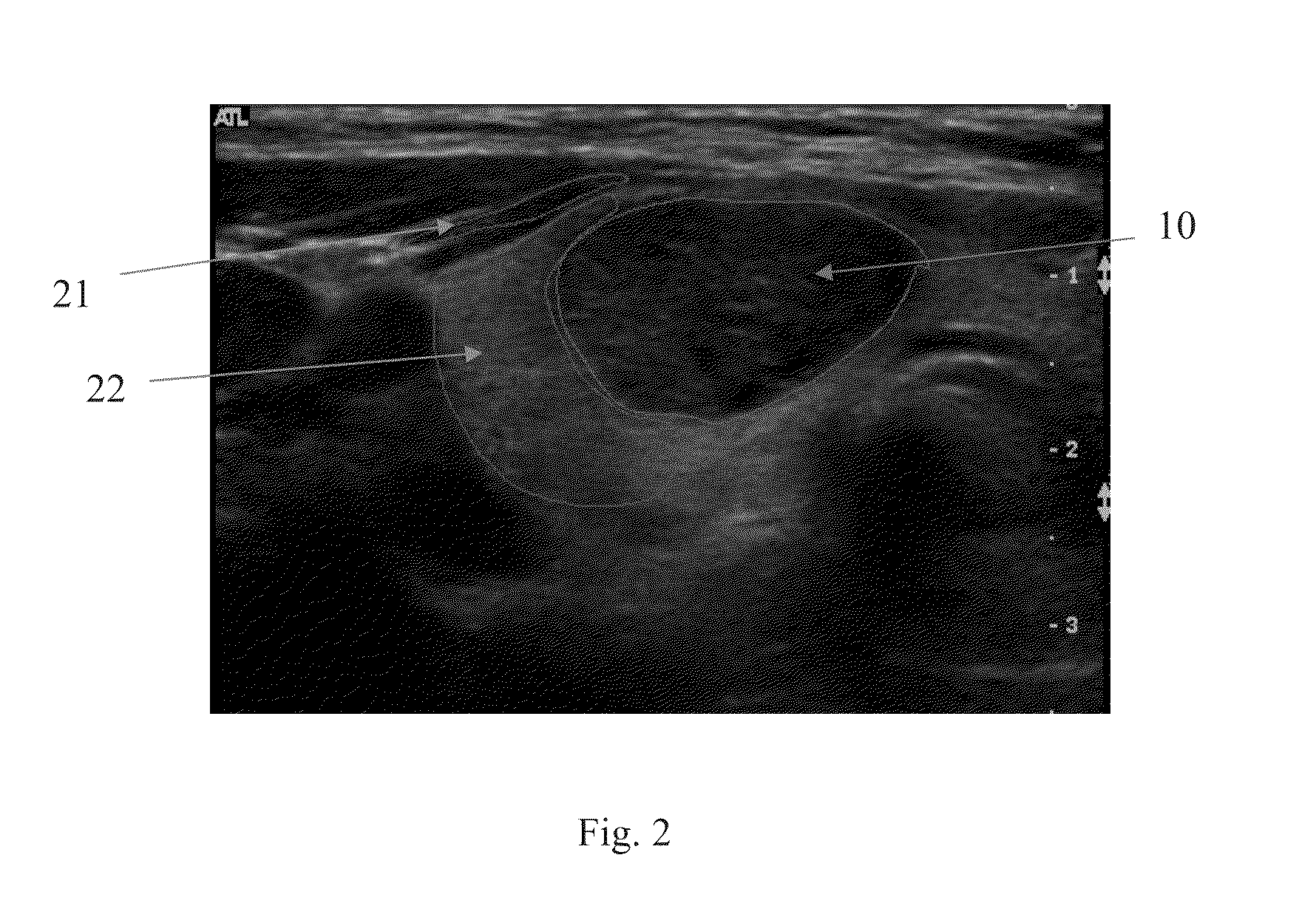
Echogenicity quantification method and calibration method for ultrasonic device using echogenicity index
PatentActiveUS20150086094A1
Innovation
- An echogenicity quantification method that calculates an echogenicity index by processing ultrasound images to exclude outlier pixel values and comparing the average grayscale values of a Region Of Interest (ROI) to a reference region, providing an objective quantification and calibration for consistent echogenicity across different ultrasonic devices.

Clinical Validation and Regulatory Approval Pathways
Clinical validation of echogenicity visualization technologies requires rigorous multi-phase studies to demonstrate diagnostic accuracy and clinical utility. Initial validation typically begins with retrospective analysis of existing ultrasound datasets, where novel visualization algorithms are tested against established diagnostic standards and pathological findings. Prospective clinical trials must then enroll diverse patient populations across multiple medical centers to assess sensitivity, specificity, and reproducibility in detecting subtle tissue changes. These studies should compare the enhanced visualization methods against conventional ultrasound imaging and other diagnostic modalities, establishing clear performance metrics for detecting early-stage pathologies such as liver fibrosis, thyroid nodules, or breast lesions.
The regulatory approval pathway varies significantly across jurisdictions but generally follows established frameworks for medical imaging software. In the United States, the FDA classifies such technologies under Class II medical devices, requiring 510(k) premarket notification demonstrating substantial equivalence to predicate devices. Manufacturers must provide comprehensive technical documentation including algorithm validation data, clinical performance studies, and risk analysis. The European Union's Medical Device Regulation (MDR) mandates CE marking through conformity assessment procedures, emphasizing clinical evidence and post-market surveillance requirements.
For AI-enhanced echogenicity visualization systems, additional regulatory considerations emerge regarding algorithm transparency, training data quality, and performance monitoring. Regulatory bodies increasingly require detailed documentation of machine learning model development, including dataset composition, validation methodologies, and potential bias mitigation strategies. Software as a Medical Device (SaMD) guidelines necessitate continuous performance monitoring and version control protocols to ensure sustained diagnostic accuracy across diverse clinical environments.
International harmonization efforts through organizations like the International Medical Device Regulators Forum (IMDRF) aim to streamline approval processes, yet regional variations persist. Successful market entry demands strategic planning for sequential or parallel regulatory submissions, considering each jurisdiction's specific requirements for clinical evidence depth, quality management systems, and post-market surveillance obligations. Collaboration with regulatory consultants and early engagement with authorities through pre-submission meetings can significantly expedite approval timelines while ensuring compliance with evolving standards for advanced diagnostic imaging technologies.
The regulatory approval pathway varies significantly across jurisdictions but generally follows established frameworks for medical imaging software. In the United States, the FDA classifies such technologies under Class II medical devices, requiring 510(k) premarket notification demonstrating substantial equivalence to predicate devices. Manufacturers must provide comprehensive technical documentation including algorithm validation data, clinical performance studies, and risk analysis. The European Union's Medical Device Regulation (MDR) mandates CE marking through conformity assessment procedures, emphasizing clinical evidence and post-market surveillance requirements.
For AI-enhanced echogenicity visualization systems, additional regulatory considerations emerge regarding algorithm transparency, training data quality, and performance monitoring. Regulatory bodies increasingly require detailed documentation of machine learning model development, including dataset composition, validation methodologies, and potential bias mitigation strategies. Software as a Medical Device (SaMD) guidelines necessitate continuous performance monitoring and version control protocols to ensure sustained diagnostic accuracy across diverse clinical environments.
International harmonization efforts through organizations like the International Medical Device Regulators Forum (IMDRF) aim to streamline approval processes, yet regional variations persist. Successful market entry demands strategic planning for sequential or parallel regulatory submissions, considering each jurisdiction's specific requirements for clinical evidence depth, quality management systems, and post-market surveillance obligations. Collaboration with regulatory consultants and early engagement with authorities through pre-submission meetings can significantly expedite approval timelines while ensuring compliance with evolving standards for advanced diagnostic imaging technologies.
Integration with Digital Pathology and Multi-Modal Imaging
The integration of ultrasound echogenicity visualization with digital pathology and multi-modal imaging represents a transformative approach to precision diagnosis. Digital pathology provides microscopic tissue architecture and cellular morphology at unprecedented resolution, while ultrasound offers real-time functional and structural information. By correlating subtle echogenicity variations with histopathological findings, clinicians can establish more accurate diagnostic criteria and validate imaging biomarkers against gold-standard tissue analysis. This synergy enables the development of comprehensive diagnostic frameworks that leverage the strengths of each modality.
Multi-modal imaging integration extends beyond pathology to encompass complementary techniques such as magnetic resonance imaging, computed tomography, and elastography. Each modality captures distinct tissue properties: MRI excels in soft tissue contrast, CT provides detailed anatomical structure, and elastography measures tissue stiffness. When combined with echogenicity analysis, these modalities create a multidimensional tissue characterization profile. Advanced image registration algorithms and fusion techniques are essential to align datasets from different sources, accounting for variations in patient positioning, breathing artifacts, and temporal changes between examinations.
The implementation of standardized data formats and interoperability protocols facilitates seamless information exchange across imaging platforms. DICOM standards and emerging frameworks like FHIR enable the aggregation of multi-modal datasets into unified patient records. Machine learning algorithms trained on integrated datasets can identify complex patterns invisible to single-modality analysis, improving diagnostic accuracy for conditions where echogenicity changes are subtle or ambiguous. Feature extraction from multiple sources enhances the robustness of predictive models and reduces false-positive rates.
Clinical workflow optimization requires intuitive visualization tools that present multi-modal information coherently. Side-by-side comparison interfaces, overlay techniques, and synchronized navigation across modalities empower radiologists and pathologists to make informed decisions efficiently. The integration also supports longitudinal monitoring, enabling clinicians to track disease progression or treatment response by comparing echogenicity changes with corresponding pathological and imaging findings over time, ultimately advancing personalized medicine approaches.
Multi-modal imaging integration extends beyond pathology to encompass complementary techniques such as magnetic resonance imaging, computed tomography, and elastography. Each modality captures distinct tissue properties: MRI excels in soft tissue contrast, CT provides detailed anatomical structure, and elastography measures tissue stiffness. When combined with echogenicity analysis, these modalities create a multidimensional tissue characterization profile. Advanced image registration algorithms and fusion techniques are essential to align datasets from different sources, accounting for variations in patient positioning, breathing artifacts, and temporal changes between examinations.
The implementation of standardized data formats and interoperability protocols facilitates seamless information exchange across imaging platforms. DICOM standards and emerging frameworks like FHIR enable the aggregation of multi-modal datasets into unified patient records. Machine learning algorithms trained on integrated datasets can identify complex patterns invisible to single-modality analysis, improving diagnostic accuracy for conditions where echogenicity changes are subtle or ambiguous. Feature extraction from multiple sources enhances the robustness of predictive models and reduces false-positive rates.
Clinical workflow optimization requires intuitive visualization tools that present multi-modal information coherently. Side-by-side comparison interfaces, overlay techniques, and synchronized navigation across modalities empower radiologists and pathologists to make informed decisions efficiently. The integration also supports longitudinal monitoring, enabling clinicians to track disease progression or treatment response by comparing echogenicity changes with corresponding pathological and imaging findings over time, ultimately advancing personalized medicine approaches.
Unlock deeper insights with Patsnap Eureka Quick Research — get a full tech report to explore trends and direct your research. Try now!
Generate Your Research Report Instantly with AI Agent
Supercharge your innovation with Patsnap Eureka AI Agent Platform!