
A chemical derivatization method and its application in detection of nucleic acid modification by lc-ms method
A derivatization and chemical technology, applied in the direction of biochemical equipment and methods, microbial determination/inspection, measuring devices, etc., can solve the problems of cumbersome operation, time-consuming and labor-intensive, insufficient detection sensitivity, etc., to avoid contamination of mass spectrometer, broad Application prospect, beneficial effect of quantitative analysis
Patent Information
- Authority / Receiving Office
- CN · China
- Patent Type
- Patents(China)
- Current Assignee / Owner
- Publication Date
- 2016-01-27
- Estimated Expiration
- Not applicable · inactive patent
Smart Images
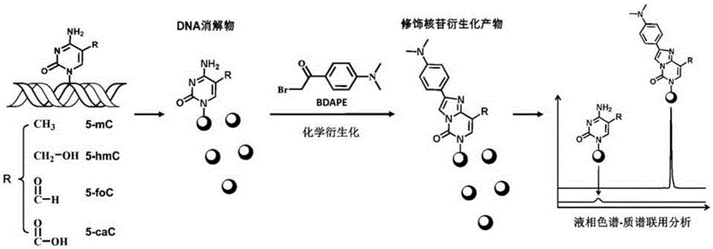
Figure 1 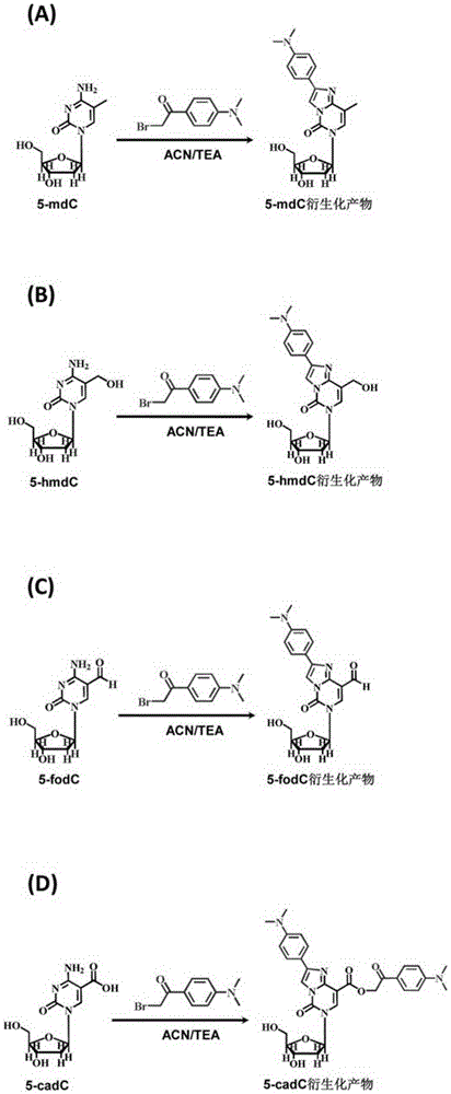
Figure 2 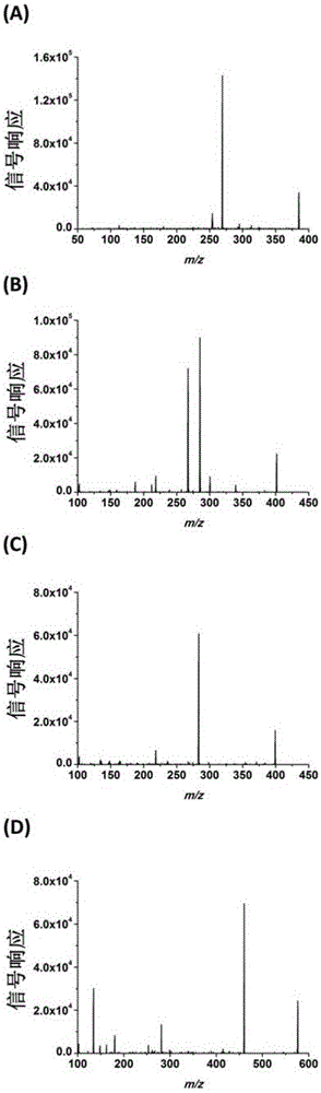
Figure 3
Abstract
Description
technical field
[0001] The invention relates to a method of chemical derivatization combined with LC-MS and its application in DNA and RNA modification analysis. Background technique
[0002] In mammalian DNA, the 5-position of cytosine will undergo methylation modification under the action of DNA methyltransferase to form 5-methylcytosine (5-mC). 5-mC participates in many important biological processes, such as genome imprinting, gene expression regulation, etc., and is an important epigenetic modification. 5-methylcytosine can be further oxidized successively by TET proteins to 5-hydroxymethylcytosine (5-hmC), 5-formylcytosine (5-foC) and 5-carboxycytosine (5-caC) , forming a new modification. These newly discovered modifiers are closely related to a variety of important physiological functions, such as cell differentiation, cell reprogramming, nervous system development, the occurrence and development of diseases, and so on. The DNA modification level of normal people ...
Examples
Embodiment 1
[0044] Example 1: Human Cell DNA Analysis
[0045] Three human cell lines: HeLa (cervical carcinoma cells), Jurkat-T (leukemic lymphocytes) and 293T (human embryonic kidney cells). Take a certain number of cultured cells and wash them with PBS buffer, put them in a 1.5mL centrifuge tube, centrifuge at 2000g for 20s to pellet the cells, and discard the supernatant. Add 0.5mL cell lysate A (320mM sucrose, 5mMMgCl 2 , 10mM Tris, 0.1mM deferoxamine, pH 7.5, 1% TritonX-100), shake vigorously with a micro-vortex mixer for 30s. The sample was centrifuged at 16000g for 20s, the supernatant was discarded, and the previous process was repeated. Then add 0.2 mL of cell lysate B (10 mM Tris, 5 mM EDTA, 0.15 mM deferoxamine, pH 8.0, 1% sodium lauryl sarcosine), and shake vigorously for 30 s with a micro-vortex mixer. Add 10 μL RNaseA (1 mg / mL) and 3 μL RNaseT1 (1 U / μL), and incubate in a water bath at 50° C. for 15 minutes to remove RNA. Then add 10 μL proteinase K (20mg / mlinH 2 O), i...
Embodiment 2
[0049] Example 2: DNA Analysis of Cancer Tissues and Paracancerous Tissues of Colorectal Cancer Patients
[0050] Take formalin-fixed, paraffin-embedded colorectal patient tissue sections (5-10 slices, about 30 mg), and use a paraffin-embedded tissue DNA extraction kit ( FFPEDNAKit, OmegaCo.) extracts DNA from tissues. The extracted DNA was dissolved in water and quantified by an ultra-micro ultraviolet spectrophotometer. Take 2-20 μg DNA, dry it in vacuum, add 17 μL water and 2 μL S1 nuclease buffer (300 mM sodium acetate, pH 4.6, 2800 mM sodium chloride, 10 mM zinc sulfate) in turn, place in 95 ° C water bath for 5 minutes, and then quickly place in Quenching in an ice-water bath for 2 minutes to untangle the DNA double strands, add 1 μL S1 nuclease (180 U / μL) and incubate in a 37° C. water bath for 12-16 hours. Then add 65 μL water, 10 μL alkaline phosphatase buffer (500 mM Tris-HCl, 100 mM magnesium chloride, pH 9.0), 1 μL alkaline phosphatase (30 U / μL), 4 μL snake veno...
Embodiment 3
[0053] Example 3: Yeast Cell DNA Analysis
[0054]Ten yeast strains: Saccharomyces cerevisiae BY4741, Saccharomyces cerevisiae W1588-4C, Debaryomyceshansenii, Schizosaccharomycespombe, Kluyveromyces marxianus , Schizosaccharomycesoctosporus, Yarrowialipolytica, Pichia pastoris, Saccharomyces boulardii and Kluyveromyceslactis were cultured, and the yeast cells The suspension was centrifuged at 4600g for 5 minutes, the collected cell pellet was washed with water, and the residual medium was removed. Disperse the yeast cells in 1 mL of cell lysate (10 mM Tris-HCl, pH8.0, 1 mM EDTA, 100 mM NaCl, 10 g / LSDS, 20 g / L TritonX-100), and then add an equal volume of phenol / chloroform (1:1, v / v , phenol saturated with 10mM Tris, 1mM EDTA, pH 8) and 1.5g glass beads (425-600μm), shake vigorously with a micro-vortex mixer for 10min. Then 1 mL LTE buffer (10 mM Tris-HCl, 1 mM EDTA, pH 8.0) was added to the solution, and centrifuged at 13500 g for 5 minutes. Transfer the upper aqueous phase...