
Minimally invasive apparatus and method for treating the retina
Patent Information
- Authority / Receiving Office
- US · United States
- Patent Type
- Applications(United States)
- Current Assignee / Owner
- Publication Date
- 2017-04-20
- Estimated Expiration
- Not applicable · inactive patent
Smart Images
Figure 1 
Figure 2 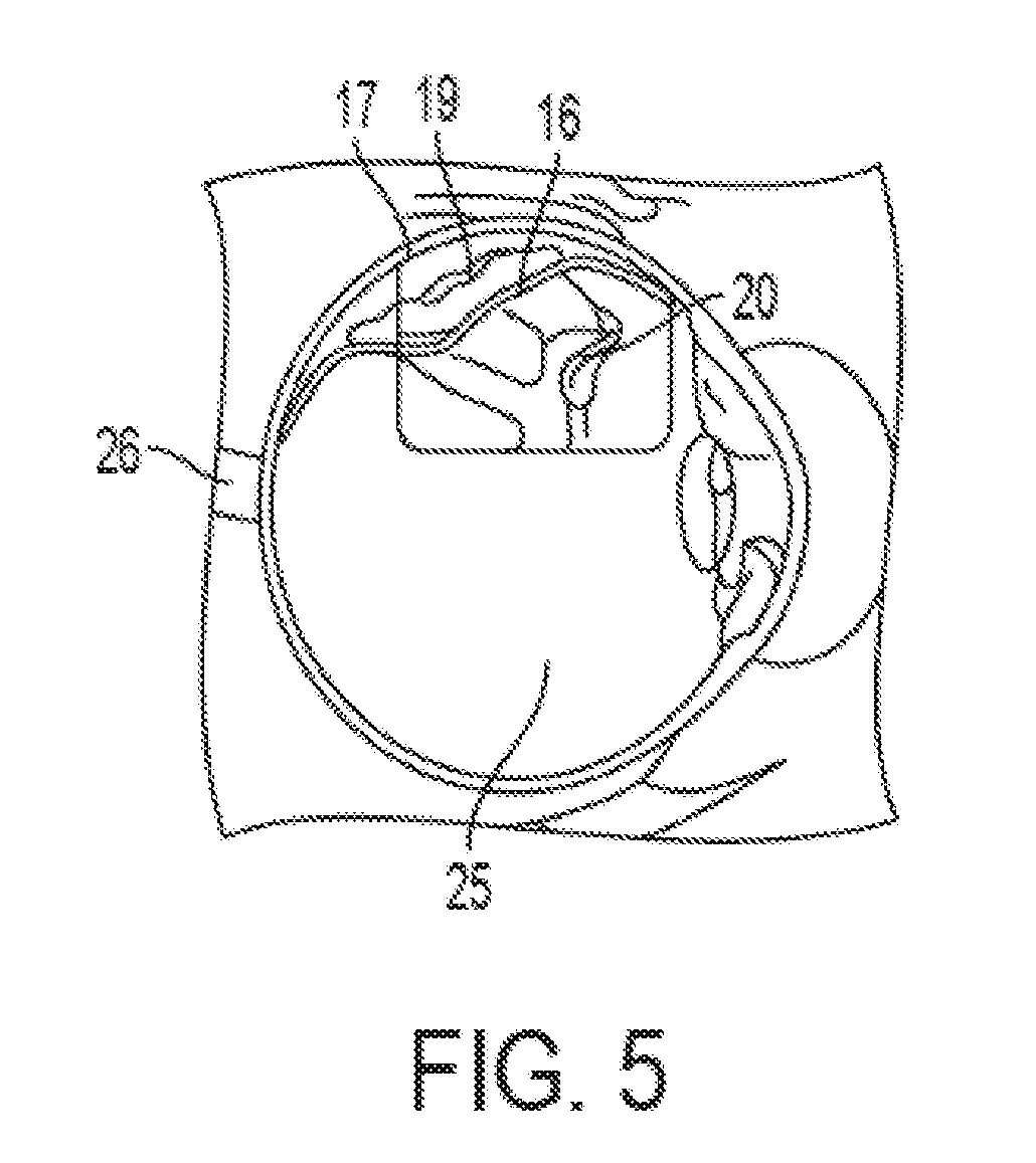
Figure 3
Abstract
Description
[0001] This application is a continuation in part of U.S. patent application Ser. No. 13 / 795,312 filed on Oct. 3, 2012, now U.S. Pat. No. 9,532,902 issued on Jan. 3, 2017 which is a national state entry from PCT / US2011 / 025686 filed on Feb. 22, 2011, which claims priority to provisional patent application No. 61 / 306,644 filed on Feb. 22, 2010, the contents of which are expressly incorporated herein by reference. All references cied herein are expressly incorporated by reference.
[0002] The retina is the interior layer of the posterior eyeball. The retina receives images as they enter through the lens, is continuous with the optic nerve, and consists of several layers, one of which contains the rod cells and cone cells that are sensitive to light.
[0003] A retinal detachment is a separation of the retina from its attachments to the underlying supporting choroid tissue within the eye. Most retinal detachments are a result of an opening that originates as a retinal break, hole, or tear. This...
Examples
Embodiment Construction
[0032]A retinal detachment is a separation of the retina from its attachments to its underlying supporting choroids tissue within the eye. Most retinal detachments are a result of an opening in the retinal wall due to a break, hole, or tear. The opening in the retina may allow the vitreous humor fluid to get in between the retina and the choroid. The fluid can force the retina away from the choroid causing blind spots where the retina has separated. Where the retina is weak, it will tear. As more of the vitreous collects behind the retina, the extent of the retinal detachment can progress and possibly involve the entire retina, leading to a total retinal detachment. A representative tear 20 is shown in FIG. 5 in which the retina 16 has separated from the underlying supporting choroid tissue 17. The tear 20 allows fluid 19 from the posterior chamber 25 to penetrate behind the retina 16 thereby separating the retina 16 from the underlying optic nerve 26. As fluid 19 builds up, the det...