
High speed spectral domain functional optical coherence tomography and optical doppler tomography for in vivo blood flow dynamics and tissue structure
a functional optical coherence and tissue structure technology, applied in the field of biomedical imaging, can solve the problems of limiting the spatial resolution to approximately 200 m, limiting the spatial resolution of flow measurements by ldf, and high spatial resolution noninvasive techniques for imaging in vivo blood flow dynamics and tissue structure are currently not available as diagnostic tools, so as to improve the system sensitivity, eliminate low frequency noise, and double the imaging range
Patent Information
- Authority / Receiving Office
- US · United States
- Patent Type
- Patents(United States)
- Current Assignee / Owner
- Publication Date
- 2008-04-15
Smart Images
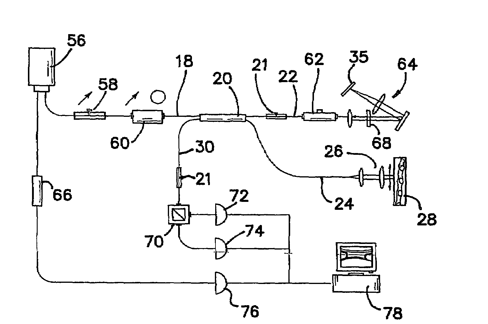
Figure 1 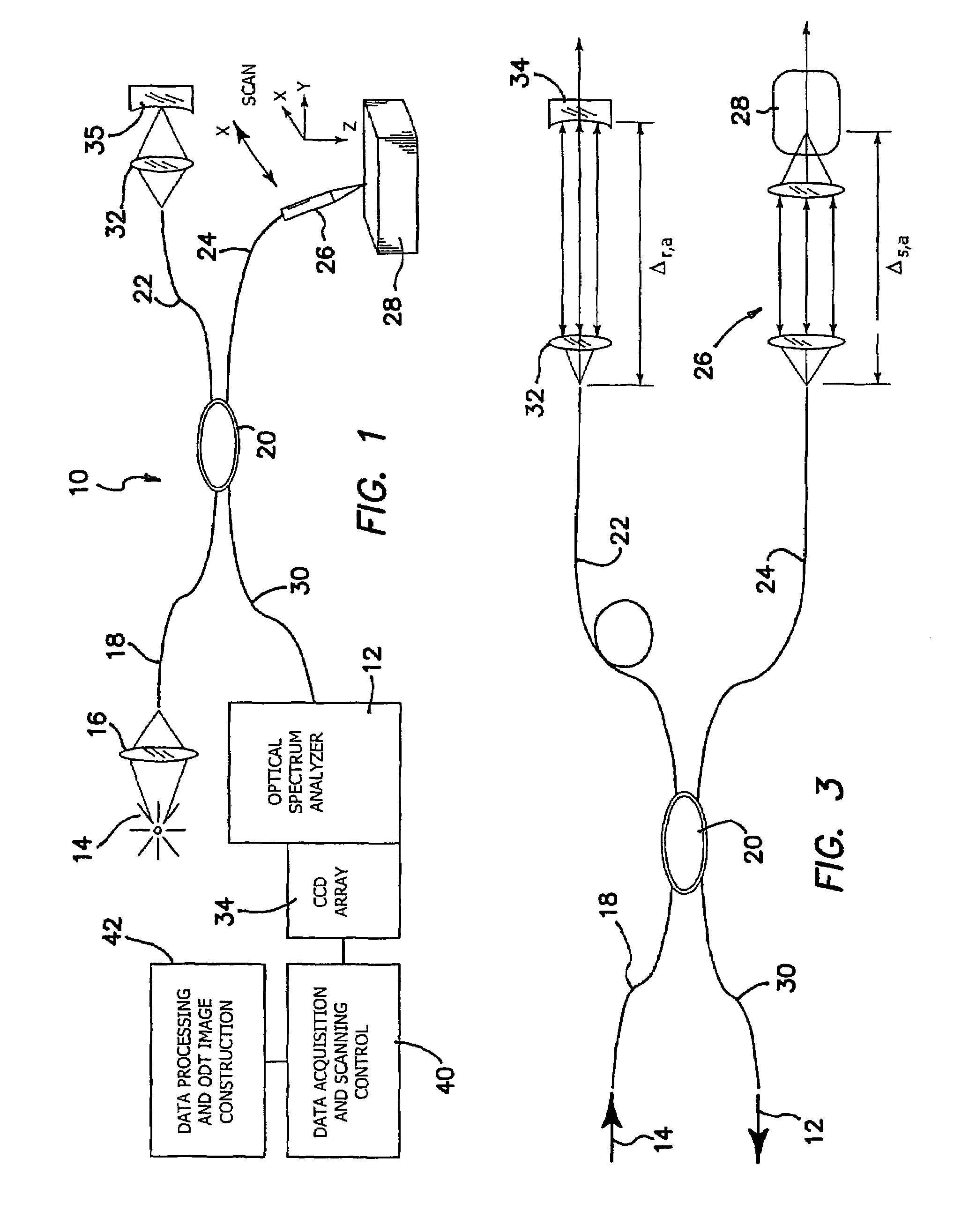
Figure 2 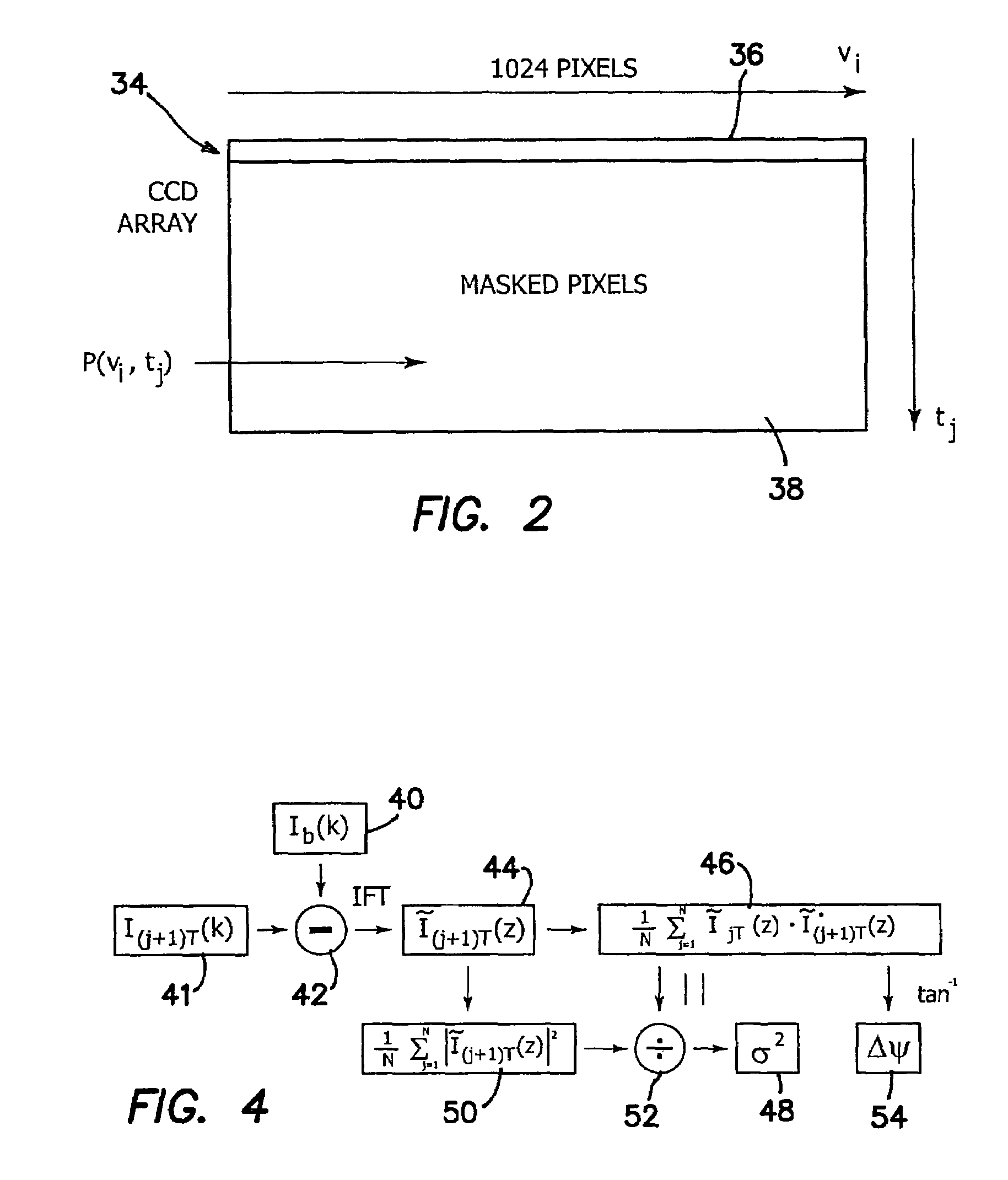
Figure 3
Abstract
Description
RELATED APPLICATIONS
[0001] The present application is related to U.S. Provisional Patent Application Ser. No. 60 / 528,024, filed on Dec. 9, 2003, which is incorporated herein by reference and to which priority is claimed pursuant to 35 USC 119.
[0002] This invention was made with Government support under Grant Nos. WF23281, GM58785, and Rr01192 awarded by the National Institutes of Health. The Government has certain rights in this invention.BACKGROUND OF THE INVENTION
[0003] 1. Field of the Invention
[0004] The invention relates to the field of biomedical imaging, and in particular to functional optical coherence tomography and functional optical Doppler tomography.
[0005] 2. Description of the Prior Art
[0006] Direct visualization of physiological anatomy provides important information to the diagnostician and therapist for the evaluation and management of disease. High spatial resolution noninvasive techniques for imaging in vivo blood flow dynamics and tissue structure are currently not avai...
Examples
Embodiment Construction
[0034]Although high resolution ODT structural and velocity images of static and moving constituents in turbid samples have been obtained using a time domain instrument, clinical application of the technique requires higher imaging speed. The invention is directed to a high speed FDOCT system for imaging in vivo blood flow. This is achieved and demonstrated according to the approach of the invention by using an FDOCT system based on spectral interferometry, an imaging construction algorithm, Monte Carlo simulation of multiple scattering and coherence gating to gain insight into and provide a model for the FDOCT imaging process.
[0035]The prior temporal domain ODT instrument acquires data for each pixel serially by a sequential two-dimensional scan, which limits imaging speed. Image acquisition time for our prototype device is given by T=NxNzΔtp, where Nx and Nz are the number of pixels in the lateral and depth dimensions, and Δtp is the pixel acquisition time. Inasmuch as detection of...