How T wave inversion aids in evaluating myocardial recovery processes
AUG 19, 20259 MIN READ
Generate Your Research Report Instantly with AI Agent
Patsnap Eureka helps you evaluate technical feasibility & market potential.
T Wave Inversion Background and Objectives
T wave inversion is a critical electrocardiographic finding that has long been recognized as a potential indicator of various cardiac abnormalities. The phenomenon occurs when the T wave, which represents ventricular repolarization, appears inverted or negative in certain leads of an electrocardiogram (ECG). Historically, T wave inversion has been associated with myocardial ischemia, infarction, and other cardiac pathologies. However, recent advancements in cardiac electrophysiology have shed new light on its significance in evaluating myocardial recovery processes.
The evolution of our understanding of T wave inversion dates back to the early 20th century when Willem Einthoven first described the ECG waveforms. Since then, numerous studies have explored the mechanisms underlying T wave inversion and its clinical implications. In the context of myocardial recovery, T wave inversion has emerged as a valuable tool for assessing the healing and remodeling of cardiac tissue following injury or stress.
The primary objective of investigating T wave inversion in myocardial recovery is to develop more accurate and non-invasive methods for monitoring cardiac healing processes. This aim aligns with the broader goals of improving patient outcomes, reducing healthcare costs, and enhancing our understanding of cardiac pathophysiology. By leveraging T wave inversion analysis, clinicians and researchers seek to gain insights into the temporal dynamics of myocardial recovery, identify potential complications, and optimize treatment strategies.
Recent technological advancements, including high-resolution ECG systems and advanced signal processing algorithms, have significantly enhanced our ability to detect and interpret subtle changes in T wave morphology. These developments have paved the way for more sophisticated analyses of T wave inversion patterns, enabling researchers to correlate these patterns with specific stages of myocardial recovery.
The current research landscape focuses on several key areas related to T wave inversion and myocardial recovery. These include the identification of specific T wave inversion patterns associated with different phases of recovery, the development of predictive models for assessing recovery trajectories, and the integration of T wave inversion data with other cardiac imaging modalities to provide a more comprehensive evaluation of myocardial health.
As we delve deeper into the role of T wave inversion in evaluating myocardial recovery processes, it is essential to consider the potential clinical applications and limitations of this approach. The ultimate goal is to translate these findings into practical tools that can guide clinical decision-making, improve risk stratification, and enhance patient care in the context of cardiac recovery and rehabilitation.
Clinical Demand for Myocardial Recovery Assessment
The clinical demand for myocardial recovery assessment has grown significantly in recent years, driven by the increasing prevalence of cardiovascular diseases and the need for more effective treatment strategies. Myocardial recovery, the process by which damaged heart tissue regains its function, is a critical aspect of patient care in cardiology. Accurate evaluation of this recovery process is essential for optimizing treatment plans, predicting outcomes, and improving patient quality of life.
T wave inversion, a common electrocardiographic finding, has emerged as a valuable tool in assessing myocardial recovery. This non-invasive method provides clinicians with crucial insights into the heart's electrical activity and structural changes during the healing process. The demand for T wave inversion analysis in myocardial recovery assessment stems from its ability to offer real-time information about the heart's condition without the need for more invasive procedures.
Healthcare providers increasingly recognize the importance of monitoring myocardial recovery to guide therapeutic interventions and adjust treatment strategies. This has led to a growing market for advanced ECG technologies and interpretation software that can accurately detect and analyze T wave inversions. The integration of artificial intelligence and machine learning algorithms in ECG analysis has further enhanced the clinical utility of T wave inversion assessment, allowing for more precise and timely detection of changes in myocardial recovery.
The aging population and the rising incidence of heart diseases have also contributed to the increased demand for myocardial recovery assessment tools. As more patients survive acute cardiac events, there is a greater need for long-term monitoring of heart function and recovery. T wave inversion analysis provides a cost-effective and easily repeatable method for tracking myocardial recovery progress over time, making it an attractive option for both healthcare systems and patients.
Furthermore, the shift towards personalized medicine has heightened the importance of individualized cardiac assessments. T wave inversion patterns can vary among patients, and their interpretation in the context of myocardial recovery requires a nuanced understanding of each patient's unique cardiac profile. This has spurred demand for more sophisticated ECG interpretation tools and training programs for healthcare professionals to accurately utilize T wave inversion data in clinical decision-making.
The pharmaceutical and medical device industries have also shown increased interest in T wave inversion assessment for myocardial recovery. These companies recognize the potential of this method in drug development and clinical trials, particularly for evaluating the efficacy of new cardiovascular therapies. The ability to track myocardial recovery through T wave inversion analysis provides valuable endpoints for clinical studies, potentially accelerating the development of novel treatments for heart diseases.
Current Challenges in T Wave Inversion Analysis
T wave inversion analysis in electrocardiograms (ECGs) plays a crucial role in evaluating myocardial recovery processes. However, several challenges persist in accurately interpreting and utilizing this information. One of the primary difficulties lies in distinguishing pathological T wave inversions from normal variants or benign causes, which can lead to misdiagnosis or unnecessary interventions.
The dynamic nature of T wave changes during the recovery process presents another significant challenge. As the myocardium heals, T wave morphology may evolve, making it difficult to establish consistent criteria for assessing recovery progress. This variability can complicate the interpretation of serial ECGs and hinder the development of standardized protocols for monitoring myocardial recovery.
Furthermore, the influence of factors such as electrolyte imbalances, medication effects, and other cardiac conditions on T wave morphology adds complexity to the analysis. Isolating the specific contribution of myocardial recovery to T wave changes from these confounding factors remains a considerable challenge in clinical practice.
The lack of comprehensive, large-scale studies correlating T wave inversion patterns with long-term outcomes in myocardial recovery also poses a significant obstacle. This gap in knowledge limits the ability to develop evidence-based guidelines for using T wave inversion as a prognostic tool in recovery assessment.
Technical challenges in ECG signal processing and interpretation further complicate T wave inversion analysis. Accurate detection and measurement of T wave characteristics, especially in the presence of noise or artifacts, require sophisticated algorithms and signal processing techniques. The development and validation of robust automated systems for T wave analysis remain ongoing challenges in the field.
Interobserver variability in T wave inversion interpretation is another persistent issue. Different clinicians may have varying thresholds for identifying significant T wave changes, leading to inconsistencies in diagnosis and treatment decisions. Standardizing the criteria for T wave inversion analysis across different healthcare settings and specialties is an ongoing challenge that impacts the reliability of myocardial recovery assessments.
The integration of T wave inversion data with other cardiac imaging modalities and biomarkers presents both opportunities and challenges. While combining multiple data sources can provide a more comprehensive view of myocardial recovery, developing integrated analysis frameworks that effectively synthesize this diverse information remains a complex task.
Addressing these challenges requires multidisciplinary efforts involving cardiologists, electrophysiologists, biomedical engineers, and data scientists. Advances in machine learning and artificial intelligence hold promise for improving the accuracy and consistency of T wave inversion analysis, but significant work remains to be done in validating and implementing these approaches in clinical practice.
Existing T Wave Inversion Evaluation Methods
01 ECG analysis for T wave inversion detection
Advanced ECG analysis techniques are used to detect and characterize T wave inversions, which can be indicative of myocardial recovery. These methods involve sophisticated signal processing and pattern recognition algorithms to accurately identify T wave morphology changes and their progression over time.- ECG analysis for T wave inversion detection: Advanced ECG analysis techniques are employed to detect and characterize T wave inversions, which can be indicative of myocardial recovery. These methods involve sophisticated signal processing and pattern recognition algorithms to accurately identify T wave morphology changes and their progression over time.
- Magnetic resonance imaging for myocardial assessment: Magnetic resonance imaging (MRI) techniques are utilized to evaluate myocardial recovery by assessing tissue characteristics, perfusion, and function. These non-invasive imaging methods provide detailed information about the myocardium, allowing for the detection of subtle changes associated with recovery processes.
- Biomarker analysis for myocardial recovery monitoring: Specific biomarkers are analyzed to monitor myocardial recovery progress. These may include cardiac troponins, natriuretic peptides, and other molecular indicators of cardiac function and repair. The biomarker profiles are used to assess the extent of recovery and guide treatment strategies.
- Wearable devices for continuous cardiac monitoring: Wearable technology is developed to provide continuous monitoring of cardiac activity, including T wave morphology. These devices allow for real-time tracking of ECG changes, enabling early detection of T wave inversion resolution and other indicators of myocardial recovery in outpatient settings.
- Machine learning algorithms for predicting myocardial recovery: Advanced machine learning and artificial intelligence algorithms are employed to analyze complex datasets combining ECG, imaging, and biomarker information. These algorithms aim to predict the likelihood and extent of myocardial recovery based on T wave inversion patterns and other clinical parameters.
02 Magnetic resonance imaging for myocardial assessment
Magnetic resonance imaging (MRI) techniques are employed to evaluate myocardial recovery, providing detailed information about tissue characteristics, perfusion, and function. These imaging methods can detect subtle changes in the myocardium that may be associated with T wave inversion and recovery processes.Expand Specific Solutions03 Biomarker analysis for myocardial recovery monitoring
Biomarker analysis is used to assess myocardial recovery in conjunction with T wave inversion observations. This approach involves measuring specific proteins or other molecules in the blood that are indicative of cardiac health and recovery, providing complementary information to ECG and imaging data.Expand Specific Solutions04 Machine learning algorithms for T wave inversion interpretation
Machine learning and artificial intelligence algorithms are developed to interpret T wave inversions and predict myocardial recovery outcomes. These advanced computational methods can analyze complex patterns in ECG data and other clinical parameters to provide more accurate and personalized assessments.Expand Specific Solutions05 Wearable devices for continuous T wave monitoring
Wearable ECG devices are designed for continuous monitoring of T wave morphology and other cardiac parameters. These devices enable long-term tracking of T wave inversions and myocardial recovery progress in real-world settings, providing valuable data for clinical decision-making and research.Expand Specific Solutions
Key Players in Cardiac Monitoring Technology
The field of T wave inversion analysis for myocardial recovery evaluation is in a mature stage of development, with a significant market size driven by the prevalence of cardiovascular diseases. The technology's maturity is evident in its widespread clinical application, supported by advanced research from institutions like Beth Israel Deaconess Medical Center and Duke University. Major players such as Siemens Healthineers AG and Medtronic, Inc. have contributed to the technological advancements, integrating T wave inversion analysis into their cardiac monitoring and diagnostic systems. The competitive landscape is characterized by a mix of established medical technology companies and specialized research institutions, fostering continuous innovation in this critical area of cardiac health assessment.
Beth Israel Deaconess Medical Center, Inc.
Technical Solution: Beth Israel Deaconess Medical Center has developed advanced ECG analysis techniques to evaluate T wave inversion for myocardial recovery assessment. Their approach combines high-resolution ECG recording with machine learning algorithms to detect subtle changes in T wave morphology[1]. The system analyzes T wave amplitude, duration, and symmetry over time to quantify the degree of myocardial recovery. Additionally, they have integrated this T wave analysis with other cardiac biomarkers and imaging data to create a comprehensive recovery evaluation platform[3].
Strengths: Comprehensive approach combining multiple data sources. Weaknesses: May require specialized equipment and expertise to implement.
Siemens Healthineers AG
Technical Solution: Siemens Healthineers has developed an AI-powered ECG analysis system that focuses on T wave inversion patterns to assess myocardial recovery. Their technology utilizes deep learning algorithms trained on large datasets of ECGs from patients with various cardiac conditions[2]. The system can detect subtle T wave changes that may not be apparent to the human eye, allowing for earlier detection of recovery trends. Siemens' approach also incorporates longitudinal analysis, tracking T wave changes over time to provide a dynamic view of the recovery process[4]. The technology has been integrated into their cardiac monitoring systems for both hospital and remote patient monitoring scenarios.
Strengths: AI-powered analysis for detecting subtle changes. Weaknesses: Dependence on large, high-quality datasets for algorithm training.
Innovative Approaches in T Wave Analysis
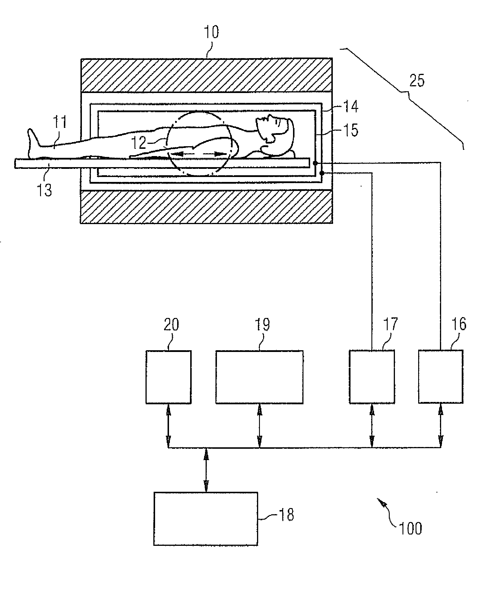
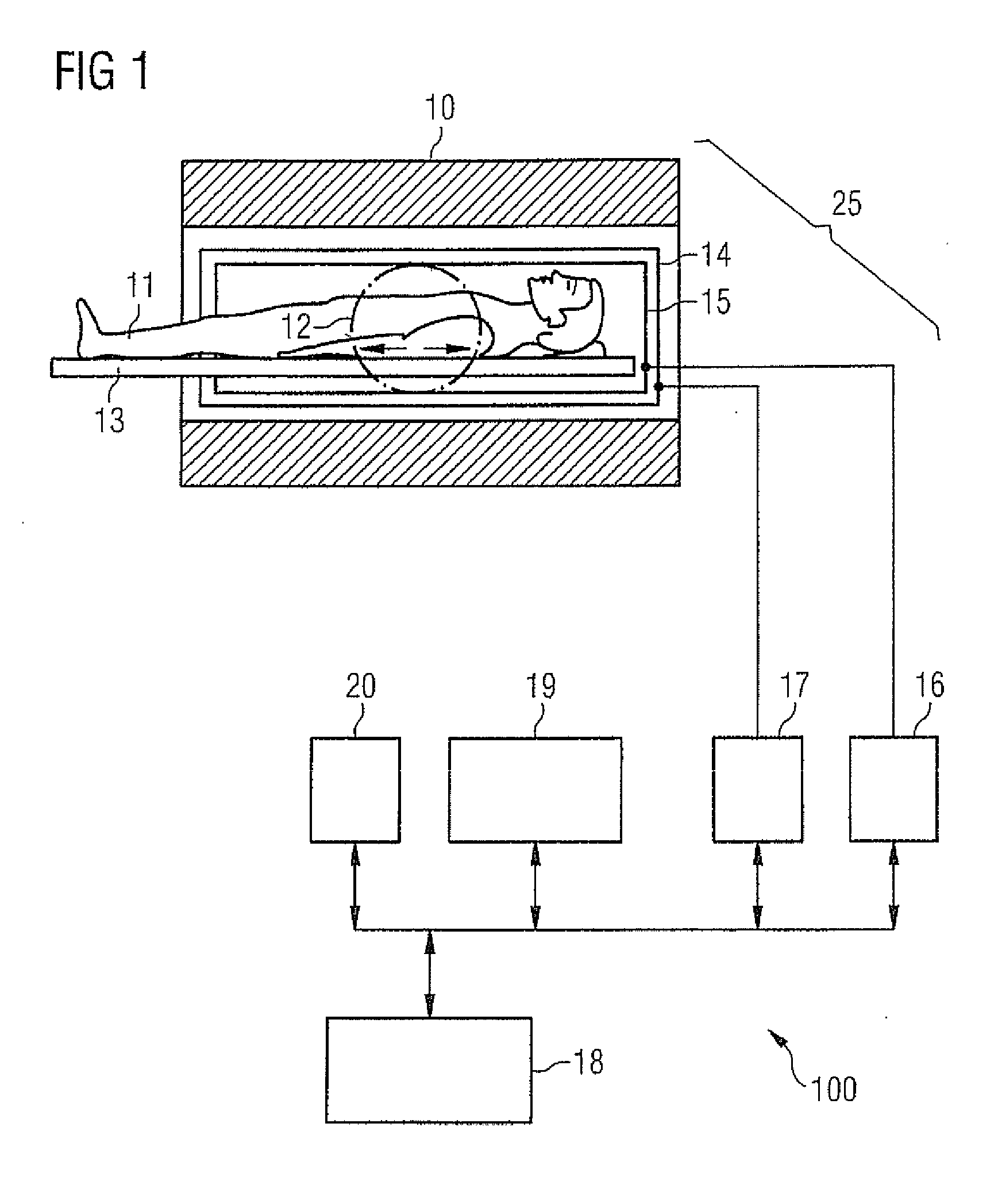
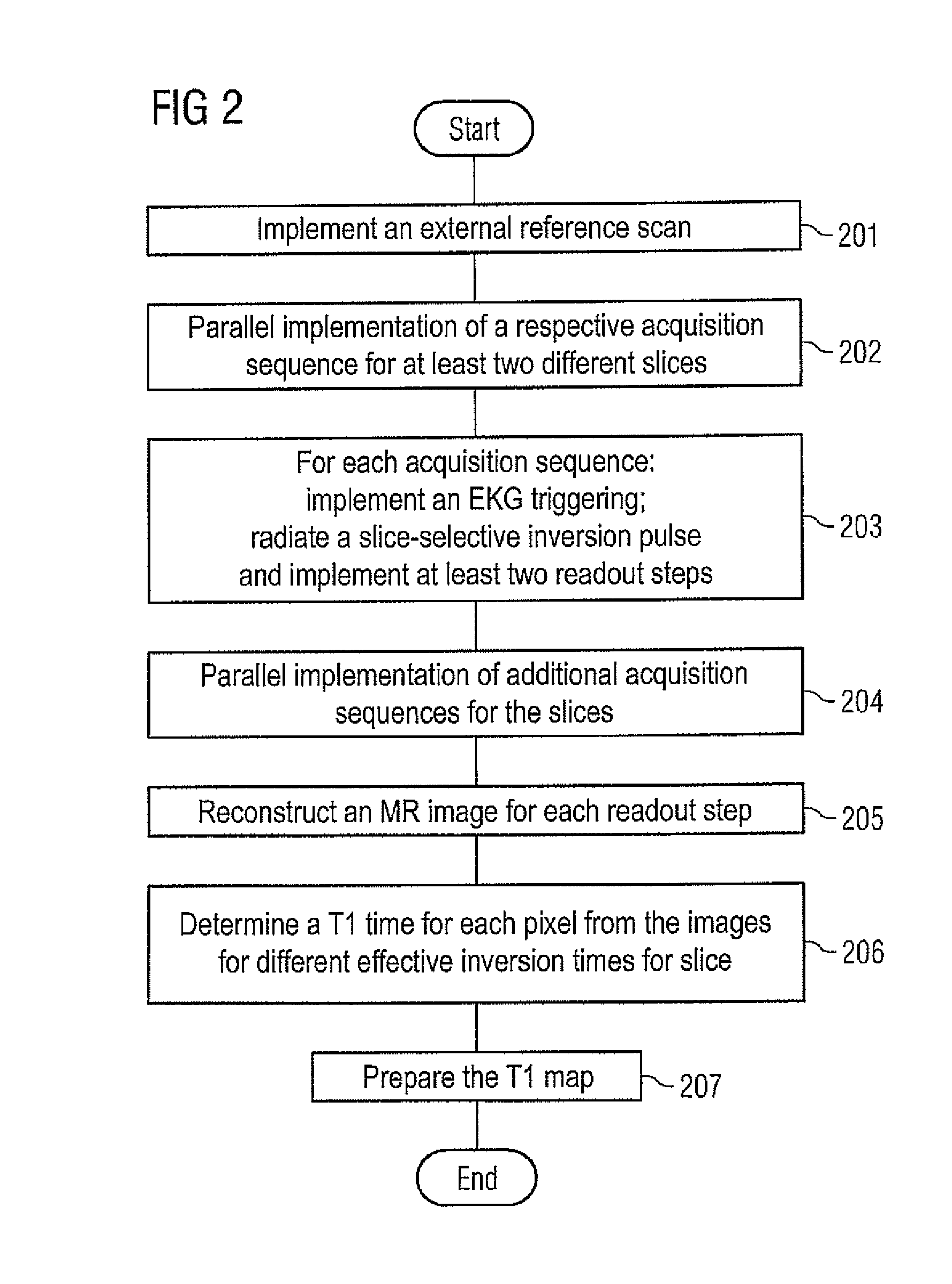
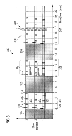
Method and apparatus for magnetic resonance imaging to create t1 maps
PatentActiveUS20110181285A1
Innovation
- A method using parallel imaging with at least two acquisition sequences for MR imaging, where slices are imaged simultaneously with a gradient echo technique, allowing for temporal overlap of up to 80% of the sequences, enabling the acquisition of multiple slices within a single breath hold phase, and utilizing slice-selective inversion pulses and balanced readout sequences to track the T1 decay curve.
Standardization of T Wave Inversion Criteria
The standardization of T wave inversion criteria is crucial for accurately evaluating myocardial recovery processes. Establishing consistent guidelines ensures reliable interpretation of electrocardiogram (ECG) results across different healthcare settings and improves the assessment of cardiac health.
One key aspect of standardization is defining the specific characteristics of T wave inversion that are clinically significant. This includes determining the depth, duration, and morphology of the inverted T waves that correlate with various stages of myocardial recovery. By establishing clear thresholds for these parameters, clinicians can more accurately differentiate between normal variants and pathological changes.
Another important consideration is the lead placement and ECG recording techniques. Standardizing the positioning of electrodes and the recording process helps minimize variability in T wave measurements. This includes specifying the optimal lead configurations for detecting T wave inversions associated with myocardial recovery, as well as defining the appropriate recording duration and frequency.
The timing of ECG recordings during the recovery process is also a critical factor in standardization efforts. Establishing guidelines for when and how often to perform ECG measurements allows for better tracking of T wave changes over time. This temporal standardization helps clinicians identify patterns of T wave inversion that are indicative of successful myocardial recovery or potential complications.
Incorporating quantitative analysis methods into the standardization process can enhance the objectivity of T wave inversion assessments. Developing algorithms and software tools that can consistently measure and analyze T wave characteristics across different ECG systems promotes more uniform interpretation of results. These automated approaches can complement visual assessments and provide additional insights into the recovery process.
Standardization efforts should also address the interpretation of T wave inversions in the context of other ECG findings and clinical data. Establishing guidelines for integrating T wave inversion criteria with other cardiac markers and patient history allows for a more comprehensive evaluation of myocardial recovery. This holistic approach helps clinicians make more informed decisions about patient management and prognosis.
Finally, the standardization of T wave inversion criteria should include provisions for ongoing validation and refinement. As new research emerges and technology advances, it is essential to periodically review and update the criteria to ensure they remain clinically relevant and accurate. This iterative process helps maintain the reliability and effectiveness of T wave inversion assessments in evaluating myocardial recovery processes.
AI Integration in ECG Interpretation
The integration of artificial intelligence (AI) in ECG interpretation has revolutionized the way healthcare professionals analyze and evaluate T wave inversion for assessing myocardial recovery processes. AI algorithms have been developed to enhance the accuracy and efficiency of ECG analysis, particularly in detecting subtle changes in T wave morphology that may indicate ongoing myocardial recovery.
Machine learning models, trained on vast datasets of ECG recordings, have demonstrated remarkable capabilities in identifying T wave inversions and correlating them with various stages of myocardial recovery. These AI-powered systems can detect patterns and anomalies that may be challenging for human interpreters to consistently recognize, especially in cases where the changes are subtle or occur over extended periods.
Deep learning architectures, such as convolutional neural networks (CNNs) and recurrent neural networks (RNNs), have shown particular promise in analyzing the temporal and spatial characteristics of T waves. These models can process large volumes of ECG data in real-time, providing rapid and accurate assessments of T wave inversions and their implications for myocardial recovery.
AI-driven ECG interpretation systems have also been developed to track the evolution of T wave inversions over time, enabling healthcare providers to monitor the progression of myocardial recovery more effectively. By analyzing sequential ECG recordings, these systems can identify trends and patterns in T wave morphology that correlate with various stages of the recovery process.
Furthermore, AI algorithms have been employed to integrate ECG data with other clinical parameters and imaging modalities, such as echocardiography and cardiac MRI. This multi-modal approach allows for a more comprehensive evaluation of myocardial recovery, with AI systems synthesizing information from various sources to provide a holistic assessment of cardiac function and tissue recovery.
The implementation of AI in ECG interpretation has also facilitated the development of predictive models that can forecast the likelihood of complete myocardial recovery based on T wave inversion patterns. These models consider various factors, including the extent and duration of T wave inversions, alongside other clinical and demographic variables, to generate personalized recovery prognoses.
As AI continues to advance, there is growing potential for the development of more sophisticated algorithms that can detect even more subtle changes in T wave morphology and provide increasingly nuanced insights into the myocardial recovery process. This ongoing evolution of AI-powered ECG interpretation promises to further enhance our understanding of cardiac recovery mechanisms and improve patient outcomes through more targeted and timely interventions.
Unlock deeper insights with Patsnap Eureka Quick Research — get a full tech report to explore trends and direct your research. Try now!
Generate Your Research Report Instantly with AI Agent
Supercharge your innovation with Patsnap Eureka AI Agent Platform!