T wave inversion's role in stress-induced cardiomyopathy detections
AUG 19, 20259 MIN READ
Generate Your Research Report Instantly with AI Agent
Patsnap Eureka helps you evaluate technical feasibility & market potential.
T Wave Inversion Background and Objectives
T wave inversion is a critical electrocardiographic (ECG) finding that has gained significant attention in the field of cardiology, particularly in the context of stress-induced cardiomyopathy detection. This phenomenon, characterized by the reversal of the normally upright T wave on an ECG, has been observed in various cardiac conditions, including acute coronary syndromes, myocarditis, and notably, stress-induced cardiomyopathy, also known as Takotsubo syndrome.
The historical context of T wave inversion research dates back to the early 20th century when Willem Einthoven developed the first practical ECG. Since then, the understanding of T wave morphology and its clinical significance has evolved substantially. In the 1980s and 1990s, researchers began to recognize the association between emotional or physical stress and transient left ventricular dysfunction, laying the groundwork for the identification of stress-induced cardiomyopathy.
The primary objective in studying T wave inversion's role in stress-induced cardiomyopathy detection is to enhance early diagnosis and improve patient outcomes. This aim is particularly crucial given that stress-induced cardiomyopathy can mimic acute myocardial infarction, potentially leading to misdiagnosis and inappropriate treatment. By refining our understanding of T wave inversion patterns specific to stress-induced cardiomyopathy, clinicians can more accurately differentiate this condition from other acute cardiac events.
Recent technological advancements have significantly contributed to the field, enabling more precise ECG analysis and interpretation. Machine learning algorithms and artificial intelligence have been employed to detect subtle T wave changes that may not be apparent to the human eye, potentially improving the sensitivity and specificity of stress-induced cardiomyopathy diagnosis.
The global prevalence of stress-induced cardiomyopathy has been increasing, possibly due to improved recognition and diagnostic capabilities. This trend underscores the importance of ongoing research into T wave inversion and its diagnostic value. Furthermore, the COVID-19 pandemic has highlighted the relevance of stress-induced cardiac conditions, as reports of Takotsubo syndrome cases have surged during this period of heightened global stress.
Looking ahead, the field aims to establish standardized criteria for T wave inversion patterns indicative of stress-induced cardiomyopathy. This standardization would facilitate more accurate and timely diagnoses across healthcare settings. Additionally, researchers are exploring the potential of combining T wave inversion analysis with other biomarkers and imaging techniques to create comprehensive diagnostic algorithms for stress-induced cardiomyopathy.
Market Need for Cardiomyopathy Detection
The market need for stress-induced cardiomyopathy detection has been growing significantly in recent years, driven by several key factors. Stress-induced cardiomyopathy, also known as Takotsubo cardiomyopathy or broken heart syndrome, is a temporary heart condition that mimics the symptoms of a heart attack. The increasing prevalence of stress-related disorders and cardiovascular diseases has heightened the importance of accurate and timely detection methods.
Cardiovascular diseases remain the leading cause of death globally, with stress being a major contributing factor. The World Health Organization reports that cardiovascular diseases account for 31% of all deaths worldwide. This staggering statistic underscores the critical need for improved diagnostic tools and early detection methods, particularly for stress-induced cardiomyopathy.
The healthcare industry has recognized the potential of T wave inversion as a valuable indicator in the detection of stress-induced cardiomyopathy. T wave inversion, observed in electrocardiograms (ECGs), can provide crucial insights into the heart's electrical activity and potential abnormalities. The market demand for advanced ECG technologies and interpretation tools that can accurately identify T wave inversions has been steadily increasing.
Healthcare providers, including hospitals, clinics, and cardiac care centers, are seeking more sophisticated diagnostic tools to improve patient outcomes and reduce healthcare costs. Early detection of stress-induced cardiomyopathy can lead to timely interventions, potentially preventing more severe cardiac events and reducing the overall burden on healthcare systems.
The aging population in many developed countries has also contributed to the growing market need for cardiomyopathy detection. As the risk of cardiovascular diseases increases with age, there is a higher demand for reliable diagnostic methods that can differentiate between various cardiac conditions, including stress-induced cardiomyopathy.
Furthermore, the COVID-19 pandemic has highlighted the importance of cardiac health monitoring, as the virus has been associated with increased cardiovascular complications. This has led to a surge in demand for remote monitoring solutions and advanced diagnostic tools that can detect subtle changes in cardiac function, including T wave inversions.
The market for stress-induced cardiomyopathy detection is also driven by the increasing awareness among both healthcare professionals and patients about the condition. As more research emerges on the role of T wave inversion in diagnosing this condition, there is a growing interest in developing and adopting technologies that can accurately interpret these ECG changes.
In conclusion, the market need for stress-induced cardiomyopathy detection, particularly focusing on T wave inversion, is substantial and continues to grow. The combination of rising cardiovascular disease prevalence, an aging population, technological advancements, and increased awareness has created a strong demand for innovative diagnostic solutions in this field.
Current Challenges in T Wave Analysis
T wave inversion analysis in stress-induced cardiomyopathy detection faces several significant challenges that hinder accurate diagnosis and interpretation. One of the primary obstacles is the variability in T wave morphology across different individuals and even within the same individual under various physiological conditions. This inherent variability makes it difficult to establish a universal standard for T wave inversion that can be reliably applied across diverse patient populations.
Another challenge lies in distinguishing pathological T wave inversions associated with stress-induced cardiomyopathy from benign or non-specific T wave changes. Stress-induced cardiomyopathy, also known as Takotsubo cardiomyopathy, can mimic acute coronary syndromes in its electrocardiographic presentation, leading to potential misdiagnosis or unnecessary interventions. The transient nature of T wave inversions in stress-induced cardiomyopathy further complicates the analysis, as the timing of ECG recording becomes crucial for accurate detection.
The influence of confounding factors on T wave morphology presents an additional challenge. Electrolyte imbalances, medication effects, and other cardiac conditions can all alter T wave appearance, making it difficult to isolate the specific changes attributable to stress-induced cardiomyopathy. This complexity necessitates a comprehensive approach to T wave analysis that considers multiple physiological and pathological factors.
Technical limitations in ECG recording and processing also contribute to the challenges in T wave analysis. Signal noise, baseline wander, and other artifacts can distort T wave morphology, potentially leading to misinterpretation. Advanced signal processing techniques are required to enhance the quality of ECG recordings and isolate T wave changes accurately.
The lack of standardized criteria for defining and quantifying T wave inversion in the context of stress-induced cardiomyopathy poses a significant challenge. While various methods have been proposed, there is no consensus on the optimal approach for measuring and interpreting T wave inversions in this specific condition. This lack of standardization hampers the comparability of research findings and the development of robust diagnostic algorithms.
Furthermore, the dynamic nature of stress-induced cardiomyopathy requires serial ECG monitoring to capture the evolution of T wave changes over time. This temporal aspect adds complexity to the analysis and necessitates the development of sophisticated algorithms capable of tracking and interpreting T wave morphology changes throughout the course of the condition.
Existing T Wave Inversion Detection Methods
01 ECG signal analysis for T wave inversion detection
Advanced algorithms are employed to analyze ECG signals, specifically focusing on the T wave morphology. These methods involve signal processing techniques to identify and characterize T wave inversions, which can be indicative of various cardiac conditions. The analysis may include feature extraction, waveform comparison, and pattern recognition to accurately detect and classify T wave inversions.- ECG signal analysis for T wave inversion detection: Methods for analyzing ECG signals to detect T wave inversion, which can be indicative of various cardiac conditions. These techniques involve processing the ECG waveform, identifying key features, and applying algorithms to determine the presence and characteristics of T wave inversion.
- Machine learning approaches for T wave inversion detection: Utilization of machine learning algorithms, including deep learning and neural networks, to improve the accuracy and efficiency of T wave inversion detection. These approaches can learn from large datasets of ECG signals to identify subtle patterns indicative of T wave inversion.
- Real-time monitoring and alert systems for T wave inversion: Development of systems for continuous, real-time monitoring of ECG signals to detect T wave inversion. These systems can provide immediate alerts to healthcare providers when abnormalities are detected, enabling prompt intervention.
- Integration of T wave inversion detection in wearable devices: Incorporation of T wave inversion detection algorithms into wearable ECG devices and smartwatches. This allows for continuous monitoring of cardiac health in non-clinical settings, providing early warning signs of potential cardiac issues.
- Multi-parameter analysis for improved T wave inversion detection: Methods that combine T wave inversion detection with other ECG parameters and physiological data to enhance the accuracy of cardiac assessments. This holistic approach can provide a more comprehensive evaluation of cardiac health and reduce false positives.
02 Machine learning approaches for T wave inversion detection
Machine learning models, including deep learning algorithms, are utilized to improve the accuracy and efficiency of T wave inversion detection. These models are trained on large datasets of ECG recordings to recognize subtle patterns and features associated with T wave inversions. The use of artificial intelligence enables more robust and adaptable detection methods, potentially reducing false positives and improving diagnostic capabilities.Expand Specific Solutions03 Real-time monitoring and alert systems for T wave inversions
Systems are developed for continuous, real-time monitoring of ECG signals to detect T wave inversions. These systems can be integrated into wearable devices or hospital monitoring equipment, providing immediate alerts to healthcare professionals when T wave inversions are detected. The real-time capability allows for prompt intervention and management of potential cardiac issues.Expand Specific Solutions04 Multi-lead ECG analysis for improved T wave inversion detection
Techniques are developed to analyze multiple ECG leads simultaneously for enhanced T wave inversion detection. By comparing and correlating T wave morphologies across different leads, these methods can provide a more comprehensive assessment of cardiac electrical activity. This multi-lead approach helps to distinguish between normal variants and pathological T wave inversions, improving diagnostic accuracy.Expand Specific Solutions05 Integration of patient data and ECG analysis for contextual T wave inversion detection
Advanced systems combine ECG analysis with patient-specific data, such as medical history, demographics, and other clinical parameters. This integrated approach provides a more comprehensive context for T wave inversion detection, allowing for personalized interpretation of ECG findings. By considering individual patient factors, these methods can improve the specificity of T wave inversion detection and aid in clinical decision-making.Expand Specific Solutions
Key Players in Cardiac Diagnostic Industry
The competitive landscape for T wave inversion's role in stress-induced cardiomyopathy detection is evolving rapidly. The market is in its growth phase, with increasing demand for advanced cardiac diagnostic tools. The global market size for cardiac monitoring devices is projected to expand significantly in the coming years. Technologically, the field is advancing, with companies like Medtronic, Philips, and Siemens Healthineers leading innovation. These firms are developing sophisticated ECG and imaging systems that can more accurately detect T wave inversions and other cardiac abnormalities. Smaller players like Contec Medical Systems and Edan Instruments are also contributing to technological advancements, particularly in portable and AI-enhanced ECG devices.
Medtronic, Inc.
Technical Solution: Medtronic has developed advanced algorithms for detecting T wave inversion in stress-induced cardiomyopathy. Their approach combines real-time ECG analysis with machine learning techniques to identify subtle changes in T wave morphology[1]. The system utilizes a multi-lead ECG monitoring device that captures high-resolution waveforms and applies advanced signal processing to isolate T waves. A neural network model trained on a large dataset of stress-induced cardiomyopathy cases is then used to classify T wave inversions and assess their clinical significance[3]. The algorithm also incorporates other ECG parameters and patient data to improve accuracy and reduce false positives.
Strengths: High sensitivity and specificity in detecting stress-induced cardiomyopathy, integration with existing cardiac monitoring systems. Weaknesses: Requires specialized hardware and may have higher costs compared to traditional ECG interpretation methods.
Koninklijke Philips NV
Technical Solution: Philips has developed an AI-powered ECG analysis platform that focuses on detecting T wave inversions associated with stress-induced cardiomyopathy. Their system employs a deep learning model trained on a vast database of ECG recordings from confirmed cases[2]. The algorithm analyzes the entire 12-lead ECG, paying particular attention to precordial leads where T wave inversions are most prominent in stress-induced cardiomyopathy. The system also considers temporal changes in T wave morphology, comparing sequential ECGs to identify evolving patterns characteristic of the condition[4]. Additionally, Philips' solution integrates with their broader cardiac care ecosystem, allowing for seamless data sharing and clinical decision support.
Strengths: Comprehensive ECG analysis, integration with existing hospital systems, and continuous learning capabilities. Weaknesses: May require significant computational resources and ongoing model updates to maintain accuracy.
Innovative Approaches in ECG Signal Processing
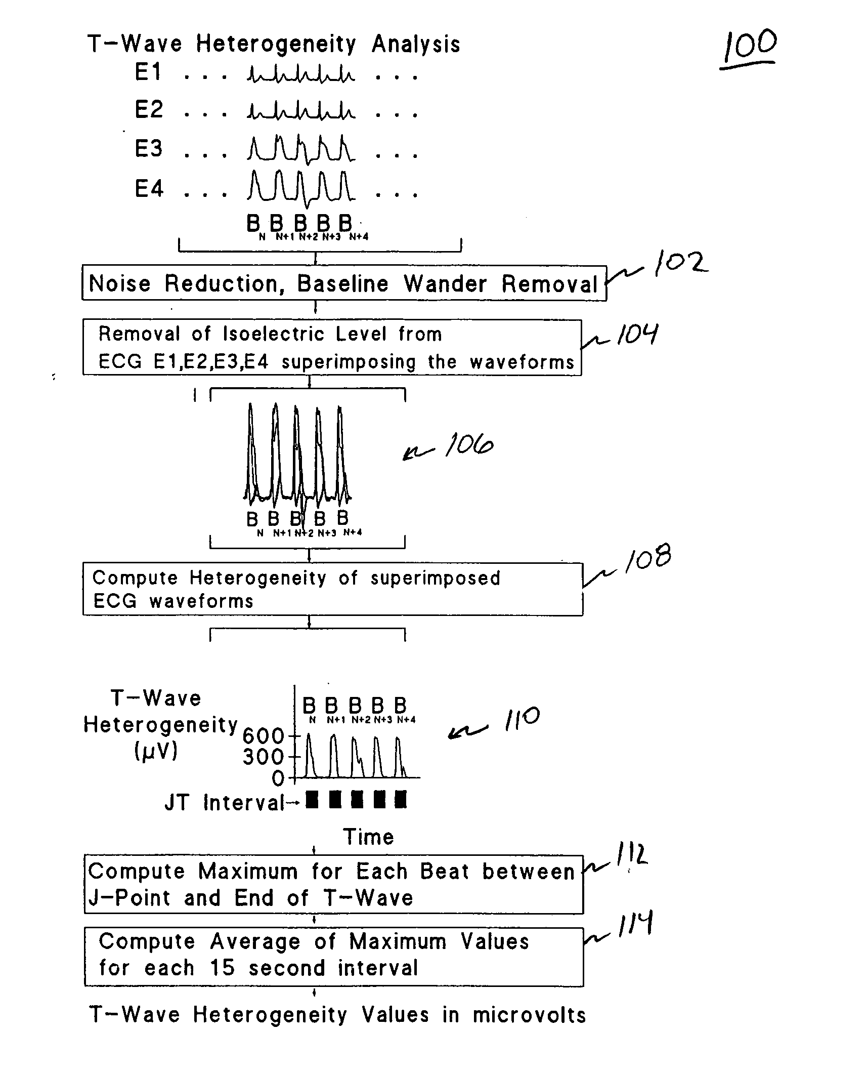
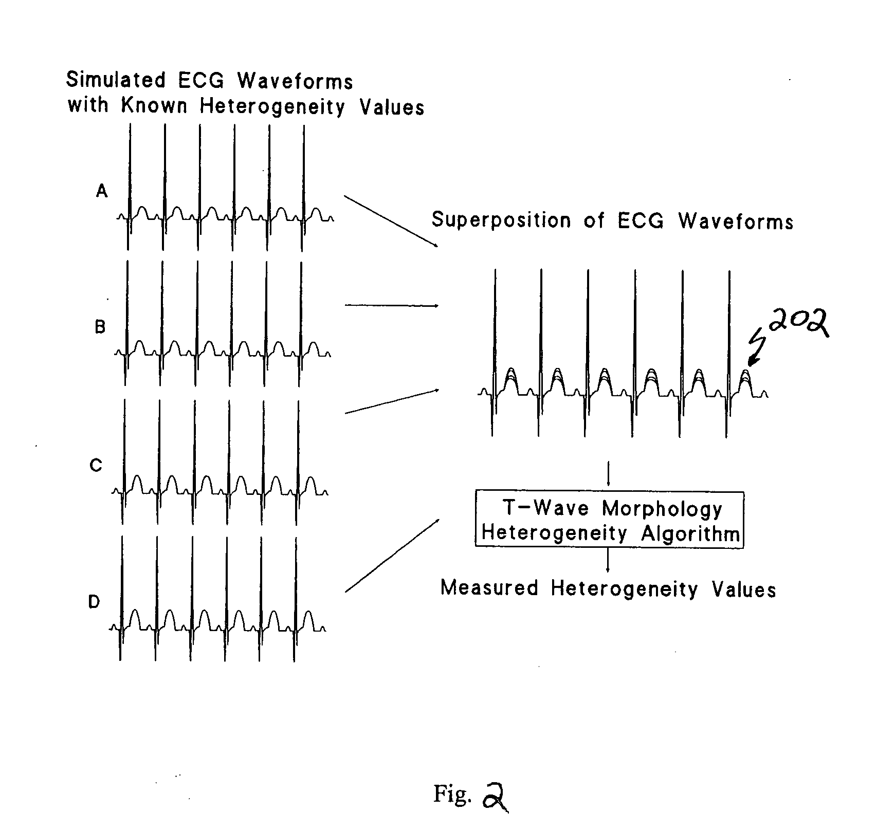
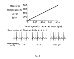
Spatial heterogeneity of repolarization waveform amplitude to assess risk of sudden cardiac death
PatentInactiveUS20050010122A1
Innovation
- A method and apparatus that quantify and track spatial heterogeneity of repolarization using second central moment analysis of T-wave morphology from multiple ECG leads, providing a complementary measure to TWA to assess cardiac electrical instability, while being resistant to artifacts such as respiratory and rhythmic motion.
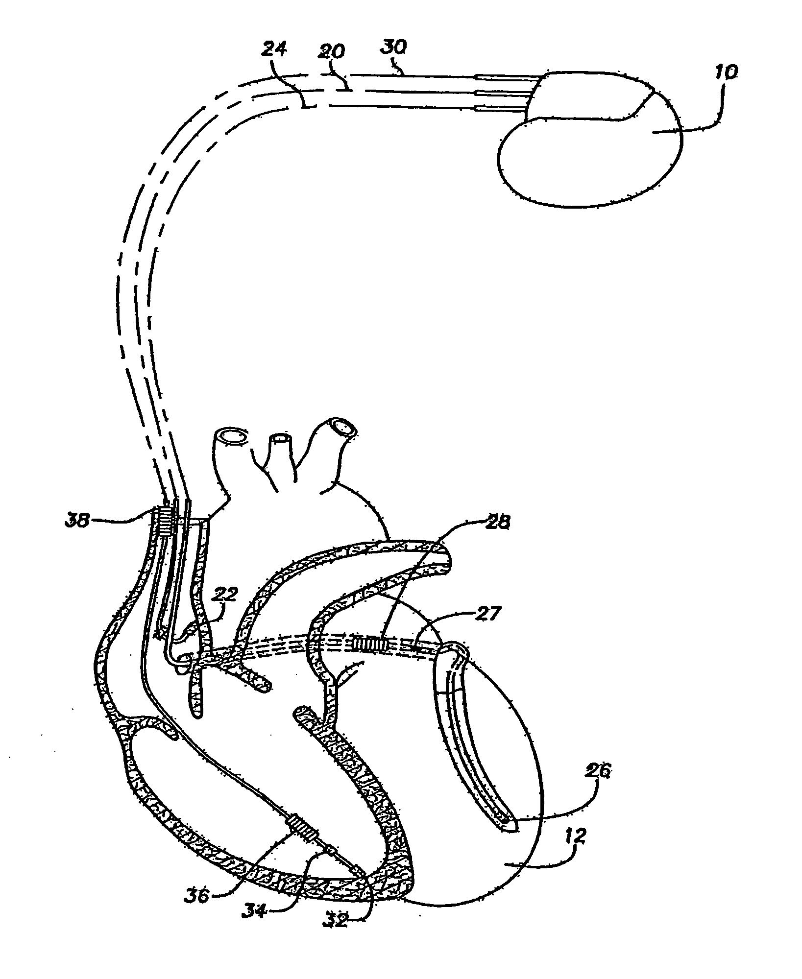
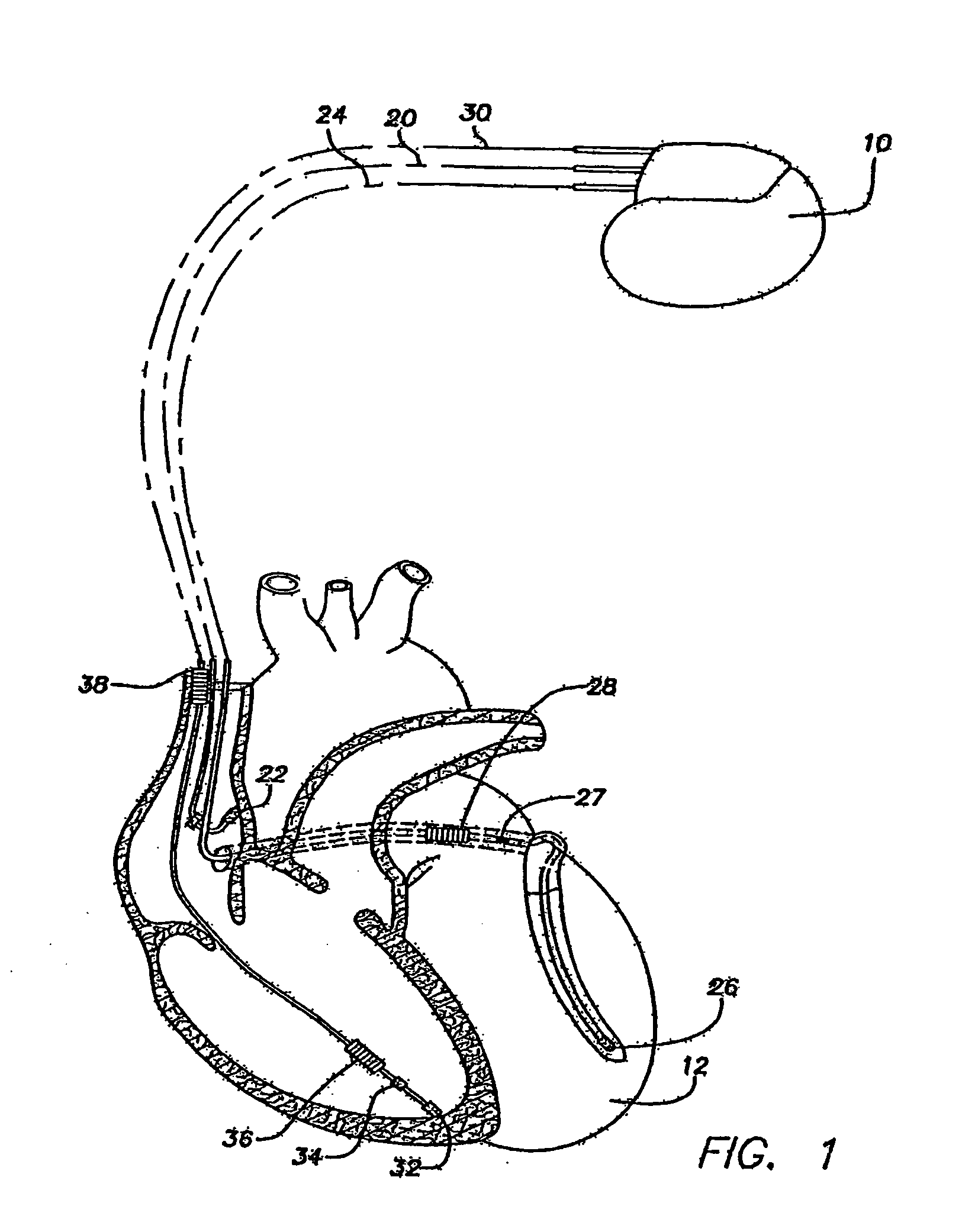
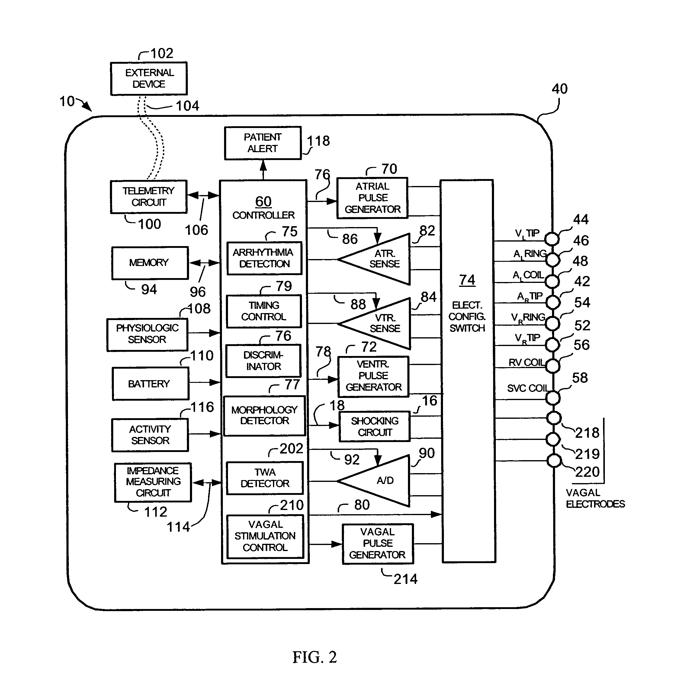
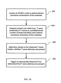
Methods and systems for detecting the presence of T-wave alternans
PatentInactiveUS7881792B1
Innovation
- An implantable cardiac device system that detects intrinsic or induced premature contractions of the ventricles and measures T-wave metrics to determine the presence of T-wave alternans, allowing for continuous monitoring without the need for surface ECGs or extensive computational resources.
Clinical Validation and Standardization
Clinical validation and standardization are crucial steps in establishing the reliability and applicability of T wave inversion as a diagnostic marker for stress-induced cardiomyopathy. To ensure the accuracy and consistency of this diagnostic approach, a comprehensive validation process must be implemented across multiple healthcare settings.
The first step in clinical validation involves conducting large-scale, multi-center studies to assess the sensitivity and specificity of T wave inversion in detecting stress-induced cardiomyopathy. These studies should include diverse patient populations to account for variations in age, gender, ethnicity, and comorbidities. By analyzing a broad range of cases, researchers can determine the robustness of T wave inversion as a diagnostic indicator across different patient subgroups.
Standardization of the measurement and interpretation of T wave inversion is essential for consistent diagnosis. This process involves developing clear guidelines for electrocardiogram (ECG) acquisition, including lead placement, recording duration, and filtering techniques. Additionally, establishing quantitative criteria for T wave inversion, such as depth, duration, and morphology, is necessary to minimize inter-observer variability.
To further enhance standardization, the creation of a centralized database of ECG recordings from confirmed stress-induced cardiomyopathy cases is recommended. This resource would serve as a reference for clinicians and researchers, facilitating the development of machine learning algorithms for automated detection and classification of T wave inversions associated with the condition.
Validation of these standardized methods should include inter-rater reliability studies, where multiple clinicians independently assess the same set of ECGs to ensure consistent interpretation. Furthermore, longitudinal studies tracking patients with T wave inversions over time can provide insights into the prognostic value of this marker and its relationship to long-term outcomes in stress-induced cardiomyopathy.
The integration of T wave inversion analysis into existing clinical decision support systems is another critical aspect of validation and standardization. By incorporating validated algorithms into electronic health records, clinicians can receive real-time alerts and recommendations based on ECG findings, potentially improving the speed and accuracy of diagnosis.
Lastly, ongoing quality assurance programs should be established to maintain the accuracy of T wave inversion interpretation in clinical practice. These programs may include regular proficiency testing for clinicians, periodic review of diagnostic accuracy, and continuous updating of guidelines based on new research findings. Through these comprehensive validation and standardization efforts, T wave inversion can become a more reliable and widely accepted tool in the detection of stress-induced cardiomyopathy.
AI Integration in ECG Analysis
The integration of artificial intelligence (AI) in ECG analysis has revolutionized the detection and interpretation of cardiac abnormalities, including stress-induced cardiomyopathy. AI algorithms have demonstrated remarkable capabilities in identifying subtle ECG changes, such as T wave inversion, which plays a crucial role in diagnosing this condition. Machine learning models, particularly deep learning neural networks, have been developed to analyze large volumes of ECG data and detect patterns associated with stress-induced cardiomyopathy.
These AI-powered systems can process ECG signals in real-time, providing rapid and accurate assessments of T wave morphology and other relevant features. By leveraging advanced signal processing techniques and pattern recognition algorithms, AI can identify even subtle T wave inversions that may be challenging for human interpreters to detect consistently. This enhanced sensitivity and specificity in T wave analysis contribute significantly to the early detection and management of stress-induced cardiomyopathy.
Furthermore, AI integration enables the development of predictive models that can assess the risk of stress-induced cardiomyopathy based on ECG patterns and other clinical data. These models can analyze historical ECG records, patient demographics, and other relevant factors to identify individuals at higher risk of developing the condition. This proactive approach allows for targeted interventions and personalized patient management strategies.
AI-driven ECG analysis also facilitates the standardization of diagnostic criteria for stress-induced cardiomyopathy. By analyzing vast datasets of ECG recordings from confirmed cases, AI algorithms can establish more precise and objective criteria for T wave inversion and other ECG changes associated with the condition. This standardization improves diagnostic accuracy and reduces inter-observer variability in ECG interpretation.
The integration of AI in ECG analysis extends beyond standalone systems, with efforts to incorporate these technologies into existing healthcare infrastructure. Cloud-based platforms and mobile applications powered by AI algorithms enable remote ECG analysis and interpretation, expanding access to expert-level diagnostics in various healthcare settings. This integration enhances the ability to detect stress-induced cardiomyopathy in diverse patient populations and clinical contexts.
As AI continues to evolve, ongoing research focuses on developing more sophisticated algorithms that can differentiate stress-induced cardiomyopathy from other cardiac conditions with similar ECG presentations. These advancements aim to further improve diagnostic accuracy and reduce false positives, ultimately leading to more targeted and effective patient care strategies in the management of stress-induced cardiomyopathy.
Unlock deeper insights with Patsnap Eureka Quick Research — get a full tech report to explore trends and direct your research. Try now!
Generate Your Research Report Instantly with AI Agent
Supercharge your innovation with Patsnap Eureka AI Agent Platform!