Investigating T wave inversion's relationship with ventricular arrhythmias
AUG 19, 20259 MIN READ
Generate Your Research Report Instantly with AI Agent
Patsnap Eureka helps you evaluate technical feasibility & market potential.
T Wave Inversion Background and Research Objectives
T wave inversion is a significant electrocardiographic finding that has long intrigued cardiologists and researchers in the field of cardiac electrophysiology. This phenomenon, characterized by the reversal of the normal T wave polarity on an electrocardiogram (ECG), has been observed in various cardiac conditions and has sparked considerable interest due to its potential implications for ventricular arrhythmias.
The study of T wave inversion dates back to the early days of electrocardiography, with initial observations made in the early 20th century. Over the decades, our understanding of this ECG abnormality has evolved significantly, driven by advancements in cardiac imaging, electrophysiology, and molecular biology. Today, T wave inversion is recognized as a complex and multifaceted phenomenon that can be both a benign variant and a marker of underlying cardiac pathology.
The relationship between T wave inversion and ventricular arrhythmias has been a subject of ongoing research and debate. While some studies have suggested a strong correlation between certain patterns of T wave inversion and an increased risk of life-threatening ventricular arrhythmias, others have found the association to be less clear-cut. This discrepancy highlights the need for further investigation to elucidate the underlying mechanisms and clinical significance of T wave inversion in the context of arrhythmogenesis.
The primary objective of this research is to conduct a comprehensive investigation into the relationship between T wave inversion and ventricular arrhythmias. This endeavor aims to bridge the gaps in our current understanding and provide valuable insights that could potentially impact clinical practice and patient management strategies.
Specifically, the research seeks to address several key questions: What are the electrophysiological mechanisms underlying T wave inversion? How do different patterns and distributions of T wave inversion correlate with the risk of ventricular arrhythmias? Are there specific patient populations or cardiac conditions in which T wave inversion is particularly predictive of arrhythmic events? Can advanced ECG analysis techniques or novel biomarkers enhance the prognostic value of T wave inversion in identifying patients at high risk for ventricular arrhythmias?
By exploring these questions, we aim to develop a more nuanced understanding of T wave inversion as a potential risk marker for ventricular arrhythmias. This research has the potential to refine risk stratification algorithms, improve the interpretation of ECG findings, and ultimately contribute to more targeted and effective strategies for preventing sudden cardiac death.
Clinical Significance of T Wave Inversion
T wave inversion on an electrocardiogram (ECG) is a significant clinical finding that has garnered considerable attention in cardiology due to its potential association with various cardiac conditions, particularly ventricular arrhythmias. The clinical significance of T wave inversion lies in its ability to serve as a marker for underlying cardiac pathology and its potential to predict adverse cardiovascular events.
In the context of ventricular arrhythmias, T wave inversion can be an important indicator of electrical instability in the heart. This abnormality in the repolarization phase of the cardiac cycle may reflect underlying structural or functional changes in the myocardium that predispose patients to dangerous rhythm disturbances. The presence of T wave inversion, especially when new or dynamic, often prompts further cardiac evaluation to assess for potential ischemia, cardiomyopathy, or other conditions that may increase the risk of ventricular arrhythmias.
Research has shown that T wave inversion in specific lead patterns can be associated with different types of cardiac pathology. For instance, anterior T wave inversion may suggest anterior wall ischemia or hypertrophic cardiomyopathy, while inferior T wave inversion might indicate inferior wall ischemia or pulmonary embolism. The distribution and persistence of T wave inversion can provide valuable clues about the underlying cardiac condition and its severity.
Moreover, T wave inversion has been linked to an increased risk of sudden cardiac death in certain populations. Studies have demonstrated that individuals with T wave inversion, particularly in the right precordial leads, may have a higher likelihood of developing life-threatening ventricular arrhythmias. This association is especially pronounced in patients with structural heart disease or inherited arrhythmia syndromes.
In clinical practice, the identification of T wave inversion often triggers a cascade of diagnostic procedures. These may include echocardiography, stress testing, cardiac MRI, or electrophysiological studies to further evaluate the heart's structure and function. The goal is to uncover any underlying pathology that may be responsible for the ECG abnormality and to assess the patient's risk for future arrhythmic events.
It is important to note that while T wave inversion can be a marker of cardiac pathology, it is not always pathological. Certain physiological conditions, such as normal variant patterns in young adults or athletes, can also present with T wave inversion. Therefore, the clinical significance of T wave inversion must always be interpreted in the context of the patient's overall clinical picture, including age, symptoms, risk factors, and other ECG findings.
Current Understanding and Challenges in T Wave Inversion Analysis
T wave inversion has long been recognized as a significant electrocardiographic finding, often associated with various cardiac conditions. Current understanding of T wave inversion's relationship with ventricular arrhythmias has evolved significantly, yet several challenges remain in its analysis and interpretation.
Recent studies have shed light on the mechanisms underlying T wave inversion and its potential link to ventricular arrhythmias. It is now widely accepted that T wave inversion can be indicative of underlying myocardial ischemia, structural heart disease, or electrolyte imbalances, all of which may predispose individuals to ventricular arrhythmias. The presence of inverted T waves, particularly in specific lead configurations, has been associated with an increased risk of life-threatening ventricular arrhythmias and sudden cardiac death.
Advances in cardiac imaging and electrophysiological mapping techniques have allowed researchers to correlate T wave inversion patterns with specific areas of myocardial dysfunction or scarring. This has led to a better understanding of the anatomical and functional substrates that may contribute to both T wave inversion and the development of ventricular arrhythmias.
However, several challenges persist in the analysis of T wave inversion and its relationship to ventricular arrhythmias. One major obstacle is the non-specific nature of T wave inversion, as it can be observed in various cardiac and non-cardiac conditions, making it difficult to determine its clinical significance in isolation. Additionally, the dynamic nature of T wave morphology and its dependence on factors such as heart rate, autonomic tone, and medication effects further complicate its interpretation.
Another significant challenge lies in the quantification and standardization of T wave inversion analysis. While visual assessment remains the primary method in clinical practice, there is a growing need for more objective and reproducible measures of T wave characteristics. Efforts to develop automated algorithms for T wave analysis have shown promise but are still limited by the complexity and variability of ECG signals.
The relationship between T wave inversion and specific types of ventricular arrhythmias also requires further elucidation. While associations have been established with conditions such as long QT syndrome and arrhythmogenic right ventricular cardiomyopathy, the predictive value of T wave inversion for different arrhythmia subtypes remains an area of active research.
Lastly, the integration of T wave inversion analysis with other risk stratification tools and biomarkers presents both an opportunity and a challenge. Developing comprehensive risk assessment models that incorporate T wave characteristics alongside other clinical, genetic, and imaging parameters could potentially improve the prediction and prevention of ventricular arrhythmias.
Existing Methods for T Wave Inversion Detection and Analysis
01 T wave inversion detection in ECG analysis
Methods and systems for detecting T wave inversion in electrocardiogram (ECG) signals. This involves analyzing the morphology and polarity of T waves to identify abnormal patterns that may indicate cardiac issues. Advanced signal processing techniques are used to isolate T waves and assess their characteristics.- T wave inversion detection in ECG analysis: Methods and systems for detecting T wave inversion in electrocardiogram (ECG) signals. This involves analyzing the morphology and polarity of T waves to identify abnormal patterns that may indicate cardiac issues. Advanced signal processing techniques are used to isolate T waves and assess their characteristics.
- Relationship between T wave inversion and cardiac conditions: Studies on the correlation between T wave inversion and various cardiac conditions, including ischemia, myocardial infarction, and structural heart diseases. This research aims to establish diagnostic criteria and risk stratification based on T wave inversion patterns observed in ECG recordings.
- Machine learning algorithms for T wave inversion analysis: Development of artificial intelligence and machine learning algorithms to automatically detect and classify T wave inversions in ECG data. These algorithms are designed to improve the accuracy and efficiency of ECG interpretation, potentially assisting healthcare professionals in diagnosis.
- T wave inversion in relation to other ECG parameters: Investigation of the relationship between T wave inversion and other ECG parameters such as QT interval, ST segment changes, and R wave amplitude. This research aims to provide a comprehensive understanding of how T wave inversion interacts with other ECG features to indicate various cardiac abnormalities.
- T wave inversion in specific patient populations: Studies focusing on T wave inversion patterns in specific patient groups, such as athletes, elderly individuals, or those with particular genetic predispositions. This research aims to establish normal variants and pathological patterns of T wave inversion in different demographics to improve diagnostic accuracy.
02 Relationship between T wave inversion and cardiac conditions
Studies on the correlation between T wave inversion and various cardiac conditions, including ischemia, myocardial infarction, and structural heart diseases. This research aims to establish diagnostic criteria and risk stratification based on T wave inversion patterns.Expand Specific Solutions03 Machine learning algorithms for T wave inversion analysis
Development of artificial intelligence and machine learning algorithms to improve the accuracy and efficiency of T wave inversion detection and interpretation. These algorithms are trained on large datasets to recognize subtle patterns and provide automated analysis of ECG signals.Expand Specific Solutions04 T wave inversion in specific patient populations
Investigation of T wave inversion characteristics in specific patient groups, such as athletes, elderly individuals, or those with genetic predispositions to cardiac disorders. This research aims to establish normal variants and pathological patterns specific to different demographics.Expand Specific Solutions05 T wave inversion in relation to other ECG parameters
Exploration of the relationship between T wave inversion and other ECG parameters, such as QT interval, ST segment changes, and R wave amplitude. This comprehensive approach helps in understanding the overall electrical activity of the heart and its implications for cardiac health.Expand Specific Solutions
Key Institutions and Researchers in Cardiac Electrophysiology
The investigation of T wave inversion's relationship with ventricular arrhythmias is in a mature stage of development, with significant market potential in the cardiovascular health sector. The technology's maturity is evident from the involvement of established medical device companies like Medtronic, Inc. and Biosense Webster, Inc., which have extensive experience in cardiac monitoring and treatment. Research institutions such as Massachusetts Institute of Technology and Beth Israel Deaconess Medical Center are contributing to the field's advancement. The market is characterized by a mix of large corporations and specialized firms like Impulse Dynamics NV, indicating a competitive landscape with opportunities for innovation and growth in ECG analysis and arrhythmia detection technologies.
Medtronic, Inc.
Technical Solution: Medtronic has developed advanced algorithms for analyzing T wave inversions in relation to ventricular arrhythmias. Their approach involves continuous monitoring of ECG signals using implantable cardioverter-defibrillators (ICDs) and external wearable devices. The company's proprietary software employs machine learning techniques to detect subtle changes in T wave morphology that may precede arrhythmic events. This system can analyze multiple ECG leads simultaneously, providing a comprehensive view of cardiac electrical activity[1]. Medtronic's technology also incorporates patient-specific baseline measurements to improve accuracy in identifying abnormal T wave patterns[3].
Strengths: Comprehensive multi-lead analysis, personalized baselines, and integration with implantable devices. Weaknesses: Potential for false positives in non-cardiac conditions affecting T waves, reliance on continuous monitoring.
Beth Israel Deaconess Medical Center, Inc.
Technical Solution: Beth Israel Deaconess Medical Center has pioneered research into the relationship between T wave inversion and ventricular arrhythmias. Their approach combines high-resolution ECG recording with advanced signal processing techniques. The center's researchers have developed a novel method for quantifying T wave heterogeneity across the myocardium, which has shown promise in predicting arrhythmic risk[2]. This method involves creating detailed T wave maps and analyzing spatial and temporal variations in repolarization patterns. Additionally, the center has conducted longitudinal studies correlating T wave inversions with clinical outcomes, providing valuable data on the prognostic significance of these ECG changes[4].
Strengths: Cutting-edge research methodologies, comprehensive clinical data integration, and focus on translational outcomes. Weaknesses: Limited to research settings, may require specialized equipment not widely available in clinical practice.
Innovative Approaches in T Wave Morphology Assessment
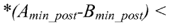
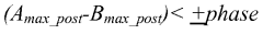
Detection of t-wave alternans phase reversal for arrhythmia prediction and sudden cardiac death risk stratification
PatentWO2011126643A2
Innovation
- An implantable medical device (IMD) system that dynamically monitors TWA by acquiring electrogram signals, using a combination of R-wave detection, signal conditioning, and microprocessor-based algorithms to assess T-wave features and detect phase reversal, enabling ambulatory monitoring and risk stratification for arrhythmias.
ECG Signal Processing Techniques for T Wave Analysis
ECG signal processing techniques play a crucial role in the analysis of T waves, particularly in investigating the relationship between T wave inversion and ventricular arrhythmias. These techniques encompass a wide range of methods designed to extract, enhance, and interpret T wave characteristics from electrocardiogram signals.
One of the fundamental techniques in T wave analysis is signal filtering. This process involves removing noise and artifacts from the ECG signal to isolate the T wave component. Various filtering methods, such as low-pass, high-pass, and band-pass filters, are employed to eliminate baseline wander, power line interference, and high-frequency noise. Advanced adaptive filtering techniques can further improve the signal quality by dynamically adjusting filter parameters based on signal characteristics.
Wavelet transform is another powerful tool in T wave analysis. This technique allows for multi-resolution analysis of the ECG signal, enabling the detection and characterization of T wave morphology changes across different frequency scales. Wavelet-based methods are particularly effective in identifying subtle T wave alterations that may be indicative of ventricular arrhythmias.
Feature extraction is a critical step in T wave analysis. Techniques such as principal component analysis (PCA) and independent component analysis (ICA) are used to extract relevant features from the T wave segment. These methods can help identify key morphological characteristics, including T wave amplitude, duration, symmetry, and area under the curve. Machine learning algorithms can then be applied to these extracted features to classify T wave patterns and predict the likelihood of ventricular arrhythmias.
Time-frequency analysis techniques, such as short-time Fourier transform (STFT) and Wigner-Ville distribution, provide insights into the temporal evolution of T wave frequency components. These methods are particularly useful in detecting dynamic changes in T wave morphology that may precede the onset of ventricular arrhythmias.
Advanced signal processing techniques, including non-linear dynamics analysis and complexity measures, have also been applied to T wave analysis. These methods can reveal subtle patterns and irregularities in T wave behavior that may not be apparent through traditional linear analysis techniques. Fractal dimension analysis and entropy-based measures have shown promise in quantifying T wave complexity and its relationship to arrhythmic risk.
Machine learning and deep learning approaches have gained significant traction in ECG signal processing for T wave analysis. Convolutional neural networks (CNNs) and recurrent neural networks (RNNs) have demonstrated impressive performance in automatically detecting and classifying T wave abnormalities, including T wave inversion. These AI-driven techniques can learn complex patterns from large datasets, potentially uncovering novel associations between T wave characteristics and ventricular arrhythmias.
Implications for Personalized Arrhythmia Risk Stratification
The investigation of T wave inversion's relationship with ventricular arrhythmias has significant implications for personalized arrhythmia risk stratification. This research direction holds promise for developing more accurate and individualized approaches to identifying patients at high risk for life-threatening arrhythmias.
T wave inversion, a common electrocardiographic finding, has long been associated with various cardiac conditions. However, its specific role in predicting ventricular arrhythmias has not been fully elucidated. By exploring this relationship in depth, clinicians and researchers can potentially refine risk assessment tools and improve patient outcomes.
One of the key advantages of focusing on T wave inversion is its non-invasive nature. Electrocardiograms (ECGs) are widely available, cost-effective, and can be easily performed in various clinical settings. Incorporating T wave inversion analysis into existing risk stratification models could enhance their predictive power without significantly increasing healthcare costs or patient discomfort.
Personalized risk stratification based on T wave inversion patterns may allow for more targeted interventions. Patients identified as high-risk could be monitored more closely, receive more aggressive medical therapy, or be considered for implantable cardioverter-defibrillator (ICD) placement earlier in their disease course. Conversely, those with lower risk profiles could potentially avoid unnecessary interventions and the associated complications.
The integration of T wave inversion analysis with other established risk factors, such as left ventricular ejection fraction, QT interval, and genetic markers, could lead to the development of comprehensive risk scores. These scores would provide a more nuanced understanding of an individual's arrhythmia risk, enabling healthcare providers to tailor management strategies more effectively.
Furthermore, the study of T wave inversion in relation to ventricular arrhythmias may reveal new insights into the underlying pathophysiology of these dangerous rhythm disturbances. This could, in turn, guide the development of novel therapeutic approaches aimed at preventing or treating arrhythmias at their source.
As personalized medicine continues to advance, the ability to stratify arrhythmia risk based on individual ECG characteristics becomes increasingly valuable. This approach aligns with the broader trend towards precision medicine, where treatments and preventive strategies are tailored to the unique characteristics of each patient.
In conclusion, the investigation of T wave inversion's relationship with ventricular arrhythmias has the potential to significantly improve personalized arrhythmia risk stratification. By leveraging this readily available ECG feature, clinicians may be able to more accurately identify high-risk patients, optimize treatment strategies, and ultimately reduce the incidence of sudden cardiac death due to ventricular arrhythmias.
Unlock deeper insights with Patsnap Eureka Quick Research — get a full tech report to explore trends and direct your research. Try now!
Generate Your Research Report Instantly with AI Agent
Supercharge your innovation with Patsnap Eureka AI Agent Platform!