T wave inversion implications in cardiac rehabilitative medicine
AUG 19, 20258 MIN READ
Generate Your Research Report Instantly with AI Agent
Patsnap Eureka helps you evaluate technical feasibility & market potential.
T Wave Inversion Background
T wave inversion is a significant electrocardiographic finding that has garnered considerable attention in the field of cardiac rehabilitative medicine. This phenomenon, characterized by the reversal of the normal T wave polarity in one or more leads of an electrocardiogram (ECG), has been a subject of extensive research and clinical interest for decades.
The T wave represents the repolarization of the ventricles, a crucial phase in the cardiac cycle. Under normal circumstances, the T wave appears as an upright deflection following the QRS complex. However, in certain conditions, this wave can become inverted, potentially indicating underlying cardiac pathology or physiological variations.
Historically, T wave inversion was first described in the early 20th century, coinciding with the development and widespread adoption of electrocardiography. Initially, it was primarily associated with acute cardiac events such as myocardial infarction. However, as our understanding of cardiac electrophysiology evolved, the implications of T wave inversion expanded to encompass a broader spectrum of cardiac conditions.
In the context of cardiac rehabilitation, T wave inversion has emerged as a valuable prognostic and diagnostic tool. Its presence can signify various cardiac abnormalities, including ischemia, cardiomyopathy, or electrolyte imbalances. Moreover, it has been observed in athletes and individuals with structurally normal hearts, adding complexity to its interpretation.
The significance of T wave inversion in cardiac rehabilitative medicine lies in its potential to guide treatment strategies and risk stratification. For patients recovering from cardiac events or undergoing rehabilitation programs, the presence or absence of T wave inversion can inform decisions regarding exercise intensity, medication adjustments, and overall management plans.
Recent advancements in cardiac imaging and molecular biology have further elucidated the underlying mechanisms of T wave inversion. These insights have led to a more nuanced understanding of its implications, moving beyond simple binary interpretations to consider factors such as the specific leads affected, the depth and symmetry of the inversion, and its dynamic changes over time.
As the field of cardiac rehabilitative medicine continues to evolve, the role of T wave inversion in patient assessment and management remains an area of active research. Ongoing studies aim to refine our understanding of its prognostic value, particularly in the context of long-term cardiac outcomes and the efficacy of rehabilitation interventions.
Clinical Significance
T wave inversion in electrocardiograms (ECGs) holds significant clinical importance in cardiac rehabilitative medicine, serving as a crucial indicator of various underlying cardiac conditions. This abnormality in the ECG waveform can provide valuable insights into the patient's cardiovascular health and guide treatment strategies in rehabilitation settings.
The presence of T wave inversion often signifies myocardial ischemia, a condition where the heart muscle receives insufficient blood supply. In the context of cardiac rehabilitation, identifying and monitoring T wave inversion patterns can help clinicians assess the severity of coronary artery disease and evaluate the effectiveness of ongoing treatment protocols. This information is vital for tailoring exercise regimens and medication adjustments to optimize patient outcomes.
Furthermore, T wave inversion can be indicative of structural heart abnormalities, such as hypertrophic cardiomyopathy or left ventricular hypertrophy. In cardiac rehabilitation programs, recognizing these underlying conditions is essential for developing appropriate exercise prescriptions and risk stratification strategies. Patients with these structural abnormalities may require modified rehabilitation approaches to ensure safe and effective recovery.
The dynamic nature of T wave inversion during cardiac rehabilitation also provides valuable prognostic information. Persistent or worsening T wave inversion may suggest inadequate myocardial recovery or the need for further interventions. Conversely, the resolution of T wave inversion over time can indicate successful rehabilitation and improved cardiac function. This temporal assessment of T wave changes allows clinicians to track patient progress and make informed decisions regarding the continuation or modification of rehabilitation protocols.
In acute coronary syndromes, T wave inversion can be an early warning sign of impending myocardial infarction. For patients in cardiac rehabilitation following such events, careful monitoring of T wave morphology is crucial for detecting potential recurrence or complications. This vigilance enables prompt intervention and adjustment of rehabilitation strategies to prevent adverse cardiac events.
T wave inversion also plays a role in assessing the efficacy of pharmacological interventions in cardiac rehabilitation. Changes in T wave patterns can reflect the impact of medications on cardiac electrophysiology and myocardial perfusion. This information aids in fine-tuning drug therapies to achieve optimal cardiac function and minimize potential side effects during the rehabilitation process.
In conclusion, the clinical significance of T wave inversion in cardiac rehabilitative medicine is multifaceted and far-reaching. It serves as a valuable tool for diagnosis, risk stratification, treatment guidance, and prognostic assessment. By carefully interpreting T wave inversion patterns, healthcare providers can enhance the precision and effectiveness of cardiac rehabilitation programs, ultimately improving patient outcomes and quality of life.
Diagnostic Challenges
T wave inversion in electrocardiograms (ECGs) presents significant diagnostic challenges in cardiac rehabilitative medicine. The interpretation of this phenomenon is complex, as T wave inversion can be indicative of various cardiac conditions, ranging from benign to life-threatening.
One of the primary challenges lies in distinguishing between pathological and physiological T wave inversions. Physiological inversions can occur in healthy individuals, particularly in certain ECG leads, making it difficult to differentiate from pathological causes. This ambiguity often leads to unnecessary anxiety for patients and potential over-investigation.
The variability in T wave morphology across different patient populations further complicates diagnosis. Factors such as age, gender, ethnicity, and athletic status can influence the appearance of T waves, requiring clinicians to have a nuanced understanding of these variations to avoid misinterpretation.
In the context of cardiac rehabilitation, the dynamic nature of T wave changes poses another challenge. As patients progress through their recovery, T wave morphology may evolve, reflecting improvements in cardiac function or, conversely, indicating potential complications. This necessitates continuous monitoring and interpretation, which can be resource-intensive and time-consuming.
The presence of confounding factors in cardiac rehabilitation settings adds another layer of complexity. Medications, electrolyte imbalances, and changes in autonomic tone can all affect T wave morphology, potentially masking or mimicking pathological inversions. Clinicians must carefully consider these factors when interpreting ECGs in rehabilitative contexts.
Furthermore, the lack of standardized criteria for interpreting T wave inversions in specific patient subgroups undergoing cardiac rehabilitation creates uncertainty. This is particularly evident in patients with pre-existing ECG abnormalities or those who have undergone cardiac interventions, where baseline ECGs may already show atypical features.
The implications of T wave inversion on exercise prescription and progression in cardiac rehabilitation programs are also challenging to navigate. Determining safe exercise parameters and knowing when to modify or halt rehabilitation based on T wave changes requires careful clinical judgment and often leads to conservative approaches that may unnecessarily limit patient progress.
Lastly, the integration of new technologies, such as wearable ECG monitors, into cardiac rehabilitation programs introduces both opportunities and challenges. While these devices offer continuous monitoring capabilities, they also generate vast amounts of data that require sophisticated interpretation algorithms and raise questions about the clinical significance of transient T wave changes detected outside of controlled clinical settings.
Current Diagnostic Methods
01 Electrocardiogram (ECG) analysis for T wave inversion detection
Advanced algorithms and machine learning techniques are employed to analyze ECG signals and detect T wave inversions. These methods can identify subtle changes in the T wave morphology, potentially indicating cardiac abnormalities or other health issues.- Electrocardiogram (ECG) analysis for T wave inversion detection: Advanced algorithms and machine learning techniques are employed to analyze ECG signals and detect T wave inversions. These methods can identify subtle changes in the T wave morphology, potentially indicating cardiac abnormalities or other health issues.
- Implications of T wave inversion in cardiac health assessment: T wave inversion can be indicative of various cardiac conditions, including ischemia, electrolyte imbalances, or structural heart abnormalities. The analysis of T wave inversions helps in risk stratification and early detection of potential cardiac issues, guiding further diagnostic and treatment decisions.
- Wearable devices for continuous T wave monitoring: Wearable technology incorporating ECG sensors enables continuous monitoring of T waves outside clinical settings. These devices can provide real-time data on T wave changes, allowing for early detection of abnormalities and timely medical intervention.
- Artificial intelligence in T wave inversion interpretation: AI-powered systems are being developed to interpret T wave inversions more accurately and efficiently. These systems can analyze large datasets of ECG recordings, identify patterns, and provide insights into the clinical significance of T wave inversions, potentially improving diagnostic accuracy.
- Integration of T wave inversion data in electronic health records: Systems and methods for integrating T wave inversion data into electronic health records are being developed. This integration allows for better tracking of cardiac health over time, facilitates communication between healthcare providers, and supports more comprehensive patient care management.
02 Implications of T wave inversion in cardiac health assessment
T wave inversion can be indicative of various cardiac conditions, including ischemia, electrolyte imbalances, or structural heart abnormalities. The analysis of T wave inversions helps in risk stratification and early detection of potential cardiac issues, guiding further diagnostic and treatment decisions.Expand Specific Solutions03 Wearable devices for continuous T wave monitoring
Wearable technology incorporating ECG sensors enables continuous monitoring of T waves in real-time. These devices can alert users and healthcare providers to significant T wave inversions, allowing for prompt medical intervention when necessary.Expand Specific Solutions04 Artificial intelligence in T wave inversion interpretation
AI-powered systems are being developed to interpret T wave inversions more accurately and efficiently. These systems can analyze large datasets of ECG recordings, potentially identifying patterns and correlations that may not be immediately apparent to human observers.Expand Specific Solutions05 Integration of T wave inversion data in electronic health records
Electronic health record systems are being enhanced to incorporate T wave inversion data, allowing for better tracking of cardiac health over time. This integration facilitates comprehensive patient care by providing healthcare providers with a more complete picture of a patient's cardiac status.Expand Specific Solutions
Key Research Institutions
The T wave inversion implications in cardiac rehabilitative medicine represent a complex and evolving field within cardiology. The market is in a growth phase, driven by increasing prevalence of cardiovascular diseases and advancements in diagnostic technologies. Key players like Medtronic, Beth Israel Deaconess Medical Center, and Impulse Dynamics are at the forefront of research and development. The technology's maturity varies, with established ECG interpretation methods coexisting alongside emerging AI-driven analytics. Companies such as Welch Allyn and Japan Lifeline are contributing to the development of more sophisticated monitoring devices, while research institutions like MIT and Shanghai Jiao Tong University are pushing the boundaries of clinical understanding and application.
Medtronic, Inc.
Technical Solution: Medtronic has developed advanced algorithms for T-wave inversion detection in their cardiac monitoring devices. Their approach combines machine learning techniques with traditional ECG analysis to improve the accuracy of T-wave inversion identification. The system uses a multi-lead ECG analysis, considering spatial and temporal relationships between T-waves across different leads[1]. This allows for better differentiation between pathological T-wave inversions and normal variants. Medtronic's technology also incorporates patient-specific baseline data to account for individual variability, enhancing the specificity of T-wave inversion detection in cardiac rehabilitation settings[3].
Strengths: High accuracy in T-wave inversion detection, personalized analysis. Weaknesses: May require more computational power, potentially increasing device cost and power consumption.
Beth Israel Deaconess Medical Center, Inc.
Technical Solution: Beth Israel Deaconess Medical Center has developed a comprehensive approach to interpreting T-wave inversions in cardiac rehabilitative medicine. Their method combines advanced ECG analysis with clinical context and imaging data. The center utilizes a machine learning algorithm trained on a large database of ECGs and corresponding clinical outcomes to identify subtle patterns in T-wave morphology[2]. This is integrated with patient-specific factors such as age, gender, and medical history to provide a more nuanced interpretation. Additionally, they have implemented a novel technique for quantifying the depth and distribution of T-wave inversions across multiple ECG leads, which has shown improved prognostic value in cardiac rehabilitation patients[4].
Strengths: Holistic approach combining ECG analysis with clinical data, potentially improving diagnostic accuracy. Weaknesses: May require extensive patient data and sophisticated software, limiting widespread adoption in smaller healthcare facilities.
Innovative T Wave Analysis
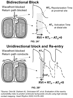
Differentiating Ischemic From Non-Ischemic T-Wave Inversion
PatentInactiveUS20070129640A1
Innovation
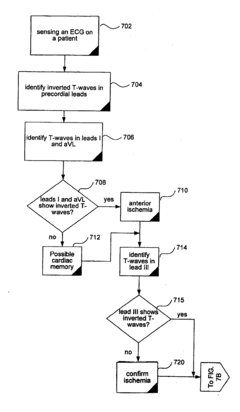
- A method and system that calculate the direction of the T-wave vector from electrocardiographic data to diagnose ischemia (vector between 75° and 200°) and cardiac memory (vector between 0° and -90°) to distinguish between the two conditions.
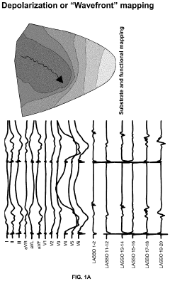
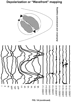
Electro-anatomic cardiac repolarization mapping
PatentPendingUS20240197236A1
Innovation
- A method and system for determining repolarization values from Electrogram (EGM) data using an electrode array, involving preprocessing steps like high-pass filtering, baseline correction, and noise reduction to obtain orthogonal bipolar compound electrograms, allowing for the creation of repolarization maps that guide cardiac procedures such as catheter ablation.

Cardiac Rehab Protocols
Cardiac rehabilitation protocols for patients with T wave inversion require careful consideration and tailored approaches. These protocols typically begin with a comprehensive assessment of the patient's cardiovascular health, including a detailed analysis of their electrocardiogram (ECG) patterns, particularly focusing on the T wave morphology.
The initial phase of cardiac rehabilitation for these patients often involves a period of closely monitored low-intensity exercise. This may include supervised walking or stationary cycling, with continuous ECG monitoring to observe any changes in T wave patterns during physical activity. The duration and intensity of these sessions are gradually increased based on the patient's tolerance and absence of adverse ECG changes.
As rehabilitation progresses, the protocols incorporate a wider range of exercises, including resistance training and flexibility exercises. However, the progression is more cautious compared to standard cardiac rehabilitation programs. Regular ECG assessments are conducted to track any improvements or changes in T wave inversion patterns over time.
Cardiac rehabilitation protocols for T wave inversion patients also emphasize education on lifestyle modifications. This includes guidance on heart-healthy diets, stress management techniques, and the importance of medication adherence. Patients are taught to recognize symptoms that may indicate cardiac stress and are provided with clear guidelines on when to seek medical attention.
The protocols often incorporate regular consultations with cardiologists to review the patient's progress and adjust the rehabilitation plan as needed. These consultations may involve periodic stress tests or other cardiac imaging studies to assess the impact of rehabilitation on cardiac function and T wave morphology.
In some cases, cardiac rehabilitation protocols for T wave inversion patients may include the use of specialized equipment or techniques. This could involve the application of external counterpulsation therapy or the use of telemetry systems for remote ECG monitoring during home-based exercise sessions.
The duration of these specialized cardiac rehabilitation protocols is typically longer than standard programs, often extending beyond the usual 12-week period. This extended timeline allows for a more gradual progression and provides ample opportunity to observe and respond to any changes in the patient's cardiac status or T wave patterns.
Patient Risk Stratification
Patient risk stratification in cardiac rehabilitative medicine, particularly concerning T wave inversion implications, is a critical aspect of managing patients with cardiovascular conditions. This process involves categorizing patients based on their likelihood of experiencing adverse cardiac events or complications during rehabilitation.
T wave inversion, a common electrocardiographic finding, plays a significant role in this stratification process. It can be indicative of various underlying cardiac conditions, ranging from benign to life-threatening. The presence and characteristics of T wave inversion help clinicians assess the severity of myocardial ischemia, identify potential arrhythmogenic substrates, and evaluate the overall cardiac health of patients.
In the context of cardiac rehabilitation, risk stratification based on T wave inversion helps tailor exercise programs and interventions to individual patient needs. Patients with more pronounced or widespread T wave inversions may require closer monitoring and a more conservative approach to rehabilitation. Conversely, those with less severe or localized inversions might be candidates for more intensive rehabilitation protocols.
The risk stratification process typically involves a comprehensive evaluation of multiple factors. These include the extent and distribution of T wave inversions, accompanying ECG abnormalities, cardiac biomarkers, imaging studies, and functional capacity assessments. By integrating these data points, clinicians can develop a more accurate risk profile for each patient.
Advanced risk stratification models incorporating T wave inversion data have been developed to enhance predictive accuracy. These models often utilize machine learning algorithms to analyze complex patterns in ECG data, providing more nuanced risk assessments than traditional methods. Such approaches enable healthcare providers to identify high-risk patients who may benefit from more aggressive interventions or closer follow-up.
It is important to note that risk stratification is not a one-time event but an ongoing process throughout the cardiac rehabilitation journey. Regular reassessment of T wave morphology and other risk factors allows for dynamic adjustment of rehabilitation strategies, ensuring optimal patient outcomes and safety.
Unlock deeper insights with Patsnap Eureka Quick Research — get a full tech report to explore trends and direct your research. Try now!
Generate Your Research Report Instantly with AI Agent
Supercharge your innovation with Patsnap Eureka AI Agent Platform!