Mechanisms of T wave inversion in left ventricular hypertrophy
AUG 19, 20259 MIN READ
Generate Your Research Report Instantly with AI Agent
Patsnap Eureka helps you evaluate technical feasibility & market potential.
T Wave Inversion Background and Objectives
T wave inversion is a significant electrocardiographic finding that has been extensively studied in the context of left ventricular hypertrophy (LVH). This phenomenon has garnered considerable attention due to its potential implications for cardiac health and its role in diagnostic and prognostic assessments. The evolution of our understanding of T wave inversion in LVH has been marked by significant advancements in cardiac electrophysiology and imaging technologies.
Historically, T wave inversion was first recognized as a potential indicator of cardiac abnormalities in the early 20th century. However, its specific association with LVH became more apparent as research in cardiovascular medicine progressed. The development of more sophisticated ECG equipment and standardized criteria for LVH diagnosis has greatly enhanced our ability to study this relationship.
The primary objective of investigating the mechanisms of T wave inversion in LVH is to elucidate the underlying electrophysiological processes that lead to this ECG pattern. This understanding is crucial for improving the accuracy of LVH diagnosis, assessing the severity of cardiac remodeling, and potentially predicting adverse cardiovascular outcomes.
One of the key areas of focus is the exploration of how structural changes in the left ventricle, such as increased wall thickness and altered myocardial fiber orientation, affect the repolarization sequence of the heart. This involves studying the complex interplay between cellular, tissue-level, and organ-level changes that occur in LVH and their impact on the cardiac electrical field.
Another important aspect of this research is to differentiate between pathological T wave inversion associated with LVH and other causes of T wave inversion, such as ischemia or electrolyte imbalances. This differentiation is critical for accurate diagnosis and appropriate patient management.
The technological advancements in cardiac imaging, including echocardiography, cardiac MRI, and advanced ECG mapping techniques, have provided new tools for correlating structural changes with electrical abnormalities. These technologies offer the potential for more precise characterization of the relationship between LVH and T wave inversion.
Furthermore, the investigation of genetic and molecular factors that contribute to both LVH development and altered repolarization patterns is an emerging area of interest. This research may provide insights into the variability of T wave inversion presentation among individuals with LVH and potentially identify new therapeutic targets.
As we delve deeper into the mechanisms of T wave inversion in LVH, the ultimate goal is to translate this knowledge into improved clinical practice. This includes developing more accurate diagnostic criteria, refining risk stratification models, and potentially guiding therapeutic interventions to prevent or reverse the electrical remodeling associated with LVH.
Clinical Significance of T Wave Inversion in LVH
T wave inversion in left ventricular hypertrophy (LVH) holds significant clinical importance in cardiovascular medicine. This electrocardiographic finding serves as a crucial marker for identifying and assessing the severity of LVH, a condition associated with increased morbidity and mortality. The presence of T wave inversion in LVH often indicates more advanced stages of cardiac remodeling and may suggest a higher risk of adverse cardiovascular events.
In clinical practice, T wave inversion in LVH is frequently observed in the lateral precordial leads (V5-V6) and limb leads (I and aVL). This pattern, known as the "strain pattern," is particularly indicative of pressure overload hypertrophy, such as that seen in hypertension or aortic stenosis. The degree and extent of T wave inversion can provide valuable insights into the severity of LVH and the underlying hemodynamic stress on the left ventricle.
The clinical significance of T wave inversion in LVH extends beyond its diagnostic value. It serves as a prognostic indicator, with more pronounced T wave inversions often correlating with worse outcomes. Studies have shown that patients with LVH and T wave inversion have a higher risk of developing heart failure, arrhythmias, and sudden cardiac death compared to those with LVH but normal T waves.
Furthermore, T wave inversion in LVH can complicate the interpretation of other cardiac conditions. For instance, it may mimic ischemic changes, making the diagnosis of coronary artery disease more challenging in patients with LVH. This underscores the importance of considering LVH when interpreting T wave abnormalities in clinical scenarios.
The presence of T wave inversion in LVH also has implications for treatment strategies. It may influence decisions regarding the aggressiveness of blood pressure control, the need for more intensive cardiac monitoring, or the consideration of additional diagnostic tests such as echocardiography or cardiac MRI to further assess ventricular structure and function.
In the context of cardiovascular risk stratification, T wave inversion in LVH is often incorporated into risk prediction models. Its presence can lead to reclassification of patients into higher risk categories, potentially altering management approaches and follow-up strategies. This highlights the importance of recognizing and appropriately interpreting this ECG finding in clinical practice.
Current Understanding and Challenges in T Wave Inversion Mechanisms
T wave inversion in left ventricular hypertrophy (LVH) is a complex electrocardiographic phenomenon that has been the subject of extensive research in cardiology. The current understanding of this mechanism involves a combination of electrical and structural changes in the heart, particularly in the left ventricle.
One of the primary factors contributing to T wave inversion in LVH is the alteration of the ventricular repolarization sequence. In a normal heart, the repolarization process typically begins from the epicardium and progresses towards the endocardium. However, in LVH, this sequence can be disrupted due to the increased myocardial mass and altered cellular properties of hypertrophied cardiomyocytes.
The thickened left ventricular wall in LVH leads to changes in the distribution of electrical forces within the heart. This redistribution affects the direction and magnitude of the cardiac electrical vector during repolarization, resulting in T wave inversion on the electrocardiogram (ECG). The increased left ventricular mass also causes a delay in the onset of repolarization in some regions of the myocardium, further contributing to the T wave abnormalities.
Another important aspect of the current understanding is the role of myocardial fibrosis in LVH. As the left ventricle hypertrophies, there is often an associated increase in interstitial fibrosis. This fibrotic tissue can disrupt the normal electrical conduction pathways within the myocardium, leading to heterogeneity in repolarization and contributing to T wave inversion.
Despite these insights, several challenges remain in fully elucidating the mechanisms of T wave inversion in LVH. One significant challenge is the variability in T wave morphology among patients with LVH. Not all individuals with LVH exhibit T wave inversion, and the degree of inversion can vary widely. This heterogeneity suggests that additional factors, possibly genetic or environmental, may influence the manifestation of T wave inversion in LVH.
Another challenge lies in distinguishing T wave inversion caused by LVH from other cardiac pathologies that can produce similar ECG changes. This differential diagnosis is crucial for accurate clinical assessment and management. Furthermore, the relationship between the severity of LVH and the degree of T wave inversion is not always linear, complicating the use of T wave inversion as a reliable marker for LVH progression.
The dynamic nature of T wave inversion in LVH also presents a challenge. Some patients may show transient T wave changes, while others exhibit persistent inversions. Understanding the factors that influence these temporal variations remains an area of active research.
Existing Models Explaining T Wave Inversion in LVH
01 Detection and analysis of T wave inversion
T wave inversion is a significant indicator in electrocardiogram (ECG) analysis. Advanced algorithms and methods are developed to accurately detect and analyze T wave inversions, which can be crucial in diagnosing various cardiac conditions. These techniques often involve signal processing, machine learning, and pattern recognition to identify abnormal T wave morphologies.- Detection and analysis of T wave inversion: T wave inversion is a significant indicator in electrocardiogram (ECG) analysis. Advanced algorithms and methods are developed to detect, measure, and interpret T wave inversions accurately. These techniques help in identifying various cardiac conditions and assessing the severity of heart abnormalities.
- Machine learning applications for T wave analysis: Machine learning and artificial intelligence techniques are increasingly used to analyze T wave morphology, including inversions. These methods improve the accuracy of ECG interpretation, enable automated diagnosis, and assist in predicting cardiac events based on T wave characteristics.
- Wearable devices for continuous T wave monitoring: Wearable ECG devices are developed to continuously monitor T waves, including inversions, in real-time. These devices allow for long-term cardiac monitoring outside clinical settings, enabling early detection of abnormalities and improving patient care.
- T wave inversion in specific cardiac conditions: Research focuses on understanding the significance of T wave inversion in various cardiac conditions such as myocardial ischemia, cardiomyopathies, and electrolyte imbalances. This knowledge aids in differential diagnosis and treatment planning for patients with specific heart disorders.
- Integration of T wave analysis in comprehensive cardiac assessment: T wave inversion analysis is integrated into comprehensive cardiac assessment systems. These systems combine multiple ECG parameters, imaging techniques, and clinical data to provide a holistic evaluation of cardiac health, improving diagnostic accuracy and treatment efficacy.
02 Wearable devices for continuous T wave monitoring
Wearable ECG devices are designed to continuously monitor T waves and other cardiac parameters. These devices incorporate miniaturized sensors and advanced data processing capabilities to detect T wave inversions in real-time, allowing for early detection of potential cardiac issues outside of clinical settings.Expand Specific Solutions03 Artificial intelligence in T wave inversion interpretation
AI-powered systems are being developed to interpret T wave inversions more accurately. These systems use deep learning algorithms and large datasets to analyze ECG patterns, potentially improving the diagnosis of cardiac conditions associated with T wave inversions and reducing false positives in ECG interpretation.Expand Specific Solutions04 T wave inversion in specific cardiac conditions
Research focuses on understanding the relationship between T wave inversions and specific cardiac conditions such as ischemia, cardiomyopathies, and channelopathies. This involves studying the mechanisms behind T wave inversions in different pathological states and developing targeted diagnostic approaches.Expand Specific Solutions05 Novel ECG lead systems for improved T wave analysis
Innovative ECG lead systems and electrode configurations are being developed to enhance the detection and analysis of T wave inversions. These systems aim to provide more comprehensive cardiac electrical activity data, potentially improving the sensitivity and specificity of T wave inversion detection.Expand Specific Solutions
Key Researchers and Institutions in Cardiac Electrophysiology
The field of T wave inversion mechanisms in left ventricular hypertrophy is in a mature stage of development, with a substantial market size driven by the prevalence of cardiovascular diseases. The technology's maturity is evident from the involvement of established medical device companies and research institutions. Edwards Lifesciences, Medtronic, and Novartis are key players, leveraging their expertise in cardiac monitoring and therapeutics. Academic institutions like Massachusetts Institute of Technology and University of Turku contribute to ongoing research, while specialized firms such as CVRx focus on innovative treatments for hypertension, indicating a competitive and diverse landscape in this niche area of cardiology.
Edwards Lifesciences Corp.
Technical Solution: Edwards Lifesciences has focused on hemodynamic monitoring solutions that indirectly address the mechanisms of T wave inversion in LVH. Their approach utilizes advanced pressure-volume loop analysis to assess left ventricular function and correlate it with ECG changes[4]. By combining real-time hemodynamic data with ECG signals, Edwards' technology provides a comprehensive view of cardiac electromechanical coupling in LVH patients. Their systems can detect subtle changes in ventricular pressure and volume that precede or accompany T wave inversions, offering early insights into the development of LVH[5]. The company has also developed algorithms that integrate ECG data with other physiological parameters to improve the specificity of LVH diagnosis and monitoring.
Strengths: Comprehensive approach combining hemodynamics and ECG, potential for early LVH detection. Weaknesses: Invasive nature of some hemodynamic monitoring techniques, which may limit widespread application.
Medtronic, Inc.
Technical Solution: Medtronic has developed advanced ECG algorithms for detecting and analyzing T wave inversion in left ventricular hypertrophy (LVH). Their approach combines machine learning techniques with traditional signal processing methods to improve accuracy in identifying LVH-related T wave changes[1]. The company's implantable cardiac monitors and external ECG devices utilize these algorithms to provide early detection of LVH progression. Medtronic's technology also incorporates multi-lead ECG analysis to differentiate between various causes of T wave inversion, enhancing diagnostic specificity for LVH[2]. Their devices can track longitudinal changes in T wave morphology, offering insights into the progression of left ventricular remodeling over time[3].
Strengths: High accuracy in LVH detection, longitudinal monitoring capabilities, and integration with implantable devices. Weaknesses: Reliance on proprietary algorithms may limit widespread adoption, and the technology may require frequent updates to maintain accuracy.
Critical Electrophysiological Studies on T Wave Inversion
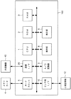
Electrocardiogram analysis device, electrocardiogram analysis method, and biological information measurement device
PatentActiveJP2018075310A
Innovation
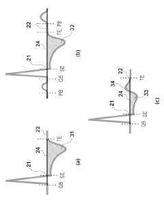
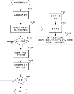
- An electrocardiogram analysis device and method that calculates an index based on the sum of electrocardiogram signals in the ST-T interval, independent of the R wave magnitude, by detecting the ST-T interval from the end point of the S wave to the end point of the T wave, using a predetermined baseline.
Diagnostic Implications for LVH Detection
The diagnostic implications of T wave inversion in left ventricular hypertrophy (LVH) are significant for accurate detection and assessment of this cardiac condition. T wave inversion, particularly in the lateral precordial leads (V5-V6), is a common electrocardiographic (ECG) finding in LVH and can provide valuable insights into the severity and progression of the condition.
One of the primary diagnostic implications is the increased sensitivity for LVH detection when T wave inversion is present in conjunction with other ECG criteria. While traditional voltage criteria alone may have limited sensitivity, the addition of T wave inversion can significantly improve the diagnostic accuracy. This is particularly important in cases where LVH may be missed by voltage criteria alone, especially in patients with obesity or chronic obstructive pulmonary disease.
The pattern and extent of T wave inversion can also provide information about the severity of LVH. More pronounced and widespread T wave inversions, especially those extending beyond the lateral precordial leads, may indicate a more advanced stage of LVH or the presence of underlying myocardial ischemia. This information can be crucial for risk stratification and treatment planning.
Furthermore, the presence of T wave inversion in LVH can help differentiate it from other cardiac conditions that may present with similar ECG findings. For instance, the combination of LVH voltage criteria and T wave inversion can help distinguish LVH from acute coronary syndromes or cardiomyopathies, which may also cause T wave abnormalities.
It is important to note that the diagnostic value of T wave inversion in LVH is enhanced when considered in conjunction with other clinical and imaging data. Echocardiography remains the gold standard for LVH diagnosis, and the correlation between ECG findings and echocardiographic measurements can provide a more comprehensive assessment of cardiac structure and function.
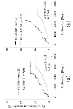
The presence of T wave inversion in LVH also has prognostic implications. Studies have shown that patients with LVH and T wave inversion have a higher risk of adverse cardiovascular events compared to those with LVH but normal T waves. This information can guide clinicians in determining the intensity of follow-up and management strategies for individual patients.
In conclusion, the diagnostic implications of T wave inversion in LVH extend beyond mere detection. They encompass improved sensitivity and specificity in diagnosis, assessment of LVH severity, differentiation from other cardiac conditions, and prognostic value. Understanding these implications is crucial for clinicians to make informed decisions about patient care and management in the context of LVH.
One of the primary diagnostic implications is the increased sensitivity for LVH detection when T wave inversion is present in conjunction with other ECG criteria. While traditional voltage criteria alone may have limited sensitivity, the addition of T wave inversion can significantly improve the diagnostic accuracy. This is particularly important in cases where LVH may be missed by voltage criteria alone, especially in patients with obesity or chronic obstructive pulmonary disease.
The pattern and extent of T wave inversion can also provide information about the severity of LVH. More pronounced and widespread T wave inversions, especially those extending beyond the lateral precordial leads, may indicate a more advanced stage of LVH or the presence of underlying myocardial ischemia. This information can be crucial for risk stratification and treatment planning.
Furthermore, the presence of T wave inversion in LVH can help differentiate it from other cardiac conditions that may present with similar ECG findings. For instance, the combination of LVH voltage criteria and T wave inversion can help distinguish LVH from acute coronary syndromes or cardiomyopathies, which may also cause T wave abnormalities.
It is important to note that the diagnostic value of T wave inversion in LVH is enhanced when considered in conjunction with other clinical and imaging data. Echocardiography remains the gold standard for LVH diagnosis, and the correlation between ECG findings and echocardiographic measurements can provide a more comprehensive assessment of cardiac structure and function.
The presence of T wave inversion in LVH also has prognostic implications. Studies have shown that patients with LVH and T wave inversion have a higher risk of adverse cardiovascular events compared to those with LVH but normal T waves. This information can guide clinicians in determining the intensity of follow-up and management strategies for individual patients.
In conclusion, the diagnostic implications of T wave inversion in LVH extend beyond mere detection. They encompass improved sensitivity and specificity in diagnosis, assessment of LVH severity, differentiation from other cardiac conditions, and prognostic value. Understanding these implications is crucial for clinicians to make informed decisions about patient care and management in the context of LVH.
Therapeutic Approaches Targeting T Wave Abnormalities
Therapeutic approaches targeting T wave abnormalities in left ventricular hypertrophy (LVH) focus on addressing the underlying causes and mitigating the associated risks. The primary goal is to reverse or prevent further progression of LVH and normalize the T wave morphology.
One of the most effective therapeutic strategies is the management of hypertension, which is a common cause of LVH. Antihypertensive medications, such as angiotensin-converting enzyme (ACE) inhibitors, angiotensin receptor blockers (ARBs), and beta-blockers, have shown promise in reducing left ventricular mass and improving T wave abnormalities. These medications not only lower blood pressure but also have direct effects on cardiac remodeling.
In cases where LVH is caused by aortic stenosis, surgical intervention through aortic valve replacement can lead to regression of LVH and improvement in T wave morphology. This approach addresses the mechanical stress on the left ventricle, allowing for structural and electrical remodeling.
For patients with hypertrophic cardiomyopathy, a genetic condition causing LVH, therapeutic options include beta-blockers, calcium channel blockers, and in some cases, surgical myectomy or alcohol septal ablation. These interventions aim to reduce left ventricular outflow tract obstruction and improve overall cardiac function, which can positively impact T wave abnormalities.
Recent research has explored the potential of novel pharmacological agents targeting specific molecular pathways involved in cardiac hypertrophy. For instance, inhibitors of the mammalian target of rapamycin (mTOR) pathway have shown promise in animal models for reducing cardiac hypertrophy and potentially normalizing T wave morphology.
Non-pharmacological approaches, such as lifestyle modifications, play a crucial role in managing LVH and associated T wave abnormalities. Regular aerobic exercise, weight management, and dietary interventions, particularly sodium restriction, can complement medical therapies and contribute to the regression of LVH.
Emerging technologies, such as cardiac resynchronization therapy (CRT), have shown potential in improving T wave abnormalities in patients with LVH and concomitant heart failure. CRT can help optimize ventricular contraction patterns, potentially leading to structural remodeling and improvements in T wave morphology.
Monitoring and follow-up are essential components of therapeutic approaches. Regular electrocardiographic and echocardiographic assessments allow for the evaluation of treatment efficacy and guide necessary adjustments to the therapeutic strategy.
As research progresses, personalized medicine approaches are gaining traction. Genetic profiling and biomarker analysis may help tailor treatments to individual patients, potentially leading to more effective management of LVH and associated T wave abnormalities.
Unlock deeper insights with Patsnap Eureka Quick Research — get a full tech report to explore trends and direct your research. Try now!
Generate Your Research Report Instantly with AI Agent
Supercharge your innovation with Patsnap Eureka AI Agent Platform!