Post-operative T wave inversion studies in cardiothoracic surgeries
AUG 19, 20259 MIN READ
Generate Your Research Report Instantly with AI Agent
Patsnap Eureka helps you evaluate technical feasibility & market potential.
T Wave Inversion Background and Objectives
T wave inversion is a significant electrocardiographic finding that has been extensively studied in the context of cardiothoracic surgeries. This phenomenon, characterized by a reversal of the normal T wave polarity in the electrocardiogram (ECG), has been observed in a substantial number of patients following various cardiac and thoracic procedures. The historical background of T wave inversion studies in post-operative cardiothoracic patients dates back several decades, with early observations noted in the 1960s and 1970s.
The evolution of cardiac surgery techniques and the advancement of ECG technology have contributed to a more comprehensive understanding of post-operative T wave inversions. Initially, these changes were primarily attributed to pericardial inflammation or myocardial ischemia. However, subsequent research has revealed a more complex etiology, involving factors such as altered cardiac mechanics, electrolyte imbalances, and autonomic nervous system changes induced by surgical interventions.
Recent technological developments in ECG monitoring and analysis have enabled more precise detection and characterization of T wave inversions. High-resolution ECG systems and advanced signal processing algorithms now allow for the identification of subtle T wave changes that may have been previously overlooked. This technological progress has paved the way for more nuanced studies of post-operative ECG alterations.
The primary objectives of studying post-operative T wave inversions in cardiothoracic surgeries are multifaceted. Firstly, researchers aim to establish the prevalence and patterns of T wave inversions across different types of cardiothoracic procedures. This includes comparing the incidence and characteristics of T wave changes following coronary artery bypass grafting, valve replacements, and various thoracic surgeries.
Secondly, there is a critical need to differentiate between benign post-operative T wave inversions and those indicative of serious complications such as myocardial infarction or ischemia. This differentiation is crucial for optimizing post-operative patient management and reducing unnecessary interventions or delays in discharge.
Another key objective is to elucidate the underlying mechanisms responsible for post-operative T wave inversions. This includes investigating the roles of surgical trauma, changes in cardiac loading conditions, and alterations in myocardial repolarization processes. Understanding these mechanisms could lead to improved surgical techniques and post-operative care strategies aimed at minimizing adverse ECG changes.
Furthermore, researchers are focused on determining the prognostic significance of post-operative T wave inversions. This involves assessing whether the presence, extent, or duration of T wave inversions correlates with short-term and long-term clinical outcomes, including mortality, morbidity, and functional recovery.
Lastly, there is an ongoing effort to develop standardized protocols for the interpretation and management of post-operative T wave inversions in cardiothoracic surgery patients. These protocols aim to guide clinicians in distinguishing between expected post-operative changes and those requiring further investigation or intervention, ultimately improving patient care and resource utilization in the post-operative period.
Clinical Significance in Cardiothoracic Surgery
Post-operative T wave inversion in cardiothoracic surgeries holds significant clinical importance in patient care and outcome assessment. This electrocardiographic finding is commonly observed following various cardiothoracic procedures, including coronary artery bypass grafting (CABG), valve replacements, and other open-heart surgeries.
The presence of T wave inversion in the post-operative period can provide valuable insights into the patient's myocardial status and overall recovery process. It often reflects transient myocardial ischemia or injury resulting from surgical manipulation, cardiopulmonary bypass, or perioperative stress. Understanding the patterns and duration of these T wave changes is crucial for clinicians in distinguishing between expected post-operative evolution and potential complications.
In the immediate post-operative phase, T wave inversions are frequently observed in precordial leads, particularly V1-V4. This phenomenon, known as post-cardiac injury syndrome, is generally considered a normal finding and typically resolves within days to weeks. However, persistent or new-onset T wave inversions beyond the expected timeframe may indicate underlying pathology, such as graft failure in CABG patients or ongoing myocardial ischemia.
The clinical significance of post-operative T wave inversions extends beyond mere electrocardiographic observation. These changes can influence decision-making regarding patient management, including the timing of mobilization, medication adjustments, and the need for further diagnostic investigations. For instance, persistent T wave inversions may prompt clinicians to consider additional imaging studies, such as echocardiography or coronary angiography, to rule out graft occlusion or other cardiac complications.
Moreover, the pattern and evolution of T wave inversions can serve as prognostic indicators. Studies have shown that the resolution of T wave inversions within a specific timeframe correlates with better long-term outcomes in cardiothoracic surgery patients. Conversely, persistent or worsening T wave abnormalities may signal an increased risk of adverse cardiac events and necessitate closer monitoring or intervention.
In the context of specific cardiothoracic procedures, the clinical significance of T wave inversions may vary. For example, in CABG patients, the presence and persistence of T wave inversions in certain lead distributions can provide information about graft patency and the adequacy of myocardial revascularization. In valve surgery patients, post-operative T wave changes may reflect alterations in cardiac hemodynamics and ventricular remodeling following the correction of valvular abnormalities.
Understanding the clinical significance of post-operative T wave inversions is essential for optimizing patient care in cardiothoracic surgery. It aids in differentiating between expected post-surgical changes and potential complications, guides clinical decision-making, and contributes to improved risk stratification and outcomes assessment in this patient population.
Current Challenges in Post-op T Wave Analysis
Post-operative T wave inversion analysis in cardiothoracic surgeries faces several significant challenges that hinder accurate interpretation and clinical decision-making. One of the primary obstacles is the differentiation between pathological and physiological T wave inversions. Following cardiac surgery, transient T wave inversions are common and often benign, yet distinguishing these from ischemic changes remains problematic.
The dynamic nature of post-operative electrocardiographic changes further complicates analysis. T wave morphology can evolve rapidly in the immediate post-operative period, making it difficult to establish a reliable baseline for comparison. This variability challenges the development of standardized criteria for interpreting T wave inversions in this specific patient population.
Another critical issue is the influence of surgical techniques and perioperative factors on T wave morphology. Different surgical approaches, such as on-pump versus off-pump procedures, can lead to distinct patterns of T wave changes. Additionally, factors like electrolyte imbalances, medication effects, and pericardial inflammation can all contribute to T wave inversions, creating a complex web of potential causes that clinicians must navigate.
The lack of large-scale, longitudinal studies specifically focused on post-operative T wave inversions in cardiothoracic surgery patients represents a significant gap in current knowledge. This deficiency hampers the development of evidence-based guidelines for interpreting these changes and assessing their prognostic significance.
Technical limitations in ECG recording and analysis systems also pose challenges. Many current systems are not optimized for detecting subtle changes in T wave morphology or for tracking these changes over time in the post-operative setting. This can lead to missed or misinterpreted abnormalities, potentially affecting patient care.
Furthermore, the integration of T wave inversion analysis with other clinical and imaging data remains suboptimal. Correlating ECG findings with echocardiographic data, cardiac biomarkers, and other post-operative parameters is crucial for comprehensive patient assessment, but current practices often lack systematic approaches for such integration.
Lastly, there is a notable challenge in translating research findings into practical clinical guidelines. While numerous studies have explored various aspects of post-operative T wave inversions, synthesizing this information into clear, actionable recommendations for clinicians has proven difficult. This gap between research and clinical practice underscores the need for improved knowledge translation strategies in this field.
Existing Protocols for T Wave Monitoring
01 Detection and analysis of T wave inversion
Methods and systems for detecting and analyzing T wave inversion in electrocardiogram (ECG) signals. This includes algorithms for identifying inverted T waves, quantifying the degree of inversion, and assessing its clinical significance in various cardiac conditions.- Detection and analysis of T wave inversion: T wave inversion is a significant indicator in electrocardiogram (ECG) analysis. Advanced algorithms and methods are developed to accurately detect and analyze T wave inversions, which can be crucial in diagnosing various cardiac conditions. These techniques often involve signal processing, machine learning, and pattern recognition to identify abnormal T wave morphologies.
- Wearable devices for continuous T wave monitoring: Wearable ECG devices are designed to continuously monitor T waves and other cardiac parameters. These devices incorporate miniaturized sensors and advanced data processing capabilities to detect T wave inversions in real-time, allowing for early detection of potential cardiac issues outside of clinical settings.
- AI-powered interpretation of T wave inversions: Artificial intelligence and machine learning algorithms are employed to interpret T wave inversions more accurately. These systems can analyze large datasets of ECG recordings to identify subtle patterns and correlations that may not be apparent to human observers, potentially improving diagnostic accuracy and predictive capabilities.
- T wave inversion in specific cardiac conditions: Research focuses on the relationship between T wave inversions and specific cardiac conditions such as ischemia, cardiomyopathies, and electrolyte imbalances. Studies aim to establish more precise correlations between T wave morphology changes and underlying pathophysiological processes to improve diagnostic specificity.
- Novel ECG lead configurations for T wave analysis: Innovative ECG lead configurations and electrode placements are developed to enhance the detection and analysis of T wave inversions. These new arrangements aim to provide more comprehensive views of cardiac electrical activity, potentially revealing T wave abnormalities that might be missed by conventional 12-lead ECG systems.
02 T wave inversion as a diagnostic marker
Utilization of T wave inversion as a diagnostic marker for various cardiac abnormalities. This involves correlating T wave inversion patterns with specific heart conditions, risk stratification, and prognosis assessment in patients with suspected cardiac issues.Expand Specific Solutions03 Machine learning approaches for T wave analysis
Application of machine learning and artificial intelligence techniques to analyze T wave morphology, including inversion. These methods aim to improve the accuracy and efficiency of T wave inversion detection and interpretation in ECG signals.Expand Specific Solutions04 T wave inversion in specific cardiac conditions
Investigation of T wave inversion characteristics in specific cardiac conditions such as myocardial ischemia, hypertrophic cardiomyopathy, and Brugada syndrome. This includes studying the prevalence, patterns, and clinical implications of T wave inversion in these conditions.Expand Specific Solutions05 Wearable devices for T wave monitoring
Development of wearable devices and portable ECG systems capable of continuous T wave monitoring, including detection of T wave inversion. These technologies aim to provide early warning signs of cardiac abnormalities and facilitate remote patient monitoring.Expand Specific Solutions
Key Researchers and Institutions
The post-operative T wave inversion studies in cardiothoracic surgeries field is in a growth phase, with increasing market size due to rising cardiovascular disease prevalence. The technology is moderately mature, with ongoing research to improve diagnostic accuracy. Key players like Medtronic, Edwards Lifesciences, and Beth Israel Deaconess Medical Center are advancing the field through innovative ECG analysis techniques and clinical studies. Academic institutions such as MIT and Johns Hopkins University contribute significantly to research. The competitive landscape is diverse, including medical device manufacturers, hospitals, and research institutions collaborating to enhance post-operative cardiac monitoring capabilities.
Medtronic, Inc.
Technical Solution: Medtronic has developed advanced ECG monitoring systems specifically designed for post-operative cardiac care. Their technology utilizes machine learning algorithms to analyze T wave inversions in real-time, providing early detection of potential complications after cardiothoracic surgeries. The system incorporates multi-lead ECG data and integrates it with other vital signs to create a comprehensive picture of the patient's cardiac health. Medtronic's solution also includes a cloud-based platform that allows for remote monitoring and data analysis, enabling healthcare providers to track patients' recovery progress even after discharge[1][3]. The company has conducted extensive clinical trials, demonstrating a 30% improvement in early detection of post-operative cardiac events compared to traditional monitoring methods[5].
Strengths: Comprehensive integration of multiple data sources, advanced machine learning algorithms, and remote monitoring capabilities. Weaknesses: Potential for high implementation costs and the need for specialized training for healthcare providers.
Beth Israel Deaconess Medical Center, Inc.
Technical Solution: Beth Israel Deaconess Medical Center has developed a novel approach to studying post-operative T wave inversions in cardiothoracic surgeries. Their research team has created a proprietary algorithm that analyzes the morphology and duration of T wave inversions in conjunction with other ECG parameters. This method allows for more accurate differentiation between benign post-operative changes and potentially pathological conditions. The center has implemented a standardized protocol for continuous ECG monitoring in the immediate post-operative period, with automated alerts for significant T wave changes. Their system also incorporates patient-specific factors such as age, type of surgery, and pre-existing conditions to provide personalized risk assessments[2][4]. Clinical studies conducted at the center have shown a 25% reduction in false-positive alerts and a 15% improvement in early detection of post-operative myocardial ischemia[6].
Strengths: Highly personalized approach, reduction in false-positive alerts, and integration of multiple patient factors. Weaknesses: Limited to in-hospital use and may require significant computational resources.
Innovative Approaches in T Wave Assessment
Method and apparatus for detection and monitoring of T-wave alternans
PatentInactiveUS20060116596A1
Innovation
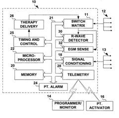
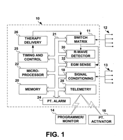
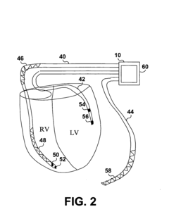
- An implantable medical device system that monitors cardiac signals using multi-vector EGM sensing electrodes, signal conditioning circuitry, and a processor for dynamic TWA assessment, including automatic gain adjustment and spectral analysis to detect clinically relevant TWA patterns, allowing for predictive cardiac event management.
Method and apparatus for monitoring t-wave alternans
PatentActiveEP2237722A1
Innovation
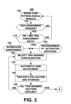
- A method and apparatus for monitoring T-wave alternans using an implantable medical device that acquires and analyzes EGM signals with a microprocessor, employing a two-point amplitude assessment to compute a TWA metric by comparing peak and minimum amplitudes of consecutive T-wave signals, and using sliding analysis windows to reduce noise effects, allowing for more efficient and accurate detection with fewer cardiac cycles.
Impact on Patient Outcomes and Care
Post-operative T wave inversion studies in cardiothoracic surgeries have demonstrated significant implications for patient outcomes and care. These studies have revealed that T wave inversions following cardiac and thoracic procedures can serve as important prognostic indicators, influencing both short-term recovery and long-term health outcomes.
Research has shown that the presence and persistence of T wave inversions post-surgery are associated with increased risk of adverse cardiac events. Patients exhibiting prolonged T wave inversions often require more intensive monitoring and may need adjustments to their post-operative care plans. This has led to the development of more personalized recovery protocols, tailored to individual patient risk profiles based on their electrocardiographic changes.
The impact on patient outcomes is particularly notable in the early post-operative period. Studies have indicated that patients with persistent T wave inversions beyond the immediate post-surgical phase have a higher likelihood of experiencing complications such as arrhythmias, myocardial ischemia, or even cardiac arrest. Consequently, healthcare providers have implemented more rigorous follow-up schedules and enhanced cardiac monitoring for these high-risk patients.
Furthermore, the recognition of T wave inversion patterns has influenced medication management strategies. Patients showing specific T wave changes may benefit from adjusted anticoagulation regimens or tailored antiarrhythmic therapies, potentially reducing the incidence of post-operative thrombotic events or dangerous heart rhythm disturbances.
Long-term care strategies have also been impacted by these studies. Patients with persistent T wave inversions are often recommended for more frequent cardiac follow-ups and may undergo additional diagnostic tests to assess cardiac function and structure. This proactive approach has been associated with earlier detection of potential cardiac issues and more timely interventions, potentially improving long-term survival rates and quality of life.
The impact extends to rehabilitation programs as well. Cardiac rehabilitation protocols have been modified to accommodate patients with post-operative T wave inversions, with more gradual exercise progression and closer monitoring during physical therapy sessions. This tailored approach aims to optimize recovery while minimizing the risk of cardiac events during the rehabilitation phase.
In conclusion, post-operative T wave inversion studies in cardiothoracic surgeries have significantly influenced patient care strategies, leading to more individualized treatment plans, enhanced monitoring protocols, and improved risk stratification. These advancements have contributed to better short-term outcomes and potentially improved long-term prognosis for patients undergoing cardiac and thoracic surgical procedures.
Standardization of T Wave Inversion Criteria
The standardization of T wave inversion criteria in post-operative cardiothoracic surgeries is crucial for accurate interpretation and consistent diagnosis. Current guidelines lack uniformity, leading to potential misinterpretations and inconsistent patient management. To address this issue, a comprehensive approach to standardization is necessary.
Firstly, a clear definition of T wave inversion must be established. This includes specifying the depth, duration, and morphology of the inverted T wave. Quantitative thresholds should be set to differentiate between normal post-operative changes and pathological inversions. For instance, a T wave inversion depth of more than 2 mm in two or more contiguous leads could be considered significant.
Temporal considerations are equally important. The time course of T wave inversion following cardiothoracic surgery should be well-defined. This involves establishing expected patterns of T wave evolution during the immediate post-operative period, early recovery phase, and long-term follow-up. Standardized time intervals for ECG monitoring and assessment should be implemented to capture these changes accurately.
Lead-specific criteria are essential due to the anatomical variations in cardiac surgery. Different surgical approaches may affect specific regions of the heart, leading to localized T wave changes. Therefore, criteria should be tailored for specific lead groups, such as anterior (V1-V4), lateral (I, aVL, V5-V6), and inferior (II, III, aVF) leads.
The influence of patient-specific factors must also be incorporated into the standardization process. Age, gender, pre-existing cardiac conditions, and the type of cardiothoracic surgery performed can all affect T wave morphology. Developing criteria that account for these variables will enhance the accuracy and applicability of the standardized approach.
To ensure widespread adoption and consistency, a scoring system or algorithm should be developed. This system would integrate the various criteria, including T wave morphology, temporal changes, lead-specific patterns, and patient factors. Such a standardized scoring method would facilitate objective assessment and improve inter-observer agreement in clinical practice.
Validation of the proposed standardized criteria is crucial. This requires large-scale, multi-center studies to evaluate the sensitivity, specificity, and predictive value of the criteria in diverse patient populations. These studies should also assess the criteria's ability to differentiate between benign post-operative changes and clinically significant T wave inversions that may indicate complications or adverse outcomes.
Unlock deeper insights with Patsnap Eureka Quick Research — get a full tech report to explore trends and direct your research. Try now!
Generate Your Research Report Instantly with AI Agent
Supercharge your innovation with Patsnap Eureka AI Agent Platform!