
Multi-threshold peripheral equalization method and apparatus for digital mammography and breast tomosynthesis
a technology of peripheral equalization and digital mammography, applied in the field of image processing, can solve problems such as unsatisfactory artifacts in images, and achieve the effects of improving image reading and interpretation efficiency, enhancing intensity, and enhancing the peripheral area of breasts
Patent Information
- Authority / Receiving Office
- US · United States
- Patent Type
- Patents(United States)
- Current Assignee / Owner
- Publication Date
- 2010-07-27
Smart Images
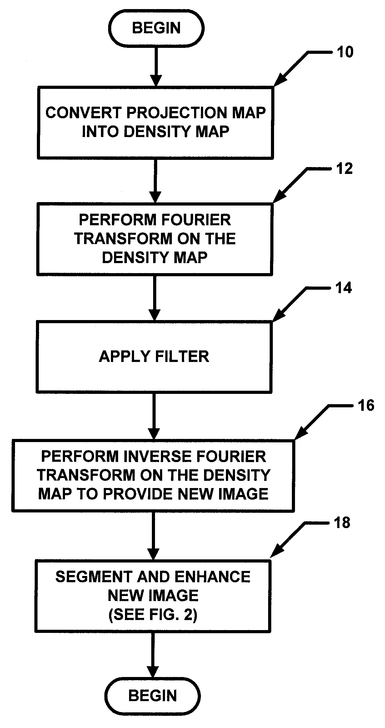
Figure 1 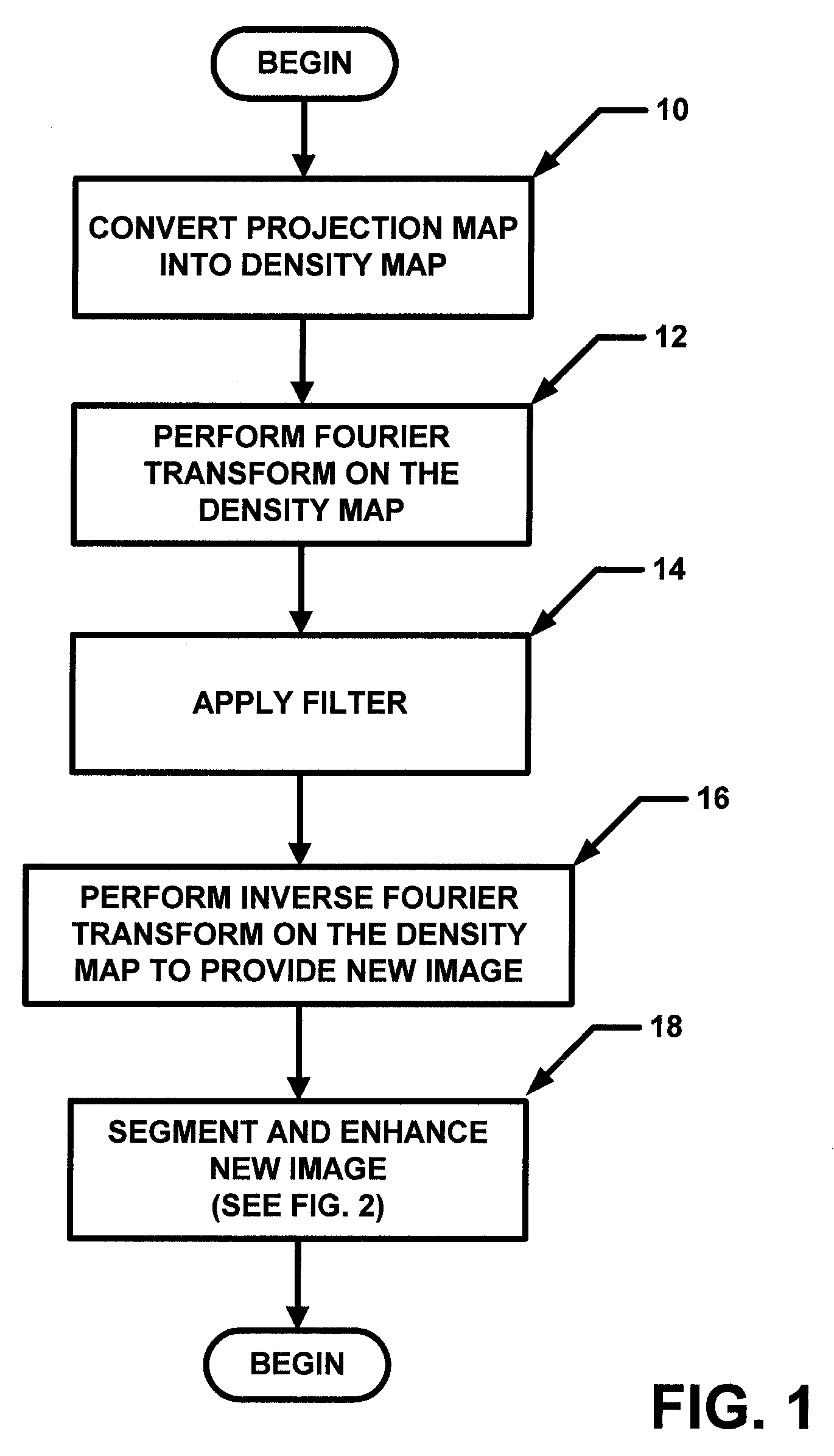
Figure 2 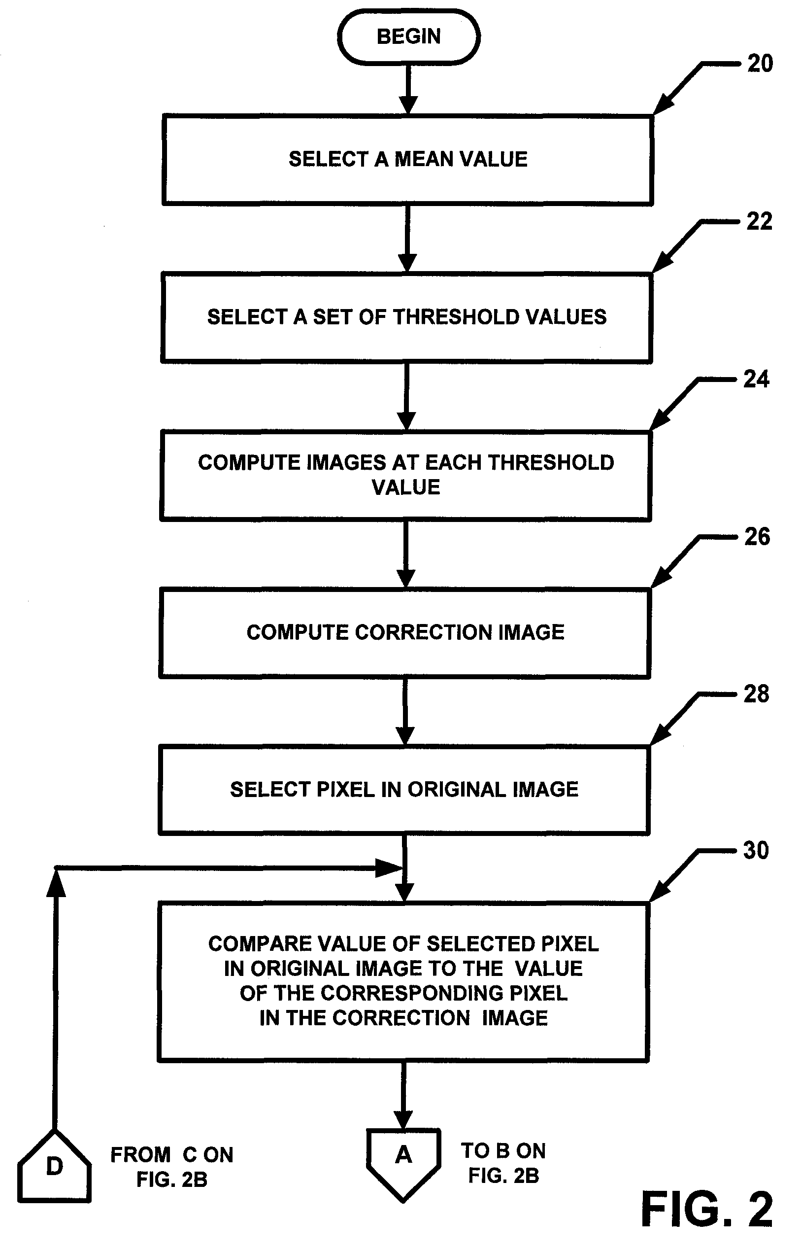
Figure 3
Abstract
Description
CROSS-REFERENCE TO RELATED APPLICATIONS
[0001] This application claims the benefit of U.S. Provisional Application No. 60 / 711,023 filed on Aug. 24, 2005, under 35 U.S.C. §119(e), which application is hereby incorporated herein by reference in its entirety.STATEMENTS REGARDING FEDERALLY SPONSORED RESEARCH
[0002] Not applicable.FIELD OF THE INVENTION
[0003] This invention relates generally to image processing and more particularly to image processing of mammography and breast tomosynthesis images.BACKGROUND OF THE INVENTION
[0004] As is known in the art, in two-dimensional (2-D) mammography and three-dimensional (3-D) breast tomosynthesis images, the peripheral area of the breast tends to have lower intensity than the central area of the breast. This is due at least in part to the thickness of a compressed breast decreasing from the central area to the peripheral area. Thus, to assess an image of the entire breast, a radiologist (or other medical professional or reader of the image) typically...
Examples
Embodiment Construction
[0019]In general overview, the general concept described herein is to estimate the normalized thickness profile (NTP) of a breast from an image (e.g. a mammogram image) and enhance the peripheral area. This can accomplished by dividing the NTP from the mammogram. In one particular but exemplary embodiment, a projection mammogram was first segmented into “breast” and “background” regions using a threshold value computed using the Otsu technique. A segmentation image (SI) was generated in which pixels were assigned a first value (e.g. value of one) in a breast region and a second value (e.g. a value of zero) in background region. The projection was then converted into an attenuation image (AI). A two-dimensional (2D) low-pass filter was applied to the AI in the spatial frequency domain to obtain a blurred image (BI), which primarily reflected variations in breast thickness. The low-pass filter used had the following filter characteristic:
F(fx,fy)=1 / {[(1−|fx| / fc)^128]*[(1−|fy| / fc)^128]...