

How does acute stress alter T wave inversion morphologies
AUG 19, 20259 MIN READ
Generate Your Research Report Instantly with AI Agent
PatSnap Eureka helps you evaluate technical feasibility & market potential.
Stress-ECG Interaction
The interaction between acute stress and electrocardiogram (ECG) patterns, particularly T wave inversion morphologies, is a complex and multifaceted phenomenon. Acute stress triggers a cascade of physiological responses, primarily mediated by the sympathetic nervous system and the hypothalamic-pituitary-adrenal (HPA) axis. These responses can significantly alter cardiac electrophysiology, leading to changes in ECG waveforms, including T wave inversions.
Stress-induced catecholamine release plays a crucial role in modifying T wave morphologies. Elevated levels of epinephrine and norepinephrine can alter ventricular repolarization patterns, potentially leading to T wave inversions. This effect is particularly pronounced in the anterior and lateral precordial leads. The mechanism involves changes in ion channel function and cellular calcium handling, which directly impact the repolarization phase of the cardiac action potential.
Furthermore, acute stress can induce regional differences in myocardial perfusion and metabolism. These heterogeneities can create electrical gradients across the myocardium, contributing to alterations in T wave morphology. Stress-induced microvascular dysfunction and endothelial impairment may exacerbate these effects, particularly in individuals with underlying cardiovascular conditions.
The autonomic nervous system imbalance during acute stress also plays a significant role in T wave inversion. Increased sympathetic activity and reduced parasympathetic tone can lead to changes in ventricular repolarization patterns. This autonomic shift can result in T wave flattening or inversion, especially in the inferolateral leads. The duration and intensity of the stress response correlate with the extent of T wave morphology alterations.
Acute mental stress has been shown to induce transient myocardial ischemia in susceptible individuals, which can manifest as T wave inversions on the ECG. This stress-induced ischemia is often silent and may occur in the absence of significant coronary artery disease. The mechanism involves coronary microvascular dysfunction and endothelial-dependent vasomotor abnormalities triggered by the acute stress response.
Hormonal changes associated with acute stress, such as cortisol elevation, can also influence T wave morphologies. Cortisol affects myocardial electrical properties through genomic and non-genomic mechanisms, potentially contributing to T wave inversions. Additionally, stress-induced changes in electrolyte balance, particularly potassium and magnesium levels, can further modulate T wave characteristics.
It is important to note that the effects of acute stress on T wave inversion morphologies can vary significantly between individuals. Factors such as age, sex, underlying cardiovascular health, and genetic predisposition all play roles in determining the extent and pattern of T wave changes in response to stress. This variability underscores the importance of considering the broader clinical context when interpreting stress-induced ECG changes.
Clinical Significance
T wave inversion morphology alterations due to acute stress have significant clinical implications in cardiovascular medicine. These changes are often observed in electrocardiograms (ECGs) and can provide valuable insights into a patient's cardiac health and stress response. The clinical significance of stress-induced T wave inversions lies in their potential to indicate underlying cardiac pathologies or predict future cardiovascular events.
In acute stress situations, such as physical exertion, emotional distress, or medical emergencies, the body's sympathetic nervous system is activated. This activation can lead to changes in cardiac repolarization, which manifests as alterations in T wave morphology. The presence of new or dynamic T wave inversions during stress can be a marker of myocardial ischemia, particularly when observed in multiple contiguous leads.
Clinicians use stress-induced T wave inversions as a diagnostic tool in various cardiac assessments. For instance, during exercise stress tests, the appearance of T wave inversions may indicate coronary artery disease or other cardiac abnormalities. These findings can prompt further investigations, such as coronary angiography or myocardial perfusion imaging, to evaluate the extent and severity of potential cardiac issues.
Moreover, the persistence or resolution of T wave inversions after the cessation of acute stress can provide additional prognostic information. Transient T wave inversions that resolve quickly may suggest a more benign condition, while persistent inversions could indicate a more serious underlying cardiac problem requiring immediate attention.
In the context of acute coronary syndromes, stress-induced T wave inversions can be an early warning sign of impending myocardial infarction. Recognizing these ECG changes in conjunction with clinical symptoms can lead to timely interventions and potentially life-saving treatments. Additionally, in patients with known coronary artery disease, the presence of new or worsening T wave inversions during periods of stress may indicate disease progression or inadequate medical management.
The clinical significance of acute stress-induced T wave inversions extends beyond coronary artery disease. These ECG changes have been associated with other cardiac conditions, such as takotsubo cardiomyopathy (stress-induced cardiomyopathy) and acute pulmonary embolism. In these cases, the T wave inversions may be more widespread and accompanied by other characteristic ECG findings.
Understanding the relationship between acute stress and T wave inversion morphologies is crucial for risk stratification in various patient populations. For example, in athletes undergoing intense physical training, distinguishing between physiological adaptations and pathological changes is essential for preventing sudden cardiac events. Similarly, in patients with pre-existing cardiac conditions, monitoring T wave changes during stress can guide treatment decisions and help assess the effectiveness of therapeutic interventions.
T Wave Inversion Basics
T wave inversion is a significant electrocardiographic finding that has been the subject of extensive research in cardiology. It refers to the reversal of the normal T wave polarity in one or more leads of an electrocardiogram (ECG). Typically, the T wave represents ventricular repolarization and is normally upright in most leads. When inverted, it can indicate various cardiac conditions, ranging from benign to life-threatening.
The morphology of T wave inversion is characterized by its depth, symmetry, and distribution across different ECG leads. Physiologically, T wave inversion occurs due to alterations in the sequence of ventricular repolarization. This can be caused by changes in the heart's electrical conduction system, myocardial ischemia, electrolyte imbalances, or structural heart abnormalities.
T wave inversion can be classified into several types based on its appearance and underlying causes. Primary T wave inversion is directly related to changes in ventricular repolarization, while secondary T wave inversion results from alterations in ventricular depolarization, such as bundle branch blocks. The clinical significance of T wave inversion varies greatly depending on its context, persistence, and associated ECG findings.
In the context of acute stress, T wave inversion can occur as a transient phenomenon. This stress-induced T wave inversion is often seen in patients experiencing acute emotional or physical stress, including those undergoing exercise stress tests. The mechanism behind stress-induced T wave inversion is complex and not fully understood, but it is believed to involve a combination of increased sympathetic activity, changes in myocardial perfusion, and alterations in cellular electrophysiology.
Understanding the basics of T wave inversion is crucial for interpreting its significance in various clinical scenarios. While it can be a normal variant in some individuals, particularly in certain ECG leads, widespread or new-onset T wave inversion often warrants further investigation. The interpretation of T wave inversion must always be done in conjunction with the patient's clinical presentation, other ECG findings, and relevant medical history.
Recent advancements in ECG technology and data analysis have enhanced our ability to detect and characterize T wave inversions more accurately. Machine learning algorithms and high-resolution ECG systems are now being employed to identify subtle changes in T wave morphology that may have diagnostic or prognostic significance. These developments are particularly relevant in the study of how acute stress alters T wave inversion morphologies, as they allow for more precise quantification of stress-induced changes.
Current Detection Methods
01 T wave inversion detection methods
Various methods and systems for detecting T wave inversion in electrocardiogram (ECG) signals. These methods may involve analyzing the morphology of T waves, using machine learning algorithms, or applying signal processing techniques to identify and characterize inverted T waves.- Detection and analysis of T wave inversion morphologies: Methods and systems for detecting and analyzing T wave inversion morphologies in electrocardiogram (ECG) signals. These techniques involve identifying specific patterns and characteristics of inverted T waves, which can be indicative of various cardiac conditions. Advanced algorithms are used to process ECG data and classify different types of T wave inversions.
- Machine learning approaches for T wave inversion classification: Application of machine learning and artificial intelligence techniques to classify and interpret T wave inversion morphologies. These approaches use large datasets of ECG signals to train models that can accurately identify and categorize different types of T wave inversions, potentially improving diagnostic accuracy and efficiency.
- Wearable devices for continuous T wave monitoring: Development of wearable ECG devices capable of continuous monitoring and real-time analysis of T wave morphologies. These devices can detect T wave inversions and other abnormalities outside of clinical settings, potentially allowing for earlier detection of cardiac issues and improved patient outcomes.
- Integration of T wave inversion analysis in cardiac imaging: Methods for integrating T wave inversion analysis with other cardiac imaging techniques, such as echocardiography or magnetic resonance imaging. This combined approach allows for a more comprehensive assessment of cardiac function and structure, potentially improving the accuracy of diagnoses related to T wave abnormalities.
- Predictive models for cardiac events based on T wave inversions: Development of predictive models that use T wave inversion morphologies to assess the risk of future cardiac events. These models analyze patterns in T wave inversions over time, along with other clinical data, to provide risk stratification and guide preventive interventions for patients with potential cardiac issues.
02 Classification of T wave inversion morphologies
Techniques for classifying different types of T wave inversion morphologies, such as symmetric, asymmetric, biphasic, or global inversions. These classifications can help in diagnosing specific cardiac conditions or assessing the severity of heart abnormalities.Expand Specific Solutions03 T wave inversion analysis in specific cardiac conditions
Studies and methods focusing on T wave inversion analysis in relation to specific cardiac conditions, such as myocardial ischemia, hypertrophic cardiomyopathy, or acute coronary syndromes. These analyses aim to improve diagnosis and risk stratification in patients with these conditions.Expand Specific Solutions04 Automated T wave inversion detection systems
Development of automated systems and algorithms for detecting and analyzing T wave inversions in ECG recordings. These systems may incorporate artificial intelligence, deep learning, or other advanced computational techniques to improve accuracy and efficiency in identifying abnormal T wave morphologies.Expand Specific Solutions05 T wave inversion monitoring in wearable devices
Integration of T wave inversion detection and analysis capabilities in wearable ECG monitoring devices. These devices aim to provide continuous, real-time monitoring of T wave morphologies for early detection of cardiac abnormalities and improved patient care in ambulatory settings.Expand Specific Solutions
Key Research Groups
The competitive landscape for acute stress-induced T wave inversion morphology analysis is in an early development stage, with a growing market driven by increasing focus on cardiovascular health. The technology is still evolving, with moderate maturity levels. Key players like Beth Israel Deaconess Medical Center, Medtronic, and Siemens Healthineers are leading research efforts, leveraging their expertise in cardiology and medical imaging. Academic institutions such as MIT and Johns Hopkins University are contributing to fundamental research, while companies like IBM and GE are exploring potential applications in AI-driven ECG analysis. The field shows promise for clinical diagnostics and stress monitoring, but further validation and standardization are needed for widespread adoption.
Medtronic, Inc.
Technical Solution: Medtronic has developed advanced algorithms for analyzing T wave inversion morphologies under acute stress conditions. Their approach utilizes machine learning techniques to process electrocardiogram (ECG) data in real-time, identifying subtle changes in T wave patterns that may indicate cardiac stress[1]. The system incorporates multiple physiological parameters, including heart rate variability and QT interval changes, to provide a comprehensive assessment of cardiac function during acute stress events[3]. Medtronic's technology also employs adaptive filtering to reduce noise and motion artifacts, ensuring accurate T wave morphology analysis even in challenging monitoring environments[5].
Strengths: Comprehensive multi-parameter analysis, real-time processing capabilities, and advanced noise reduction. Weaknesses: May require specialized hardware and extensive clinical validation.
Siemens Healthineers AG
Technical Solution: Siemens Healthineers has developed a sophisticated ECG analysis platform that focuses on detecting and characterizing T wave inversion morphologies during acute stress. Their approach combines high-resolution ECG recording with advanced signal processing techniques to isolate and analyze T wave changes[2]. The system utilizes a proprietary algorithm that considers the spatial and temporal characteristics of T wave inversions, allowing for more accurate differentiation between physiological and pathological changes[4]. Siemens' technology also incorporates stress-specific normative data to contextualize T wave alterations, enhancing the clinical relevance of the findings[6].
Strengths: High-resolution ECG analysis, sophisticated algorithms for T wave characterization, and integration of stress-specific normative data. Weaknesses: May be complex to implement in all clinical settings and could require specialized training for optimal use.
Molecular Mechanisms
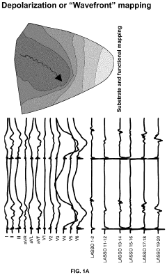
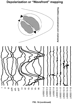
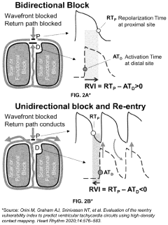
Electro-anatomic cardiac repolarization mapping
PatentPendingUS20240197236A1
Innovation
- A method and system for determining repolarization values from Electrogram (EGM) data using an electrode array, involving preprocessing steps like high-pass filtering, baseline correction, and noise reduction to obtain orthogonal bipolar compound electrograms, allowing for the creation of repolarization maps that guide cardiac procedures such as catheter ablation.

Diagnostic Implications
The diagnostic implications of acute stress-induced T wave inversion morphologies are significant in clinical cardiology. These alterations in electrocardiogram (ECG) patterns can provide valuable insights into the patient's cardiovascular health and stress response mechanisms.
Acute stress-induced T wave inversions often mimic ischemic changes, potentially leading to misdiagnosis of acute coronary syndromes. This similarity poses a challenge for clinicians in differentiating between stress-induced and pathological T wave inversions. Accurate interpretation is crucial to avoid unnecessary invasive procedures or treatments.
The presence of stress-induced T wave inversions may indicate an increased susceptibility to stress-related cardiac events. This finding can be used as a prognostic marker, helping identify patients who may benefit from stress management interventions or closer cardiovascular monitoring.
In some cases, stress-induced T wave inversions may unmask underlying cardiac pathologies that are not apparent under normal conditions. This phenomenon can aid in the early detection of subclinical cardiovascular diseases, allowing for timely intervention and prevention of adverse outcomes.
The duration and extent of T wave inversions can provide information about the severity and persistence of the stress response. Prolonged or widespread inversions may indicate a more pronounced stress reaction, potentially requiring further evaluation or intervention.
Understanding the specific morphological changes in T waves during acute stress can help in developing more refined diagnostic criteria. This knowledge can improve the accuracy of stress test interpretations and enhance the overall diagnostic capabilities of ECG in stress-related cardiac conditions.
The reversibility of stress-induced T wave inversions is another important diagnostic consideration. The time course of T wave normalization after stress resolution can offer insights into the individual's cardiac recovery capacity and overall cardiovascular health.
Lastly, the pattern and distribution of T wave inversions across different ECG leads can provide information about the specific areas of the heart affected by stress. This localization can be valuable in understanding the physiological impact of stress on different regions of the myocardium and guiding further diagnostic investigations if necessary.
Stress Management Impact
The impact of stress management on T wave inversion morphologies is a critical area of study in cardiovascular health. Effective stress management techniques have shown promising results in mitigating the adverse effects of acute stress on cardiac function, particularly in relation to T wave inversions.
Research has demonstrated that implementing stress reduction strategies can lead to significant improvements in T wave morphologies during periods of acute stress. Techniques such as deep breathing exercises, progressive muscle relaxation, and mindfulness meditation have been found to modulate the autonomic nervous system response, potentially reducing the likelihood and severity of stress-induced T wave inversions.
Studies have shown that regular practice of stress management techniques can enhance heart rate variability, a key indicator of cardiac health. This improved variability may contribute to more stable T wave patterns, even in the face of acute stressors. Furthermore, individuals who engage in consistent stress management practices often exhibit lower baseline levels of stress hormones, which may help maintain normal T wave morphologies during challenging situations.
The integration of stress management programs in clinical settings has yielded positive outcomes for patients with a history of stress-related cardiac issues. These programs typically combine cognitive-behavioral therapy with relaxation techniques, providing patients with tools to better cope with acute stress. As a result, many participants have shown reduced frequency and severity of T wave inversions during follow-up electrocardiograms.
Workplace stress management initiatives have also demonstrated potential in preserving normal T wave morphologies among employees in high-stress occupations. Companies that have implemented comprehensive stress reduction programs, including mindfulness training and work-life balance policies, have reported lower incidences of stress-related cardiac abnormalities, including T wave inversions, among their workforce.
Emerging research is exploring the use of biofeedback and neurofeedback techniques as targeted interventions for managing acute stress and its impact on T wave morphologies. These approaches allow individuals to gain conscious control over physiological processes, potentially enabling them to maintain more stable cardiac electrical activity during stressful events.
While the evidence supporting the positive impact of stress management on T wave inversions is growing, further research is needed to fully elucidate the mechanisms by which these interventions influence cardiac electrical activity. Long-term studies are particularly important to assess the sustained effects of stress management practices on T wave morphologies and overall cardiovascular health.
Unlock deeper insights with PatSnap Eureka Quick Research — get a full tech report to explore trends and direct your research. Try now!
Generate Your Research Report Instantly with AI Agent
Supercharge your innovation with PatSnap Eureka AI Agent Platform!