
Method and equipment arrangement for presenting information in radiology
Patent Information
- Authority / Receiving Office
- US · United States
- Current Assignee / Owner
- Publication Date
- 2007-02-01
- Estimated Expiration
- Not applicable · inactive patent
Smart Images
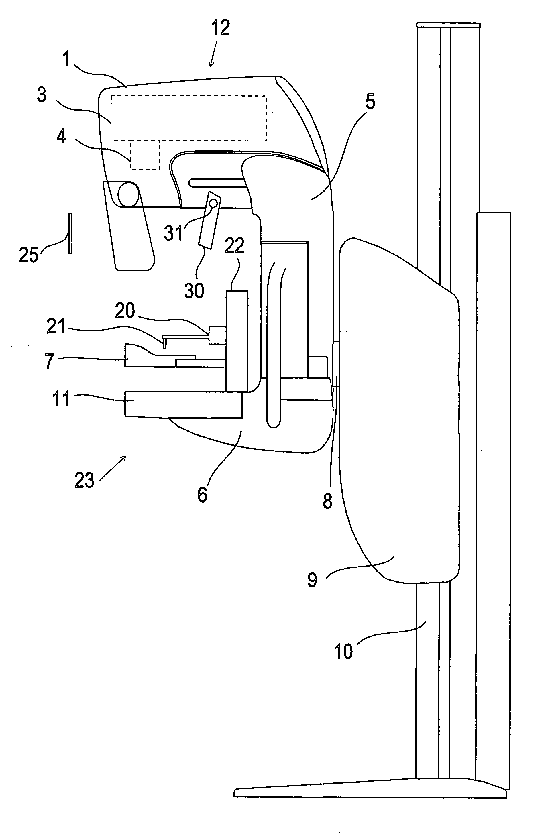
Figure 1 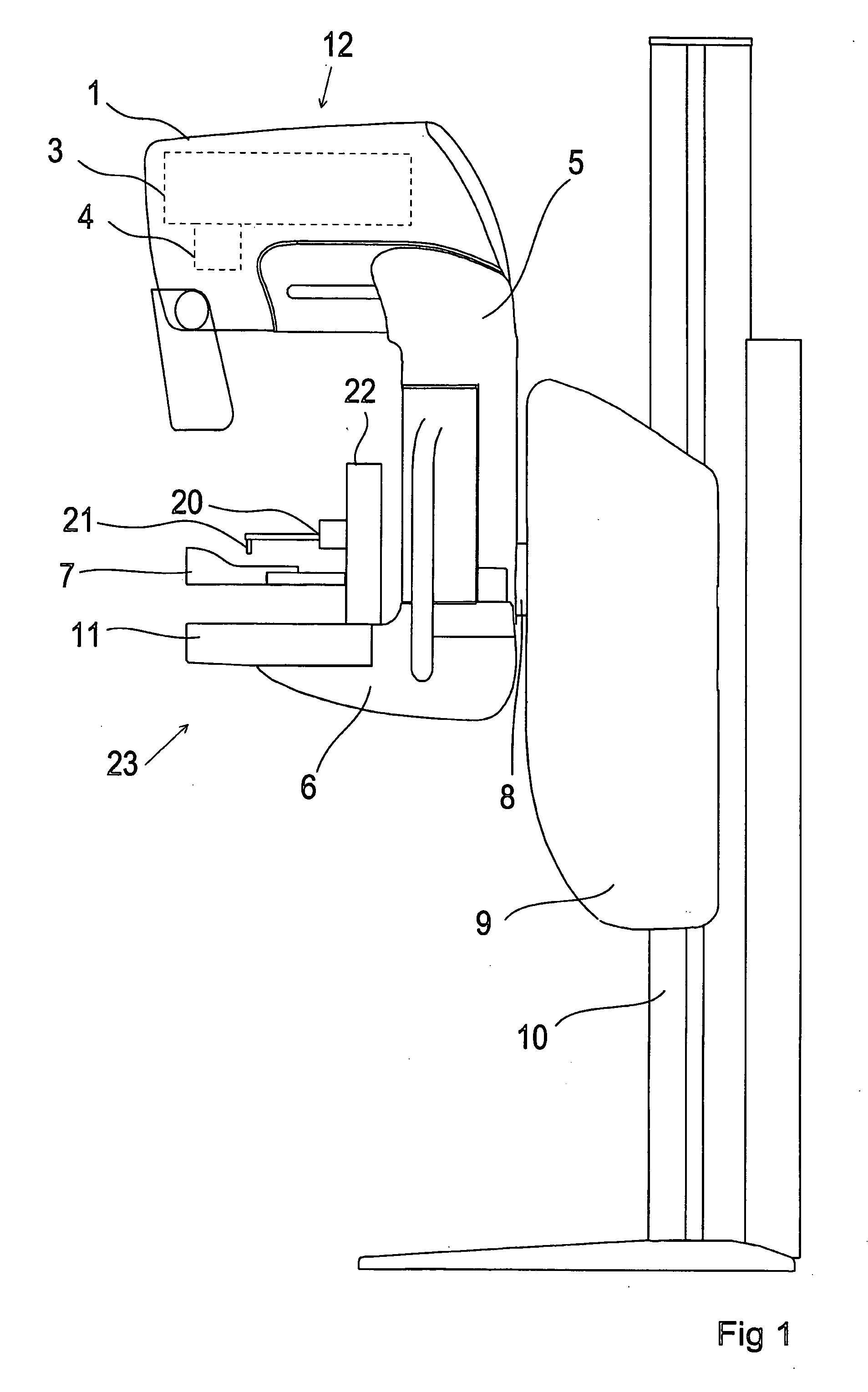
Figure 2 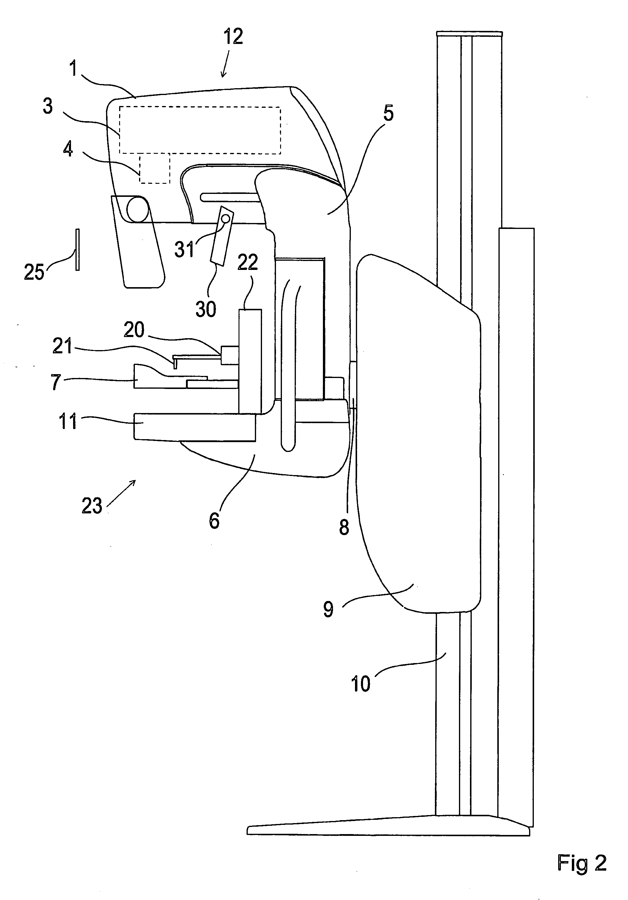
Figure 3
Abstract
Description
TECHNICAL FIELD
[0001] In radiology it is typical to perform invasive procedures. In these procedures accuracy is of primary importance and a desirable characteristic. An objective is also to interpret the radiological images as correctly as possible. PRIOR ART
[0002] Image information obtained by radiology is utilised in performing invasive procedures. In order to obtain a sufficiently high accuracy of the invasive procedures the person performing the invasive procedures must be very focused on the performance, but still there exists a risk of an inaccurate invasive procedure.
[0003] In the following we discuss the subject with regard to mammography. For instance, the western manner of life (fatty diet etc.) has contributed to an ever more common occurrence of women's breast cancer. As this is a disease of a serious category, much attention is paid to a sufficiently early detection of breast cancer in more and more countries. Such detection is realised with mammography equipment. [...
Examples
Embodiment Construction
[0012]FIG. 1 shows as an example of radiology equipment arrangements an X-ray imaging device used in X-ray mammography examination. The mammography device shown in FIG. 1 comprises a C-formed arm 1, 5, 6, in which an X-ray tube end 12 and the image generating section 23 comprising a full-field detector 11 are attached at the opposing branches 1, 6 of the console. The console further includes a movable compression plate 7 for compressing a breast. The console is mounted in bearings so that it can be turned around a shaft 8, which is attached so that it is substantially in parallel with the branches 1, 6 and substantially perpendicular to the bar 5 connecting the branches. The shaft 8 is also attached to a sliding body 9 movable along a vertical post 10, which provides the height adjustment of the device.
[0013] In the first phase of breast examination an image is taken of the whole breast. The devices in current use are to an ever-increasing degree digital mammography devices, where ...