
Quantification of contrast-enhanced ultrasound parameteric maps with a radiomics-based analysis
a radiomics-based analysis and contrast-enhanced ultrasound technology, applied in image enhancement, instruments, recognition of medical/anatomical patterns, etc., can solve problems such as complex patient management, no rapid and efficient methods to determine which treatment regimens, and anatomical changes in lesion siz
Patent Information
- Authority / Receiving Office
- US · United States
- Current Assignee / Owner
- Publication Date
- 2022-06-23
Smart Images
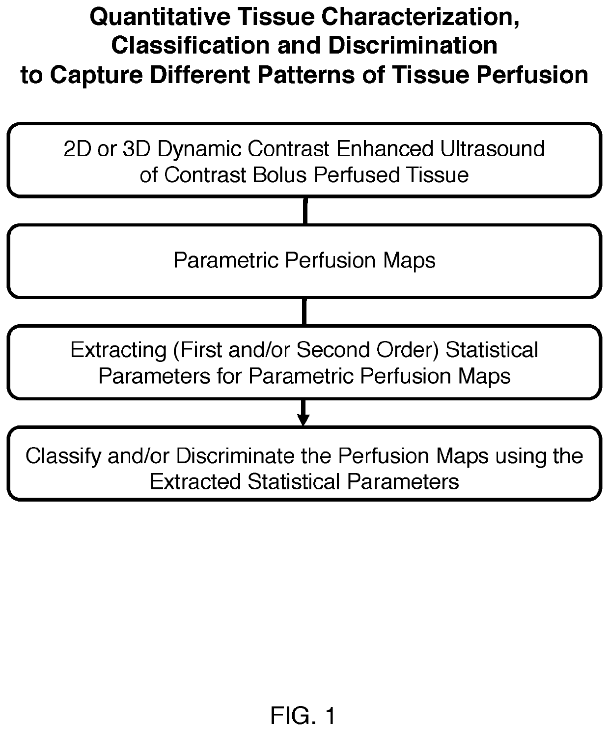
Figure 1 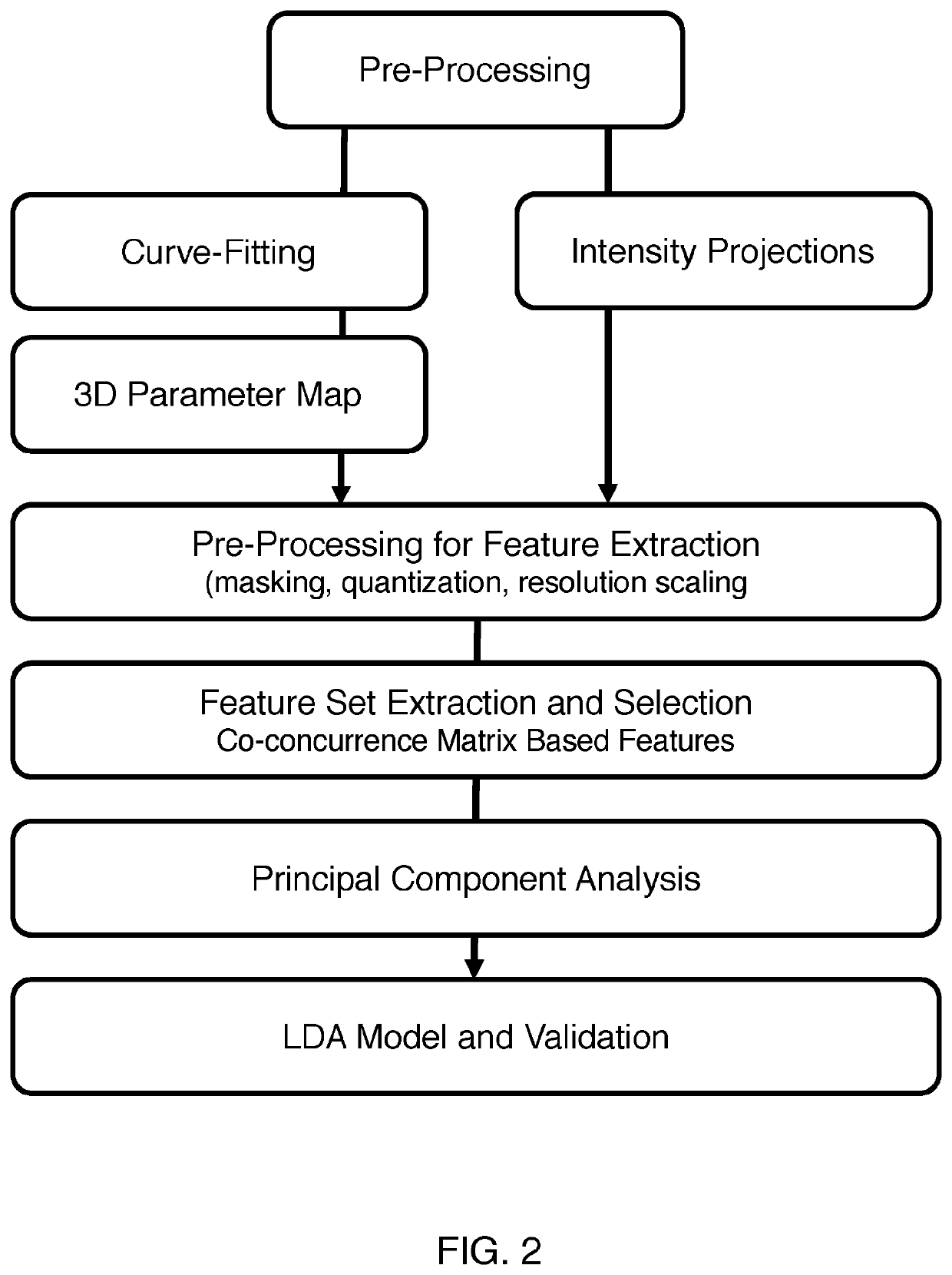
Figure 2 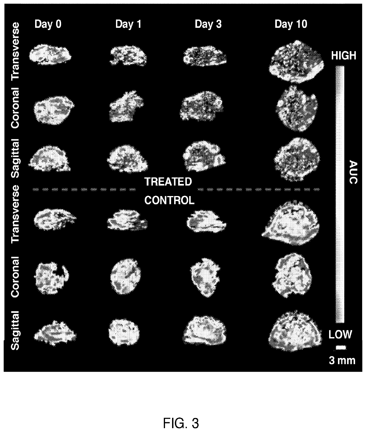
Figure 3
Abstract
Description
FIELD OF THE INVENTION
[0001] This invention relates to methods, devices and systems to determine cancer treatment responses using volumetric dynamic contrast-enhanced ultrasound (DCE-US).BACKGROUND OF THE INVENTION
[0002] Advances in anti-cancer agents have significantly enriched the therapeutic armamentarium available to clinicians for managing disease, but have further complicated patient management because not all patients respond to treatments similarly. There are currently no rapid and efficient methods to determine which treatment regimens will be effective on a patient-by-patient basis at baseline or within weeks of starting treatment.
[0003] Conventional anatomical-based assessments with the Response Evaluation Criteria in Solid Tumors (RECIST 1.1) are performed at earliest 2-3 months after treatment start and do not account for acute cytostatic effects that do not always result in anatomical changes in lesion size. Thus, there is a significant need for tools to rapidly assess or...
Examples
Embodiment Construction
Experimental Design
[0030]In an exemplary and illustrative embodiment, experiments were designed to extract image features from 3D-DCE US longitudinal perfusion maps from liver metastases and to combine these as multi-parametric biomarkers that can differentiate responders from non-responders. To identify reliable features from parametric maps, repeatable histogram and texture features (image features) were isolated to discriminate between responsive and non-responsive tumors treated with the anti-angiogenic agent Bevacizumab on a subject-by-subject basis.
[0031]As a proof of concept, the use of two approaches was investigated to generate multi-parametric biomarkers for treatment assessment; i) a statistical approach, and ii) a GLMNET approach, developed on pre-clinical data and tested on pre-clinical test and human test data. Pre-clinical test data included Bevacizumab-treated and control animals, as well as a cohort for feature repeatability assessment. In addition, tested in pre-cl...