
Manufacturing method of model of object to be imaged and individual imaging method
An imaging method and manufacturing method technology, applied in the field of medical imaging, can solve problems such as low CT performance, rough calibration and correction, increase patient radiation dose, etc., and achieve the effect of reducing radiation dose
Patent Information
- Authority / Receiving Office
- CN · China
- Current Assignee / Owner
- Publication Date
- 2015-11-25
Smart Images
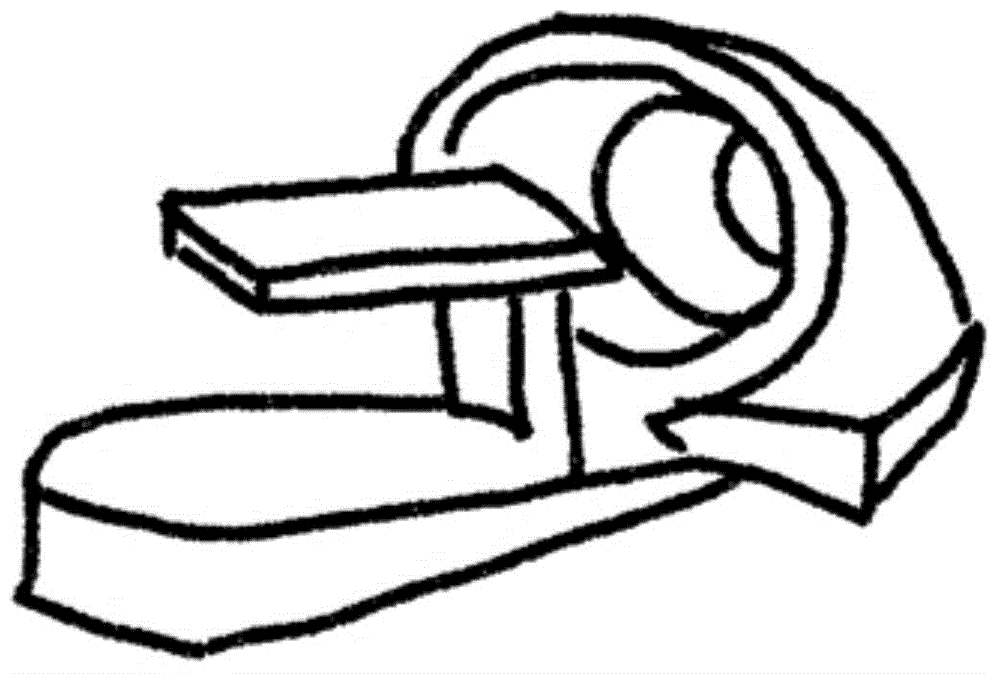
Figure 1 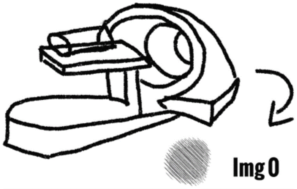
Figure 2 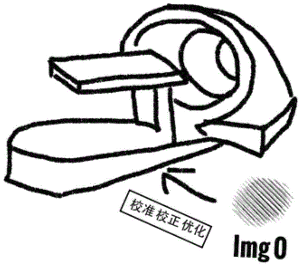
Figure 3
Abstract
Description
Technical field
[0001] The present invention relates to the technical field of medical imaging, in particular to a method for manufacturing an object to be imaged and an individualized imaging method. Background technique
[0002] Such as Figure 1 to Figure 4 As shown, in the existing medical imaging technology, the prior knowledge of the object to be imaged has not been fully utilized to obtain higher diagnostic accuracy, less radiation damage and lower cost. For example, when doing PET imaging, a cylinder is used as a prosthesis to obtain image data information Img0, and the image data information Img0 is used for system calibration and system correction, and then the human body is imaged through the calibrated and corrected imaging system , To obtain image data information Img1. This process of calibrating and correcting the imaging system using a cylinder as a prosthesis is rough, and the imaging system itself has not been optimized.
[0003] In addition, for some combined im...
Examples
Embodiment 1
[0071] Such as Figure 7 to Figure 10 As shown, an individualized imaging method disclosed in the present invention includes the steps:
[0072] S1: Acquire image data information Img0 of the mold of the object to be imaged;
[0073] S2: Calibrate, correct, and optimize the imaging system through the image data information Img0, scan and image the imaging object with the calibrated, corrected, and optimized imaging system to obtain image data information Img1;
[0074] S3: By optimizing the image data information Img0 and the image data information Img1, higher quality imaging results and data analysis results Img10 are obtained.
[0075] Such as Image 6 As shown, in this embodiment, before step S1, a mold is manufactured by a 3D module manufacturing device and method such as manual manufacturing, 3D printing, injection molding, or die-casting molding.
[0076] The image data information Img0 in the step S1 is obtained by obtaining the data of the mold, and then extracting parameters f...
Embodiment 2
[0081] The invention discloses an individualized imaging method, which comprises the steps:
[0082] S1: Acquire image data information Img0 of the mold of the object to be imaged;
[0083] S2: Image the object to be imaged to obtain image data information Img1, and obtain higher-quality imaging results and data analysis results Img10 by optimizing the image data information Img0 and image data information Img1;
[0084] S3: Optimize the model by using the higher-quality imaging result and the data analysis result Img10 obtained in S2.
[0085] In this embodiment, the image data information Img0 in step S1 is obtained by obtaining model data and then extracting parameters from the data, or directly obtained by imaging with an imaging system.
[0086] The difference between this embodiment and the first embodiment is that the image data information Img0 is not used to calibrate, correct, and optimize the imaging system. This embodiment is aimed at some occasions where the imaging system...
Embodiment 3
[0088] Such as Figure 13 , 14 As shown in, 17, and 18, an individualized imaging method disclosed in the present invention includes the steps:
[0089] S1: The mold of the object to be imaged is imaged by an A imaging system to obtain image data information Img0;
[0090] S2: Calibrate, correct, and optimize a B imaging system through the image data information Img0, and use the calibrated, corrected and optimized B imaging system to scan and image the object to be imaged to obtain image data information Img1;
[0091] S3: By processing the image data information Img0 and the image data information Img1, the A-B fusion imaging result and the data analysis result Img10 are obtained.
[0092] Such as Picture 12 As shown, in this embodiment, before step S1, a mold is manufactured by a 3D module manufacturing device and method such as manual manufacturing, 3D printing, injection molding, or die-casting molding.
[0093] This embodiment is mainly aimed at obtaining combined imaging data. I...