
A pathogenic mutation of hereditary central halo retinopathy and its detection reagent
A retinopathy and hereditary technology, applied in the field of the pathogenic mutation of hereditary central halo retinopathy and its detection reagent, can solve the problems of unreported, cone and rod dystrophy, cone cell degeneration, etc.
Patent Information
- Authority / Receiving Office
- CN · China
- Patent Type
- Patents(China)
- Current Assignee / Owner
- Publication Date
- 2019-03-15
- Estimated Expiration
- Not applicable · inactive patent
Smart Images
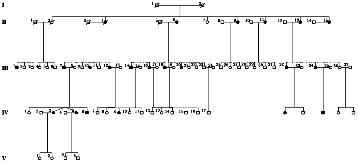
Figure 1 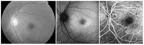
Figure 2 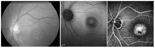
Figure 3
Abstract
Description
technical field
[0001] The invention belongs to the field of biomedicine and relates to a pathogenic mutation of hereditary central halo retinopathy and a detection reagent thereof. Background technique
[0002] Central areolar choroidal dystrophy (CACD) is a group of progressive retinal degenerative diseases caused by genetic defects, such diseases are mainly caused by the irreversible atrophy of retinal pigment epithelium (RPE) and choroidal blood vessels pathological features. CACD patients mainly harm the macular area of the patient, causing a progressive decline in central vision, and severe cases can cause blindness. CACD is a clinically common and serious hereditary blinding disease, which can be divided into four stages according to its clinical features. Early CACD patients are difficult to make an accurate clinical diagnosis because their phenotypes are easily confused with other fundus disease patients. Therefore, it is particularly important to find other po...
Examples
Embodiment 1
[0049] A five-generation family with central areolar choroidal dystrophy (CACD) was tested for GUCA1A mutations.
[0050] experimental method:
[0051] 1. Collection of clinical resources of the family and establishment of a genetic resource bank:
[0052] The clinical data and blood samples of each member of the family were collected, see the family diagram figure 1 . Clinical data mainly include personal medical history, family history, best corrected visual acuities (BCVAs), slit lamp examination, fundus photography, color vision examination, visual field examination (Humphrey perimetry), visual evoked potential detection (visual-evokedpotentials); VEP), full field electrophysiology (electroretinography; ERG), fundus fluoresceinangiography (FFA) and optical coherence tomography (optical coherencetomography; OCT), etc. The blood genomic DNA of each family member was extracted with a blood genomic DNA extraction kit (Qiagen, Hilden, Germany).
[0053] 2. Discover the path...
Embodiment 2
[0083] A functional study was conducted on the pathogenic mutation GUCA1A p.Arg120Leu detected in Example 1.
[0084] experimental method:
[0085] 1. Conservative analysis:
[0086] The NCBI HomoloGene database (http: / / www.ncbi.nlm.nih.gov / homologene) was used to evaluate and predict the conservation of the screened mutations in multiple species.
[0087] 2. Research on protein crystal structure changes:
[0088] SWISS MODEL (http: / / swissmodel.expasy.org / ) prediction software was used to predict the structure of GUCA1A wild-type protein and the mutant protein carrying the p.Arg120Leu mutation, and evaluate the protein structure changes caused by the mutation.
[0089] 3. Zebrafish experiment:
[0090] Synthesize the full-length cDNA of wild type and mutant human GUCA1A gene respectively and clone it into pxT7 vector to construct pxT7 tagged GUCA1A WT and GUCA1A MU Plasmids from which to synthesize the corresponding GUCA1A mRNA, including GUCA1A WT and GUCA1A MU mRNA. ...