
A pathogenic mutation of hereditary usher syndrome and its detection reagent
A syndromic and genetic technology, applied in the field of biomedicine, which can solve the problems of less research and no reports.
Patent Information
- Authority / Receiving Office
- CN · China
- Patent Type
- Patents(China)
- Current Assignee / Owner
- Publication Date
- 2019-10-01
Smart Images
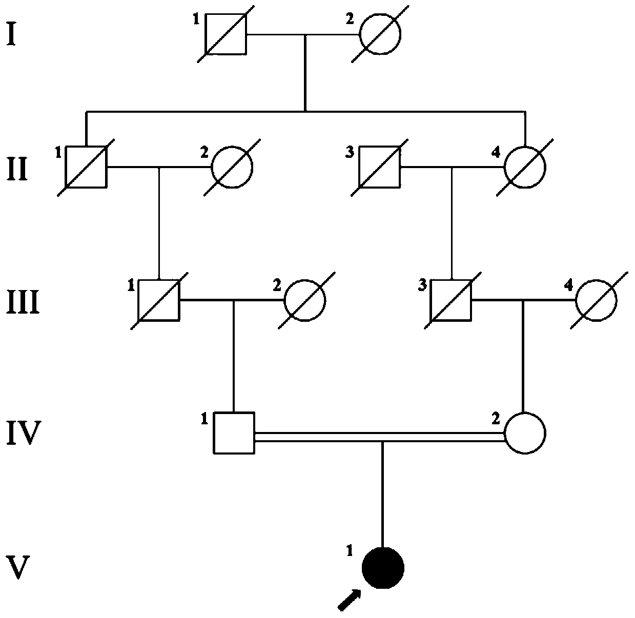
Figure 1 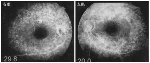
Figure 2 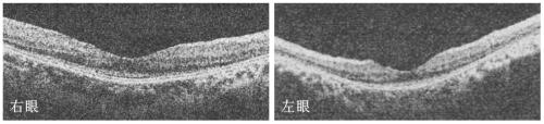
Figure 3
Abstract
Description
technical field
[0001] The invention belongs to the field of biomedicine and relates to a pathogenic mutation of hereditary Usher syndrome and a detection reagent thereof. Background technique
[0002] Usher syndrome, also known as hereditary deafness retinitis pigmentosa syndrome, retinitis pigmentosa sensorineural deafness syndrome, is an autosomal recessive genetic disease. The main manifestations of Usher syndrome are congenital sensorineural deafness, progressive retinitis pigmentosa, and vision loss. Clinically, Usher syndrome is mainly divided into the following three types: type Ⅰ is congenital severe deep sensorineural deafness, the patient's vestibular response disappears, and retinitis pigmentosa occurs around the age of 10; type Ⅱ is congenital moderate to severe sensorineural deafness Deafness, the vestibular response is normal, and retinitis pigmentosa occurs at the age of 18 to 20; type III is progressive sensorineural deafness, the vestibular response is unc...
Examples
Embodiment 1
[0040] Detection of CEP78 gene mutation in a consanguineous Usher syndrome family.
[0041] experimental method:
[0042] 1. Collection of clinical resources of the family and establishment of a genetic resource bank:
[0043] The clinical data and blood samples of the patients in this family were collected, see the family chart figure 1. Clinical data mainly include personal medical history, family history, best corrected visual acuities (BCVAs), slit lamp examination, fundus photography, color vision examination, visual field examination (Humphrey perimetry), full field electroretinography (ERG) ), fundus fluorescein angiography (FFA) and optical coherence tomography (optical coherence tomography; OCT), etc. The blood genomic DNA of each family member was extracted with a blood genomic DNA extraction kit (Qiagen, Hilden, Germany).
[0044] 2. Discover the disease-causing mutation of the patient with the help of high-throughput next-generation sequencing:
[0045] 2.1 De...
Embodiment 2
[0075] Prediction of splicing abnormalities that may be caused by the pathogenic mutation CEP78 c.1629-2A>G detected in Example 1.
[0076] experimental method:
[0077] 1. Prediction of cut site changes:
[0078] The mutation we found is located near the receptor splice site of the intron between exon 14 and exons 13 and 14. For the relationship between the mutation and the normal splice site, see Figure 4 . We used a series of splice site prediction websites to predict possible splice site changes, including:
[0079] (1) Human Splicing Finder, website: http: / / www.umd.be / HSF3 / HSF.html ;
[0080] (2) NetGene2 Server, URL: http: / / www.cbs.dtu.dk / services / NetGene2 / ;
[0081] (3) Splicing Regulation Online Graphical Engine, URL: http: / / sroogle.tau.ac.il / ;
[0082] (4) Berkeley Drosophila Genome Project, website: http: / / www.fruitfly.org / seq_tools / splice.html ;
[0083] (5) SplicePort, URL: http: / / spliceport.cbcb.umd.edu / .
[0084] Experimental results:
...