
A urinary sediment microscopic image visible component recognition method based on deep learning
A microscopic image and deep learning technology, applied in the field of medical microscopic image processing, can solve the problem of insufficient samples of urine sediment microscopic images, and achieve the effects of rich image features, simple operation and excellent efficiency
Patent Information
- Authority / Receiving Office
- CN · China
- Current Assignee / Owner
- Publication Date
- 2019-05-10
Smart Images
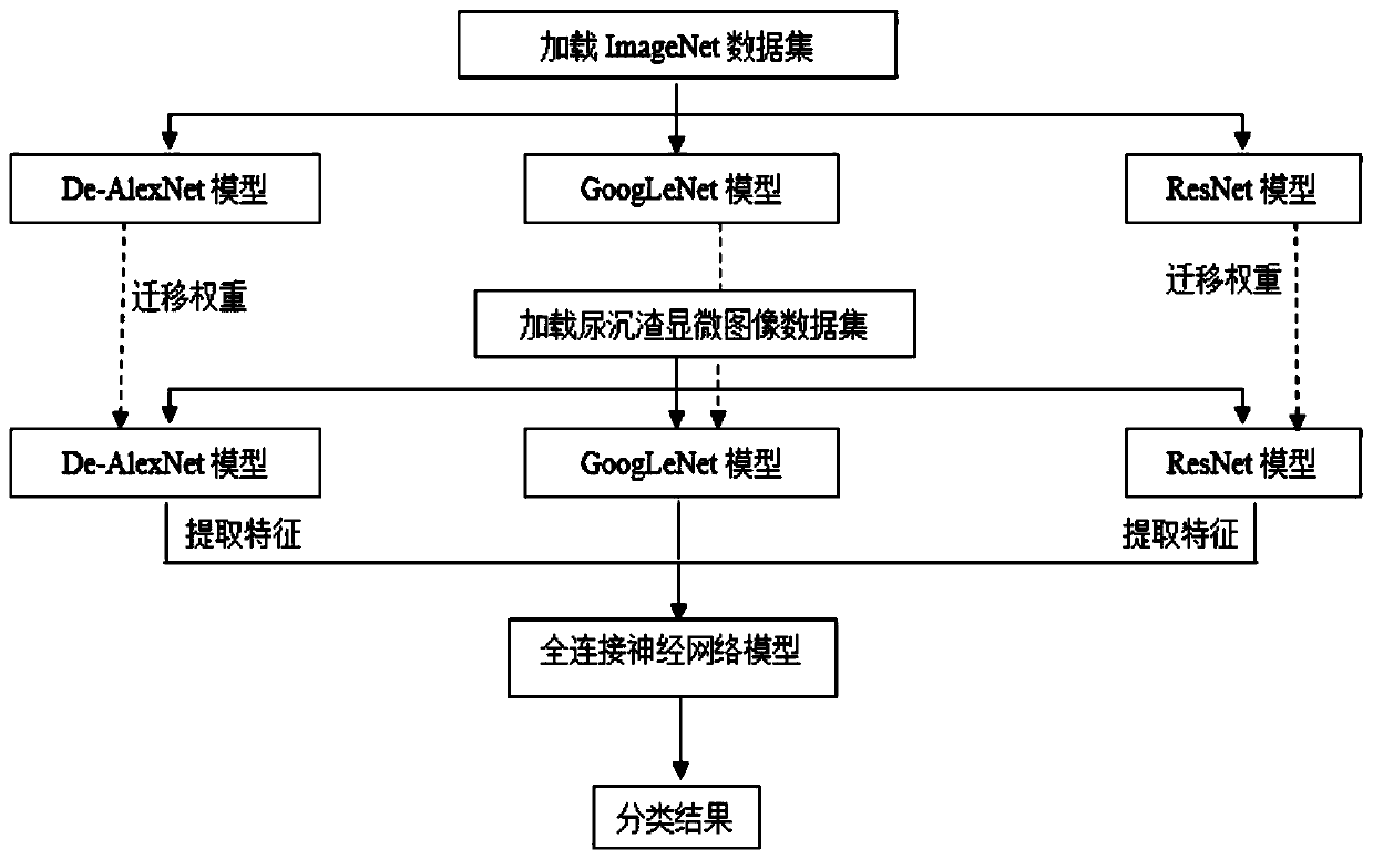
Figure 1 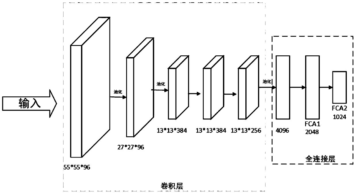
Figure 2 
Figure 3
Abstract
Description
technical field
[0001] The invention relates to the technical field of medical microscopic image processing, in particular, a deep learning-based recognition method for formed components in microscopic images of urinary sediment. Background technique
[0002] Urine sediment examination plays an important role in the diagnosis and differentiation of kidney diseases, urinary system diseases, circulatory system diseases and infectious diseases, and is one of the routine inspection items in hospitals. At present, urine sediment inspection can be carried out in three ways: dry chemical method, flow cytometry method, and image microscope detection and analysis method, among which image microscope detection and analysis method is currently a relatively common and reliable method for urine sediment inspection. Due to the heavy inspection workload in the hospital every day and the low inspection efficiency, the camera urine sediment automatic analyzer has become an ideal choice for m...
Examples
Embodiment Construction
[0032] The specific implementation manner and working principle of the present invention will be further described in detail below in conjunction with the accompanying drawings.
[0033] This method consists of four parts: improving the AlexNet model to the De-AlexNet model, transferring the weights of the CNN model, fine-tuning the learning rate and cascading features, and integrating the features extracted by the three convolutional neural network models and designing a classifier.
[0034] The first part removes the fully connected layer of the FC7 layer of the AlexNet model with a dimension of 4096, and adds two layers of FCA1 and FCA2 layers with fully connected layers of dimensions 2048 and 1024 respectively.
[0035] In the second part, the De-AlexNet model, GoogLeNet model and ResNet model are pre-trained on the ImageNet dataset to obtain weights, and then the weights are transferred to the urine sediment microscopic image dataset to continue training.
[0036] In the ...