
Methods and systems to detect and classify retinal structures in interferometric imaging data
a technology of interferometric imaging and classification methods, applied in image analysis, image enhancement, instruments, etc., can solve the problems of inconvenient or even impossible manual segmentation of human operators in high-throughput clinical environments, high computational and execution time requirements, and high difficulty in segmentation, so as to reduce the number of redundant processing and reduce the segmentation error
Patent Information
- Authority / Receiving Office
- US · United States
- Patent Type
- Applications(United States)
- Current Assignee / Owner
- Publication Date
- 2019-04-11
Smart Images
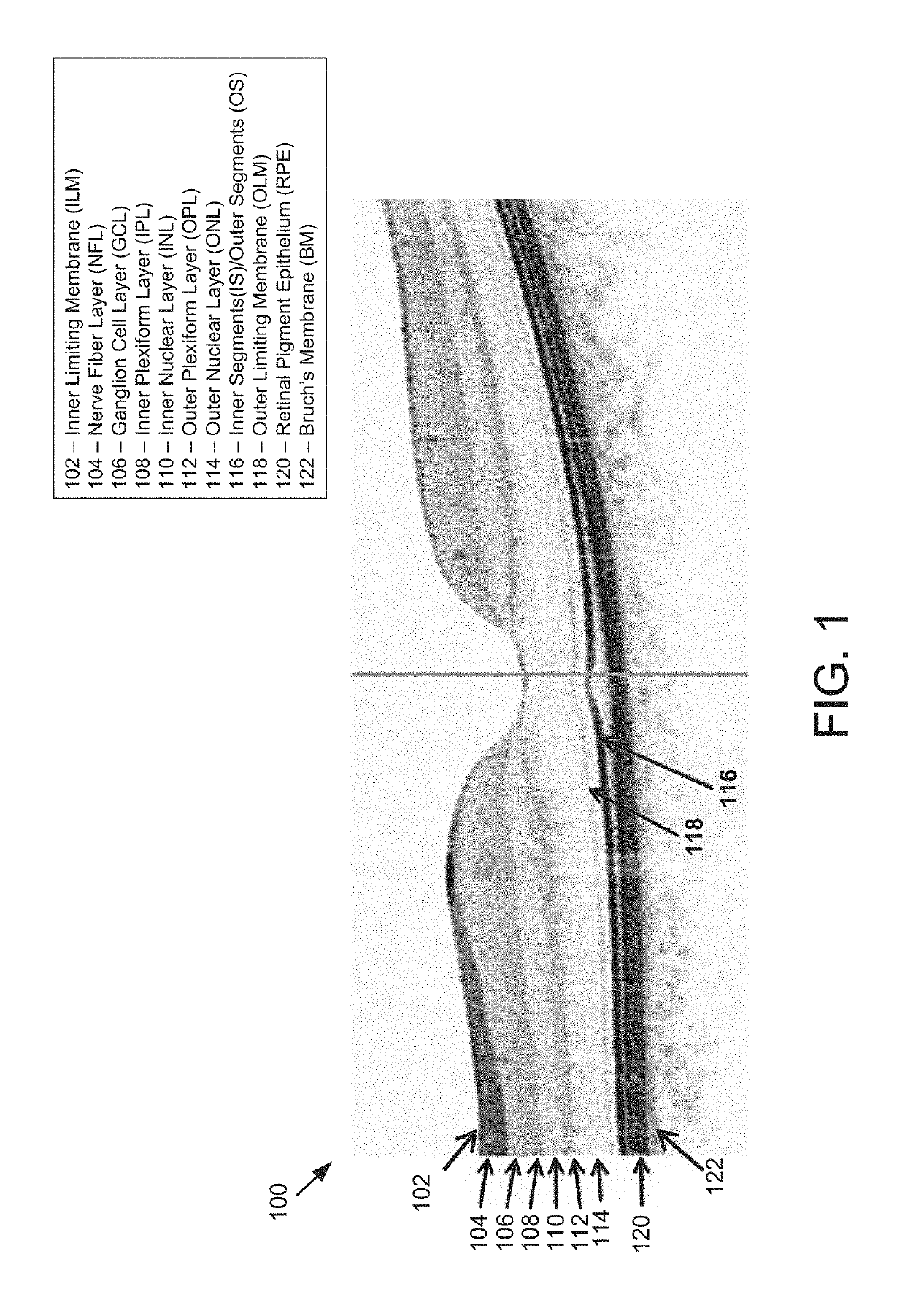
Figure 1 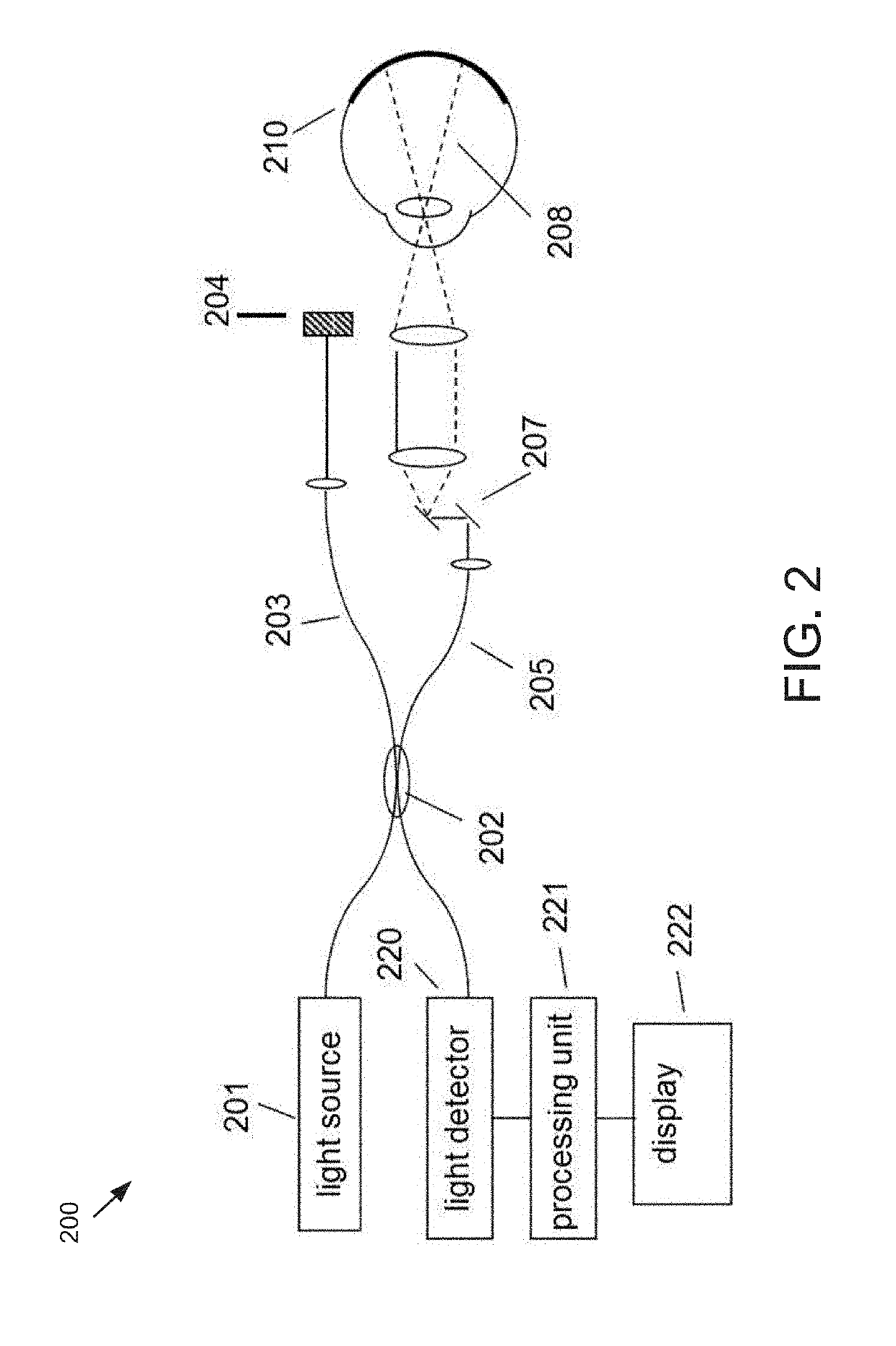
Figure 2 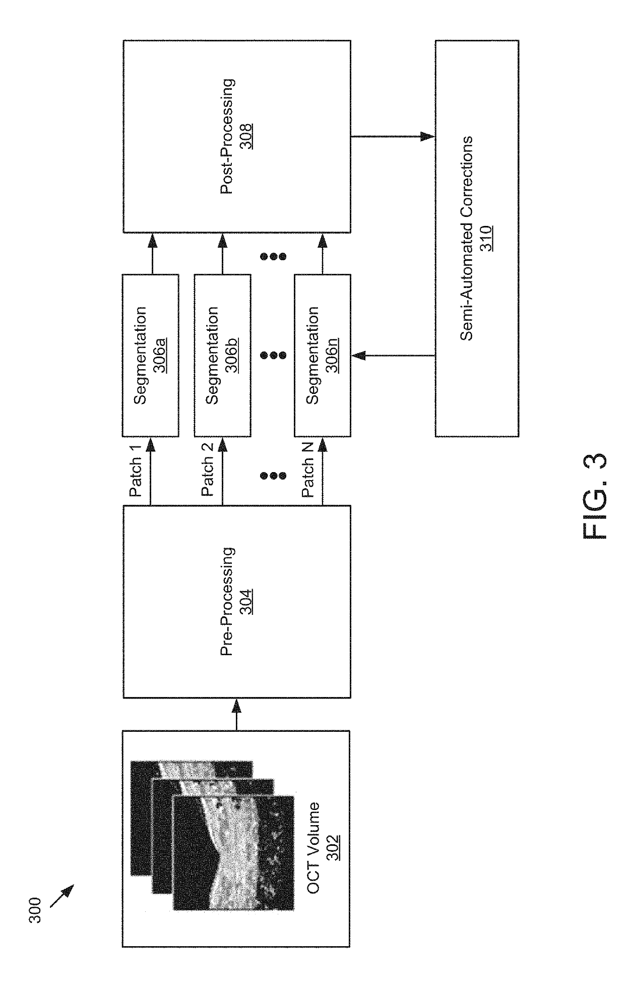
Figure 3
Abstract
Description
PRIORITY
[0001] This application is a divisional application of U.S. patent application Ser. No. 15 / 246,036, filed Aug. 24, 2016, which claims priority to U.S. Provisional Application Ser. No. 62 / 210,953, filed Aug. 27, 2015, the contents of each of which is hereby incorporated by reference.TECHNOLOGICAL FIELD
[0002] The present application concerns improved methods and systems for detecting and identifying structures in images obtained with interferometric imaging systems.BACKGROUND
[0003] A wide variety of interferometric imaging techniques have been developed to provide high resolution structural information in a wide range of applications. Optical Coherence Tomography (OCT) is a technique for performing high-resolution cross-sectional imaging that can provide images of samples including tissue structure on the micron scale in situ and in real time. OCT is an interferometric imaging method that determines the scattering profile of a sample along the OCT beam by detecting light reflecte...
Examples
Embodiment Construction
[0031]All patent and non-patent references cited within this specification are herein incorporated by reference in their entirety to the same extent as if the disclosure of each individual patent and non-patient reference was specifically and individually indicated to be incorporated by reference in its entirely.
[0032]FIG. 2 illustrates a generalized Fourier Domain optical coherence tomography (FD-OCT) system 200 that is used to collect an OCT dataset suitable for use with the present set of embodiments. As depicted, the FD-OCT system 200 includes a light source 201, typical sources including, but not limited to, broadband light sources with short temporal coherence lengths or swept laser sources. Light from source 201 is routed, typically by optical fiber 205, to illuminate the sample 210, which could be any of the tissues or structures within an eye. The light is scanned, typically with a scanner 207 between the output of the fiber and the sample, so that the beam of light (dashed...