
Radiological imagery method and device
a radiological imaging and method technology, applied in the direction of x-ray apparatus, radiation intensity measurement, electrical apparatus, etc., can solve the problems of inability to achieve the effect of x-ray imaging in industrial level, slow image acquisition speed of detectors, and insufficient global output, so as to reduce notably the dose d of x-ray photons, the effect of simple design and operation mod
Patent Information
- Authority / Receiving Office
- US · United States
- Patent Type
- Patents(United States)
- Current Assignee / Owner
- Publication Date
- 2007-08-07
- Estimated Expiration
- Not applicable · inactive patent
Smart Images
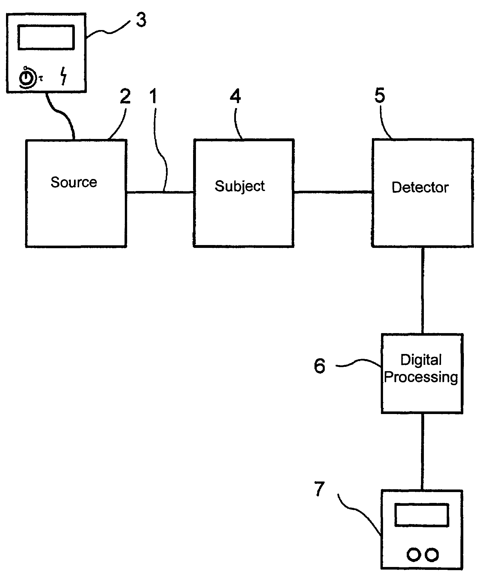
Figure 1 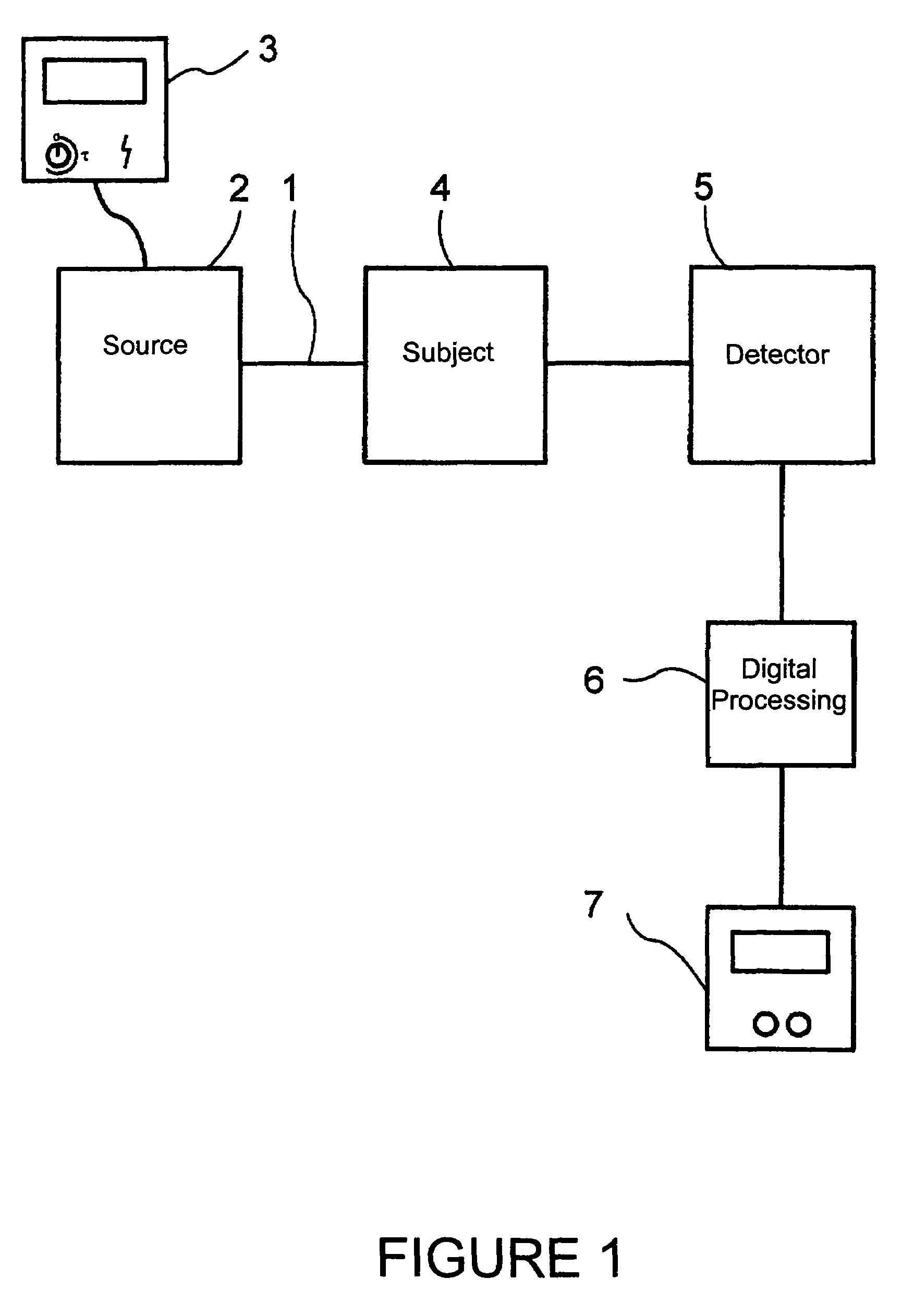
Figure 2 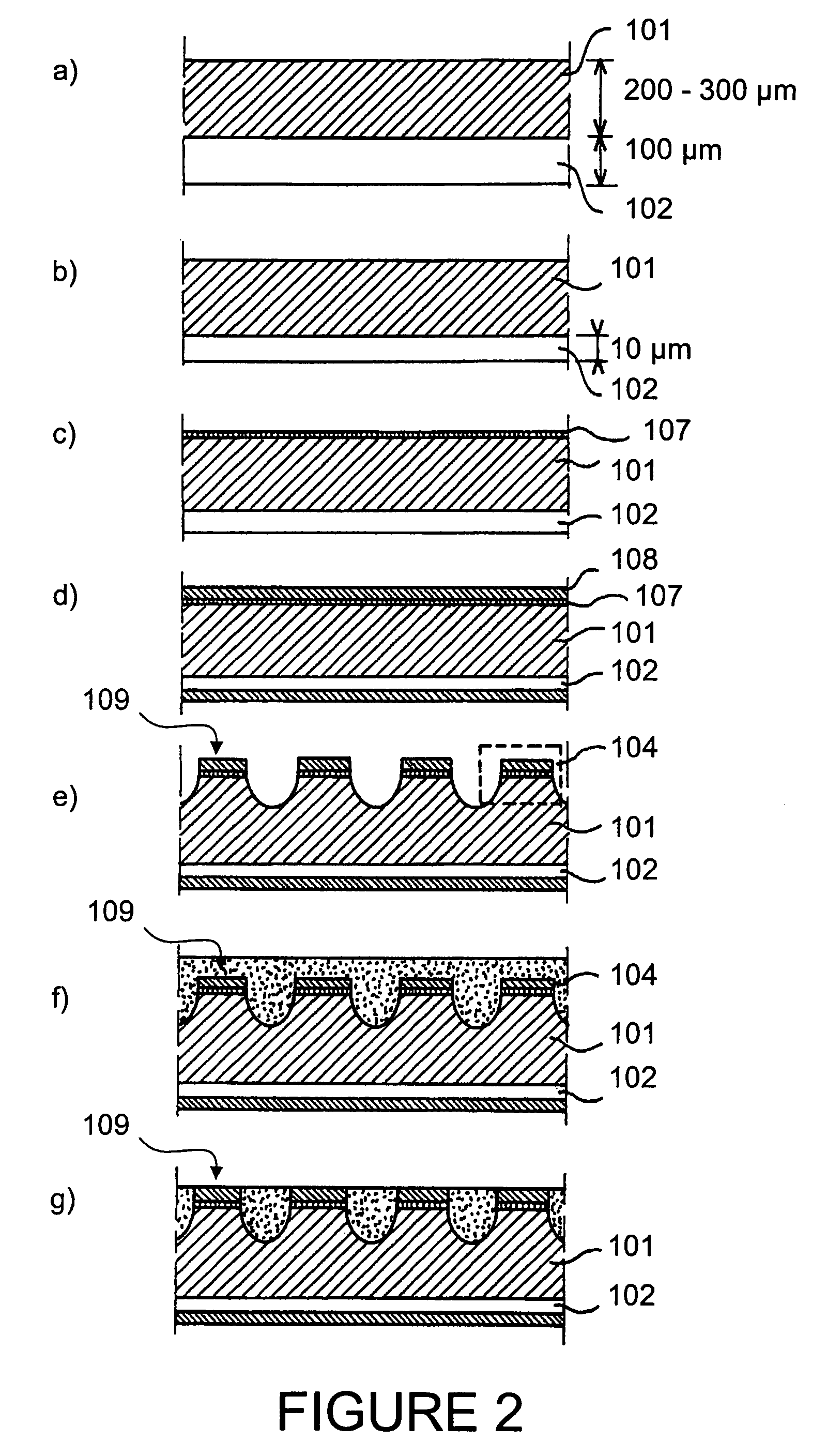
Figure 3
Abstract
Description
CROSS-REFERENCE TO RELATED APPLICATION
[0001] This application is a National Stage entry of International Application No. PCT / FR02 / 03904, filed Nov. 14, 2002, the entire specification claims and drawings of which are incorporated herewith by reference.BACKGROUND OF THE INVENTION
[0002] 1. Field of the Invention
[0003] This invention relates to a method and a radiological imaging device.
[0004] Since its first use at the beginning of the 20th century, radiography has vastly transformed and simplified the diagnostic of numerous diseases. This technique is based upon the absorption, by the organ examined, of a portion of the X-rays emited by a source and the impression of the complementary X-rays having traversed the organ in question, on a screen sensitive to the latter. A negative image of the organ examined is thus obtained. Although other imaging techniques (nuclear magnetic resonance (NMR), Echography, . . . ) have appeared more recently, this conventional radiology still represents a ver...
Examples
Embodiment Construction
[0045]The instant signal S generated by a current detector under a flux F of X-ray photons is weak and of continuous type at a few ms-scale. This signal S is therefore difficult to extract from the background noise measured by the detector, itself continuous at this time scale. In order to obtain a response R of the detector which is significant and processable, the signal S should be integrated for an exposure time Te sufficient to imply that the patient receives, during the acquisition of this response R, the dose D=F×Te. The aim of the present invention is to replace the response R of the detector with a pulse signal. By measuring the instant signal Si for a short duration Ti before Te, with a flux of photons Fi, and in sensing said signal Si with an X-ray detector having very quick response time tr, the dose may then be reduced. Indeed, one switches thus from the dose D to a dose Di=Fi×Ti where Ti / Te is smaller than Fi / Fe so that Di is shorter than D. Fi may be greater than F bu...