
Inhibitors of nitric oxide synthase for treatment of melanoma
a technology of nitric oxide and synthase, which is applied in the direction of chemical treatment enzyme inactivation, artificial cell constructs, drug compositions, etc., can solve the problems of increased proliferation, dna damage, and limited characterization of the effect of no stress and its molecular mechanisms, and achieve the effect of reducing nitric oxide stress
Patent Information
- Authority / Receiving Office
- US · United States
- Patent Type
- Patents(United States)
- Current Assignee / Owner
- Publication Date
- 2014-02-04
Smart Images
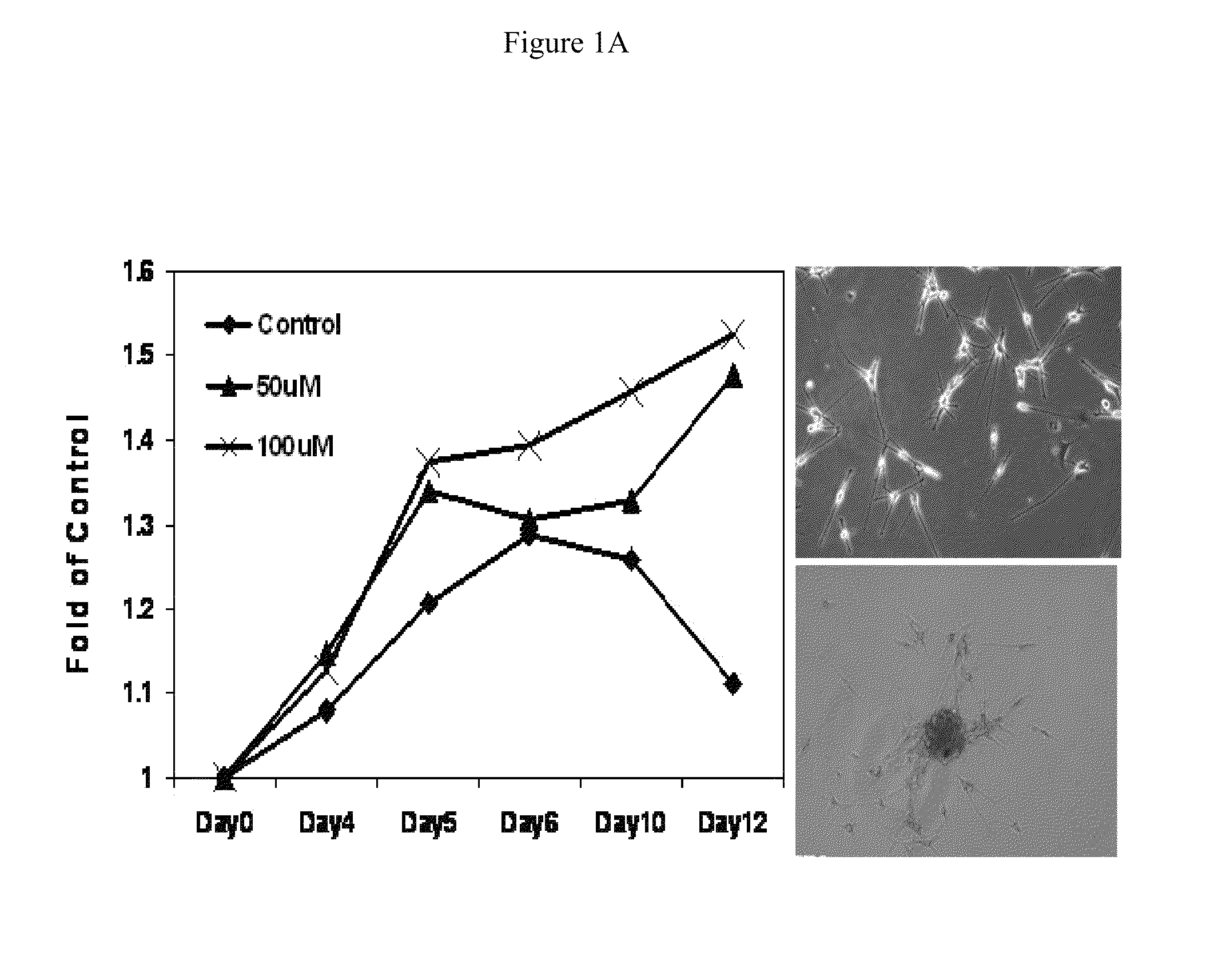
Figure 1 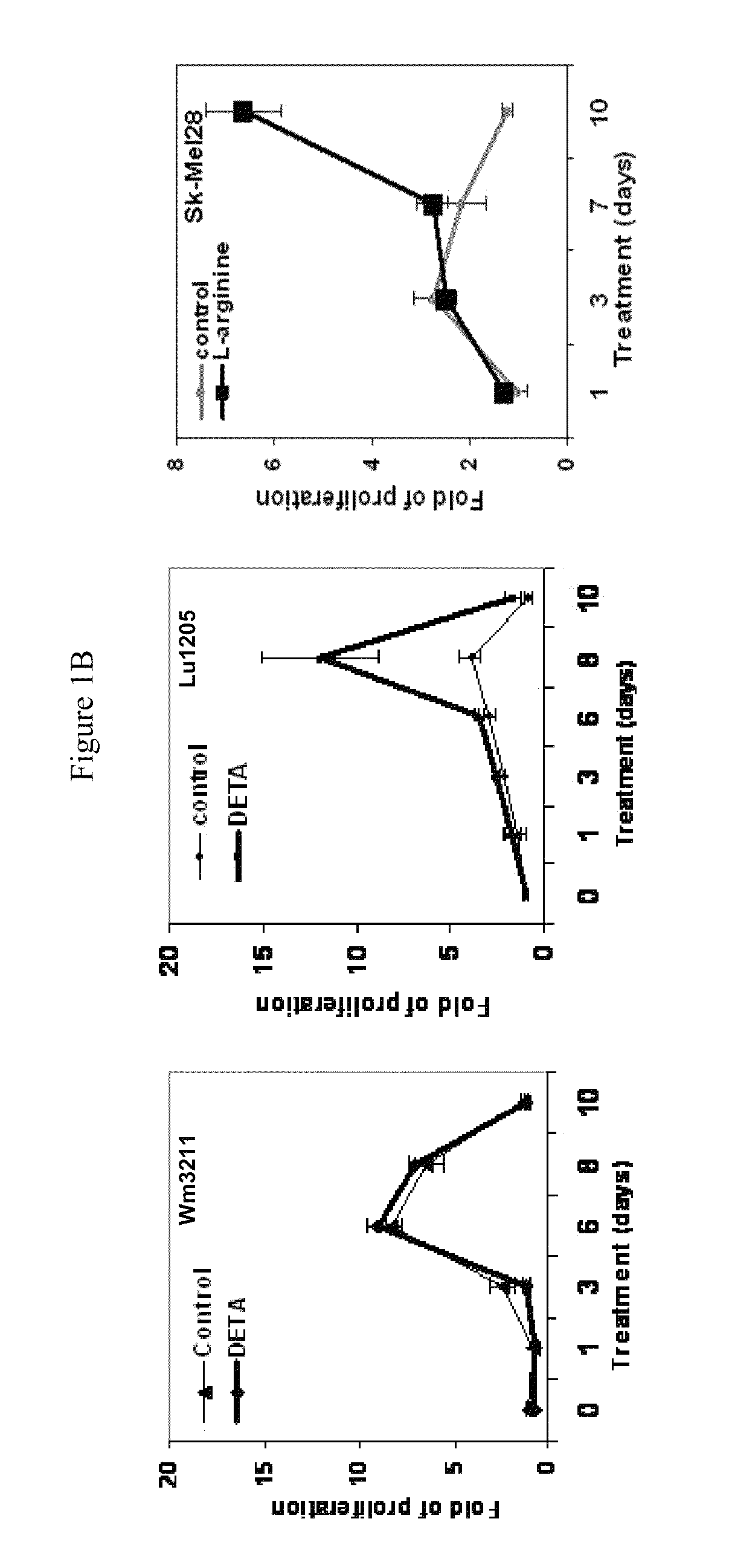
Figure 2 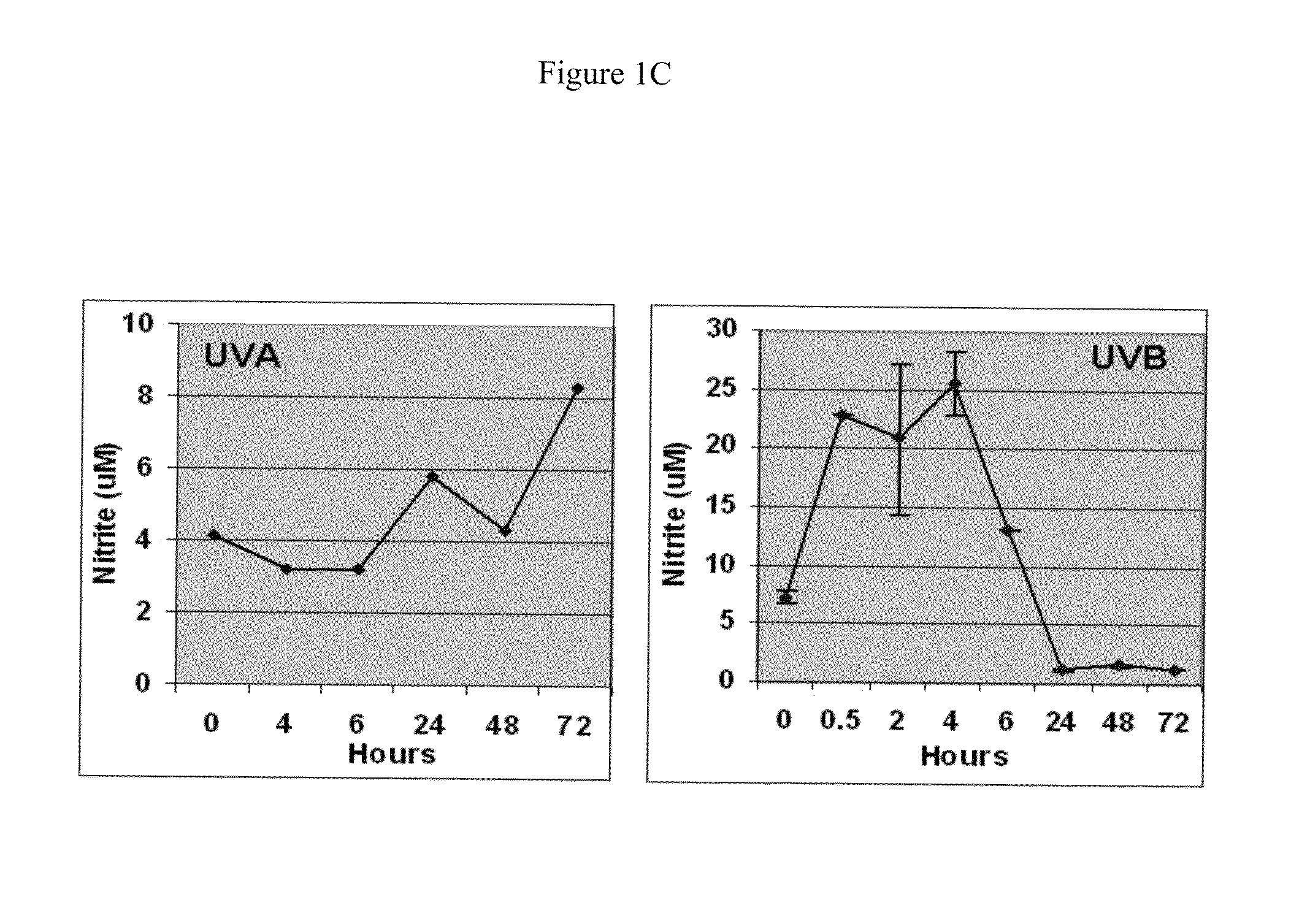
Figure 3
Abstract
Description
[0001] This application claims priority benefit of application Ser. No. 61 / 461,029 filed Jan. 12, 2011, the entirety of which is incorporated herein by reference.
[0002] This invention was made with government support under GM049725 awarded by the National Institutes of Health. The government has certain rights in the invention.BACKGROUND OF THE INVENTION
[0003] Ultraviolet (UV) radiation has been implicated as a major environmental contributor to the development of most cutaneous melanomas. Sunscreens and sun awareness behavior have been used for the prevention of cutaneous melanoma, but their clinical utility remains mixed. The mechanistic role of UV radiation in melanomagenesis needs to be more comprehensively defined. However, it is known that in human skin, UV radiation not only generates reactive oxygen species (ROS), but also produces a marked increase of nitric oxide (NO). (Russo P. A., Halliday G. M. Inhibition of nitric oxide and reactive oxygen species production improves the ...
Examples
example 1
[0075]NO stress stimulates the proliferation of melanoma cells in vitro. NO donor DETA / NO (1-[N-(2-Aminoethyl)-N-(2-ammonioethyl)amino]diazen-1-ium-1,2-diolate) was utilized to mimic NO stress in cell culture media. As shown in FIG. 1A, in human primary melanocytes, cell proliferation measured by the MTT colorimetric assay in a 12-day period was significantly stimulated by DETA / NO exposure compared to control. At the end of the experiment, melanocytes incubated with DETA / NO were still proliferative, while the proliferation of control cells peaked by day 6 and started to drop. When extending the exposure time of DETA / NO in melanocytes to 10 weeks, it was observed that many foci formed in culture dishes and cells exhibited notable vertical growth potential: these foci were sub-cloned and re-seeded in a low density and showed evidence of continued foci formation. Marked stimulation of proliferation was also evident in human metastatic Lu1205 and SK-Mel28 cells with DETA / NO (100 μM) tre...
example 2
[0078]nNOS expression is elevated melanoma compared to normal melanocytes, which is sensitive to UV radiation and growth factors. First, as shown in FIG. 2A, an immunoblotting assay revealed that the nNOS expression levels in human melanoma cell lines were much higher compared to levels in primary normal human melanocytes. Similar experiments were also carried out with mouse cells since UVB-induced melanoma in the HGF / SF transgenic mouse melanoma model is well characterized and recapitulates fairly well the etiology and histopathology of human melanoma. (Noonan F. P., Otsuka T., Bang S., Anver M. R., Merlino G. Accelerated ultraviolet radiation-induced carcinogenesis in hepatocyte growth factor / scatter factor transgenic mice. Cancer Res. 2000; 60:3738-43.) Three mouse melanoma cell lines established from the developed lesions have been used as the screening panel for translational or mechanistic studies. Similar to human cells, immunoblotting study also showed marked elevated nNOS e...
example 3
[0082]nNOS silencing was associated with reduced invasion potential in metastatic melanoma cells. Using siRNA transfection, nNOS was transiently knocked down in two metastatic melanoma cells (A375 and Lu1205) (FIG. 3A). Immunoblotting analysis showed that with nNOS depletion, the expression levels of JunD, MMP-1, APE / Ref-1, and Bcl-2 were significantly reduced. Consistently, it was found that the invasion potential was also decreased in nNOS-depleted melanoma cells (FIG. 3B).