How to Select Optimal Staining Methods in Gel Electrophoresis?
JUN 30, 20259 MIN READ
Generate Your Research Report Instantly with AI Agent
Patsnap Eureka helps you evaluate technical feasibility & market potential.
Gel Electrophoresis Staining Evolution and Objectives
Gel electrophoresis has been a cornerstone technique in molecular biology since its inception in the 1960s. The evolution of staining methods for this technique has been driven by the need for increased sensitivity, reduced toxicity, and improved convenience. Initially, researchers relied on radioactive labeling, which offered high sensitivity but posed significant safety concerns. The introduction of ethidium bromide in the 1970s marked a significant milestone, providing a non-radioactive alternative with good sensitivity.
As the field progressed, the objectives for gel electrophoresis staining methods expanded beyond mere visualization. Researchers sought quantitative analysis capabilities, compatibility with downstream applications, and the ability to detect multiple targets simultaneously. These evolving needs led to the development of fluorescent dyes like SYBR Green and SYBR Gold in the 1990s, which offered improved sensitivity and reduced mutagenicity compared to ethidium bromide.
The turn of the millennium saw a surge in the development of novel staining techniques, driven by advancements in fluorescence technology and growing environmental concerns. Cyanine dyes, such as SYBR Safe and GelRed, emerged as safer alternatives with comparable or superior performance to traditional stains. Concurrently, the rise of proteomics fueled the development of protein-specific stains like Coomassie Blue and silver staining methods, each offering unique advantages in terms of sensitivity and compatibility with mass spectrometry.
Recent years have witnessed a shift towards more specialized staining methods tailored to specific applications. For instance, the development of in-gel activity assays has enabled researchers to visualize enzyme activities directly within polyacrylamide gels. Additionally, the advent of multiplexed staining techniques has allowed for the simultaneous detection of different biomolecules within a single gel, greatly enhancing the information obtained from a single experiment.
Looking forward, the objectives for gel electrophoresis staining continue to evolve. There is a growing emphasis on developing eco-friendly stains that maintain high sensitivity while minimizing environmental impact. Researchers are also exploring ways to integrate staining methods with advanced imaging technologies, such as super-resolution microscopy, to push the boundaries of detection limits and spatial resolution. As the field of molecular biology continues to advance, the quest for optimal staining methods in gel electrophoresis remains an active area of research, driven by the ever-increasing demands for sensitivity, specificity, and versatility in biomolecular analysis.
Market Analysis for Gel Electrophoresis Staining Solutions
The gel electrophoresis staining solutions market has experienced steady growth in recent years, driven by increasing demand in molecular biology research, genetic testing, and forensic applications. This market segment is an integral part of the broader electrophoresis market, which was valued at approximately $2.7 billion in 2020 and is projected to reach $3.9 billion by 2025, growing at a CAGR of 7.6%.
Gel staining solutions play a crucial role in visualizing DNA, RNA, and protein samples separated by electrophoresis. The market is primarily segmented into fluorescent dyes, silver staining, and Coomassie blue staining solutions. Fluorescent dyes, particularly ethidium bromide and SYBR Green, dominate the market due to their high sensitivity and ease of use.
The increasing adoption of personalized medicine and advancements in proteomics and genomics research are key factors driving market growth. Additionally, the rising prevalence of genetic disorders and cancer has led to increased demand for gel electrophoresis techniques in diagnostic applications, further fueling market expansion.
North America currently holds the largest market share, followed by Europe and Asia-Pacific. The United States, in particular, is a major contributor to market growth due to its well-established research infrastructure and significant investments in life sciences research. However, the Asia-Pacific region is expected to witness the highest growth rate in the coming years, attributed to increasing research activities and government initiatives to boost biotechnology sectors in countries like China and India.
Key players in the gel electrophoresis staining solutions market include Thermo Fisher Scientific, Bio-Rad Laboratories, Merck KGaA, and Lonza Group. These companies are focusing on product innovations and strategic collaborations to maintain their market positions. For instance, the development of safer alternatives to traditional ethidium bromide stains has gained traction in recent years.
The COVID-19 pandemic has had a mixed impact on the market. While it initially disrupted supply chains and research activities, the increased focus on molecular diagnostics and vaccine development has subsequently driven demand for gel electrophoresis products, including staining solutions.
Looking ahead, the market is expected to continue its growth trajectory, with emerging trends such as the integration of artificial intelligence in image analysis and the development of environmentally friendly staining solutions shaping its future. The increasing adoption of automated electrophoresis systems and the growing demand for point-of-care diagnostic tools are also anticipated to create new opportunities in the gel electrophoresis staining solutions market.
Current Staining Techniques and Challenges
Gel electrophoresis staining techniques have evolved significantly over the years, with several methods currently available to researchers. Each technique offers unique advantages and challenges, making the selection process crucial for optimal results. The most widely used staining methods include ethidium bromide, SYBR Green, silver staining, and Coomassie Blue.
Ethidium bromide remains a popular choice due to its high sensitivity and ease of use. However, its mutagenic properties pose significant safety concerns, leading to increased restrictions in many laboratories. This has prompted the development of safer alternatives, such as SYBR Green, which offers comparable sensitivity without the associated health risks.
SYBR Green has gained traction as a safer alternative to ethidium bromide, providing excellent sensitivity for DNA detection. Its main advantages include lower toxicity and compatibility with various imaging systems. However, it is more expensive than ethidium bromide and may exhibit some background fluorescence, potentially affecting result interpretation in certain applications.
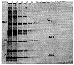
Silver staining is highly regarded for its exceptional sensitivity in protein detection, capable of visualizing nanogram quantities of protein. This technique is particularly valuable for proteomic studies and when working with low-abundance proteins. However, it is a more complex and time-consuming process compared to other staining methods, requiring multiple steps and careful handling to achieve optimal results.
Coomassie Blue staining remains a staple in many laboratories for protein visualization. It offers a good balance between sensitivity and ease of use, making it suitable for routine protein analysis. While not as sensitive as silver staining, it provides reliable results for most standard protein electrophoresis applications. The main challenges include potential background staining and the need for destaining steps to improve contrast.
Fluorescent dyes, such as SYPRO Ruby and Deep Purple, have emerged as powerful alternatives for protein staining. These dyes offer high sensitivity, broad linear dynamic range, and compatibility with mass spectrometry. However, they require specialized imaging equipment and can be more expensive than traditional staining methods.
One of the primary challenges in gel electrophoresis staining is achieving an optimal balance between sensitivity, safety, and cost-effectiveness. Researchers must consider factors such as the type of biomolecule being analyzed (DNA, RNA, or protein), the required detection limit, downstream applications, and available equipment when selecting a staining method.
Another significant challenge is the reproducibility of staining results across different laboratories and experiments. Variations in staining protocols, gel composition, and environmental conditions can affect staining efficiency and consistency. Standardization efforts and the development of more robust staining protocols are ongoing to address these issues.
Prevalent Gel Staining Protocols and Applications
01 Fluorescent dye staining methods
Fluorescent dyes are widely used for staining DNA and proteins in gel electrophoresis. These dyes bind to nucleic acids or proteins and emit light when excited, allowing for visualization of separated molecules. Common fluorescent dyes include ethidium bromide, SYBR Green, and Coomassie Blue. This method offers high sensitivity and can detect even small amounts of biomolecules.- Fluorescent dye staining methods: Fluorescent dyes are widely used in gel electrophoresis for staining nucleic acids and proteins. These dyes bind to the molecules and emit light when excited, allowing for visualization of the separated bands. Common fluorescent dyes include ethidium bromide, SYBR Green, and SYPRO Ruby. This method offers high sensitivity and can detect nanogram quantities of DNA or protein.
- Silver staining techniques: Silver staining is a highly sensitive method for detecting proteins in polyacrylamide gels. It involves the reduction of silver ions to metallic silver on the protein bands. This technique can detect as little as 0.1 ng of protein per band and is compatible with mass spectrometry analysis. The process typically involves fixing, sensitizing, silver impregnation, and development steps.
- Coomassie Blue staining: Coomassie Brilliant Blue is a popular protein stain used in gel electrophoresis. It binds to proteins through ionic interactions between the dye's sulfonic acid groups and the protein's amino groups. This method is less sensitive than silver staining but is simpler to perform and more compatible with downstream applications. Coomassie staining can detect as little as 50 ng of protein per band.
- UV-based detection methods: UV-based detection methods utilize ultraviolet light to visualize DNA or protein bands in gels. This can be achieved through the use of UV-absorbing dyes or by exploiting the natural UV absorbance of nucleic acids. UV transilluminators are commonly used to excite fluorescent dyes or to directly visualize DNA bands. This method allows for real-time monitoring of electrophoresis and is non-destructive to the samples.
- Automated gel imaging and analysis systems: Advanced automated systems have been developed for gel imaging and analysis. These systems integrate various staining methods with high-resolution cameras and image processing software. They can automatically detect and quantify bands, generate reports, and perform comparative analysis across multiple gels. Some systems also incorporate features for excising specific bands for further analysis, enhancing the efficiency and reproducibility of gel electrophoresis experiments.
02 Silver staining techniques
Silver staining is a highly sensitive method for detecting proteins and nucleic acids in gels. It involves the reduction of silver ions to metallic silver, which forms a visible precipitate on the gel. This technique is particularly useful for detecting low abundance proteins and is often used in proteomics research. Silver staining can be more sensitive than traditional Coomassie staining but may require more steps in the protocol.Expand Specific Solutions03 UV-based detection methods
Ultraviolet (UV) light is used to visualize DNA and protein bands in gels, often in conjunction with UV-sensitive dyes or intrinsic fluorescence of certain biomolecules. This method is non-destructive and allows for real-time monitoring of electrophoresis progress. UV transilluminators are commonly used equipment for this purpose, and some systems incorporate digital imaging for analysis and documentation.Expand Specific Solutions04 Automated gel staining systems
Automated systems have been developed to streamline the gel staining process, improving reproducibility and reducing hands-on time. These systems can handle multiple staining protocols, including pre- and post-electrophoresis staining, and often integrate washing and destaining steps. Some advanced systems also incorporate imaging capabilities for immediate analysis of stained gels.Expand Specific Solutions05 Novel staining agents and methods
Researchers are continually developing new staining agents and methods to improve sensitivity, specificity, and ease of use in gel electrophoresis. These innovations include environment-friendly alternatives to traditional toxic stains, reversible staining techniques for downstream applications, and multiplex staining methods for simultaneous detection of different biomolecule types. Some novel approaches also aim to reduce background staining and increase the dynamic range of detection.Expand Specific Solutions
Key Manufacturers in Gel Electrophoresis Staining Industry
The gel electrophoresis staining methods market is in a mature stage, with a steady global market size estimated in the hundreds of millions of dollars. The technology is well-established, with major players like Life Technologies, Bio-Rad Laboratories, and Agilent Technologies offering a range of staining solutions. These companies have developed advanced, highly sensitive stains and imaging systems, indicating a high level of technical maturity. However, there is ongoing innovation in areas like fluorescent stains and rapid, automated staining processes. Smaller specialized firms and academic institutions continue to contribute improvements, suggesting potential for further refinement and niche applications in this essential life science technique.
Life Technologies Corp.
Technical Solution: Life Technologies Corp. has developed advanced staining methods for gel electrophoresis, focusing on fluorescent dyes and sensitive detection systems. Their SYBR Safe DNA gel stain offers a safer alternative to ethidium bromide, with comparable sensitivity[1]. They have also introduced the Invitrogen SYPRO Ruby protein gel stain, which provides high sensitivity for protein detection, with a linear dynamic range over three orders of magnitude[2]. Additionally, their Molecular Probes line includes a variety of specialized stains for specific applications, such as glycoprotein detection and phosphoprotein staining[3].
Strengths: Wide range of specialized stains, high sensitivity, and safer alternatives. Weaknesses: Some stains may be more expensive than traditional options.
Bio-Rad Laboratories, Inc.
Technical Solution: Bio-Rad Laboratories has developed a comprehensive suite of staining solutions for gel electrophoresis. Their Stain-Free technology eliminates the need for staining steps in protein gels, allowing for rapid visualization and quantification[4]. For nucleic acids, they offer the EZ-Vision line of fluorescent dyes, which are non-mutagenic and can be viewed with standard UV transilluminators[5]. Bio-Rad also provides traditional stains like Coomassie Blue and silver stain kits, optimized for high sensitivity and low background[6]. Their approach focuses on offering a range of options to suit different experimental needs and budget constraints.
Strengths: Innovative Stain-Free technology, diverse product range, and optimized traditional stains. Weaknesses: Some advanced technologies may require specialized equipment.
Innovative Staining Approaches for Gel Electrophoresis
Method of separating protein, method of staining protein and liquid protein-staining agent and protein-staining kit to be used in these methods
PatentInactiveUS20090223823A1
Innovation
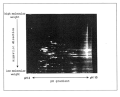
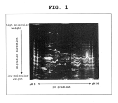
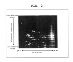
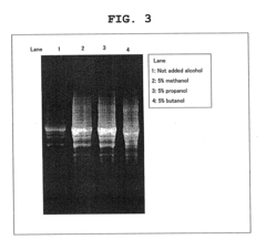
- A method using a staining liquid containing a staining agent, a surfactant such as sodium dodecylsulfate, and a buffer solution for gel electrophoresis, which allows for simultaneous staining and surfactant treatment, enabling quick and sensitive protein separation without the need for extensive washing.
Method of silver staining for bio-related substance
PatentWO2006035781A1
Innovation
- A method involving sequential immersion of gels in aqueous sodium thiosulfate, ethanol-acetic acid, and ethanol-silver nitrate solutions without washing, optimized to enhance detection sensitivity and minimize chemical modification, using polyacrylamide or agarose gels for electrophoresis-separated bio-related substances.
Safety and Environmental Considerations in Gel Staining
Safety and environmental considerations play a crucial role in gel electrophoresis staining methods. The selection of optimal staining techniques must balance effectiveness with potential risks to researchers and the environment. Traditional staining methods often involve hazardous chemicals, necessitating careful handling and disposal procedures.
Ethidium bromide, a commonly used fluorescent dye, is highly mutagenic and potentially carcinogenic. Laboratories employing this stain must implement strict safety protocols, including personal protective equipment (PPE) usage and specialized waste management. Alternative stains, such as SYBR Safe and GelRed, offer reduced toxicity while maintaining comparable sensitivity. These safer options minimize health risks and simplify disposal processes.
Environmental impact is another critical factor in staining method selection. Many conventional stains contain heavy metals or other pollutants that can harm ecosystems if improperly disposed of. Laboratories must adhere to local and national regulations regarding chemical waste management. Some institutions have implemented recycling programs for certain stains, reducing environmental burden and operational costs.
The development of non-toxic, biodegradable stains represents a significant advancement in gel electrophoresis safety. These eco-friendly alternatives, such as methylene blue and Coomassie Brilliant Blue, offer reduced environmental impact without compromising staining quality. However, their sensitivity may be lower compared to fluorescent dyes, necessitating careful consideration of experimental requirements.
Proper ventilation and containment systems are essential when working with volatile staining agents. Fume hoods and specialized imaging systems help minimize researcher exposure to potentially harmful vapors. Additionally, automated staining systems can further reduce direct contact with hazardous materials, enhancing laboratory safety.
Training and education on safe staining practices are paramount. Researchers must be well-versed in proper handling, storage, and disposal techniques for all staining agents used in their work. Regular safety audits and updates to laboratory protocols ensure ongoing compliance with evolving safety standards and environmental regulations.
In conclusion, the selection of optimal staining methods in gel electrophoresis must carefully weigh safety and environmental considerations against experimental needs. By prioritizing less toxic alternatives, implementing robust safety measures, and considering environmental impact, laboratories can maintain high-quality results while minimizing risks to personnel and the ecosystem.
Cost-Effectiveness Analysis of Staining Methods
When selecting optimal staining methods for gel electrophoresis, cost-effectiveness is a crucial factor to consider. This analysis examines the economic aspects of various staining techniques, balancing their performance with associated costs.
Ethidium bromide (EtBr) has long been a popular choice due to its low cost and high sensitivity. The reagent itself is inexpensive, and the staining process is relatively quick. However, the total cost must account for proper disposal of EtBr-contaminated materials and potential health risks, which may increase long-term expenses.
SYBR Green offers improved safety over EtBr and comparable sensitivity. While the initial cost of SYBR Green is higher, its increased photostability allows for extended imaging times, potentially reducing the need for repeated experiments. This factor should be considered when calculating overall costs.
Silver staining, known for its high sensitivity, particularly for protein gels, involves a more complex and time-consuming process. The reagents are more expensive than fluorescent dyes, and the multi-step procedure increases labor costs. However, its ability to detect very low amounts of protein may reduce the need for sample concentration or gel replication, potentially offsetting some expenses.
Coomassie Brilliant Blue is a cost-effective option for protein staining, with relatively inexpensive reagents and a straightforward protocol. While less sensitive than silver staining, its reliability and ease of use make it a economical choice for routine protein analysis.
Fluorescent dyes like SYPRO Ruby provide high sensitivity and a wide linear range for protein quantification. The initial cost of these dyes is high, but their ability to detect a broad range of proteins and compatibility with mass spectrometry may justify the expense in certain research contexts.
When considering cost-effectiveness, it's essential to factor in not only the direct costs of reagents but also indirect expenses such as equipment requirements, labor time, and potential need for repeated experiments. For instance, while some methods may have higher upfront costs, they might prove more economical in the long run by providing more reliable or sensitive results, reducing the need for replications.
The choice of staining method should also consider the specific requirements of the experiment. For high-throughput applications, a faster and more automated staining process may be more cost-effective despite higher reagent costs. Conversely, for less frequent analyses, a more labor-intensive but cheaper method might be preferable.
In conclusion, the most cost-effective staining method depends on various factors including the frequency of use, required sensitivity, safety considerations, and available resources. A comprehensive analysis should weigh these factors against the specific needs of the research to determine the optimal balance between cost and performance.
Unlock deeper insights with Patsnap Eureka Quick Research — get a full tech report to explore trends and direct your research. Try now!
Generate Your Research Report Instantly with AI Agent
Supercharge your innovation with Patsnap Eureka AI Agent Platform!
