Method and system for eliminating geometrical artifacts in CT (Computerized Tomography) image

A CT image and geometric artifact technology, applied in computer tomography scanners, echo tomography, etc., can solve problems such as irreversibility, influence on correct interpretation of CT images, and rough correction methods, so as to eliminate geometric artifacts and artificial The error cannot be recovered and the effect of eliminating the existence of manual adjustment error

CN102652674AInactive Publication Date: 2012-09-05CAPITAL NORMAL UNIVERSITY
1 Cites 23 Cited by

Patent Information

Authority / Receiving Office
CN · China
Current Assignee / Owner
Publication Date
2012-09-05
Estimated Expiration
Not applicable · inactive patent

Smart Images

  • Figure 1
    Figure 1
  • Figure 2
    Figure 2
  • Figure 3
    Figure 3
Patent Text Reader

Abstract

The invention relates to a method and system for eliminating geometrical artifacts in a CT (Computerized Tomography) image. The method comprises the following steps of: performing CT scanning on a die body to obtain the projection coordinate of the mass center of the die body on a CT detector; defining geometrical parameters according to the projection coordinate of the mass center of the die body on the CT detector, and substituting the geometrical parameters in a reconstruction formula, wherein the geometrical parameters include: a distance h of a projection point of the turntable rotating center on the detector and a center of the detector, an included angle alpha between an ideal position of a straight line where the detector id and an actual position, and a distance SDD between the projection point of the turntable rotating center on the detector and an X ray source focus; performing CT scanning on an object to be imaged to obtain fan-beam projection parameters of the object to be imaged; and processing the fan-beam projection parameters of the object to be processed by the reconstruction formula to obtain CT image data of the object to be imaged and further obtain a CT image with the CT image data of the object to be imaged serving as a gray level. According to the technical scheme, the method and the system can be used for accurately obtaining the CT image without the geometrical artifacts.
Need to check novelty before this filing date? Find Prior Art

Description

technical field

[0001] The invention relates to the field of CT applications, in particular to a method and system for eliminating geometric artifacts in CT images. Background technique

[0002] CT (X-ray computed tomography, computed tomography) is an important non-destructive testing technology. Its working principle is: using different objects to absorb and transmit X-rays differently, using X-ray sources to emit X-rays to penetrate object, and then use a highly sensitive detector to detect the X-ray intensity after penetrating the object, and process the acquired intensity data by the processing device to obtain a cross-sectional or three-dimensional image of the object. By observing the image, you can Learn about the internal structure of objects. In the medical field, by using CT to detect the human body, a cross-sectional or three-dimensional image of the inspected part of the human body can be obtained, and then small lesions in any part of the body can be found. ...

Examples

Embodiment Construction

[0062] The principles and features of the present invention are described below in conjunction with the accompanying drawings, and the examples given are only used to explain the present invention, and are not intended to limit the scope of the present invention.

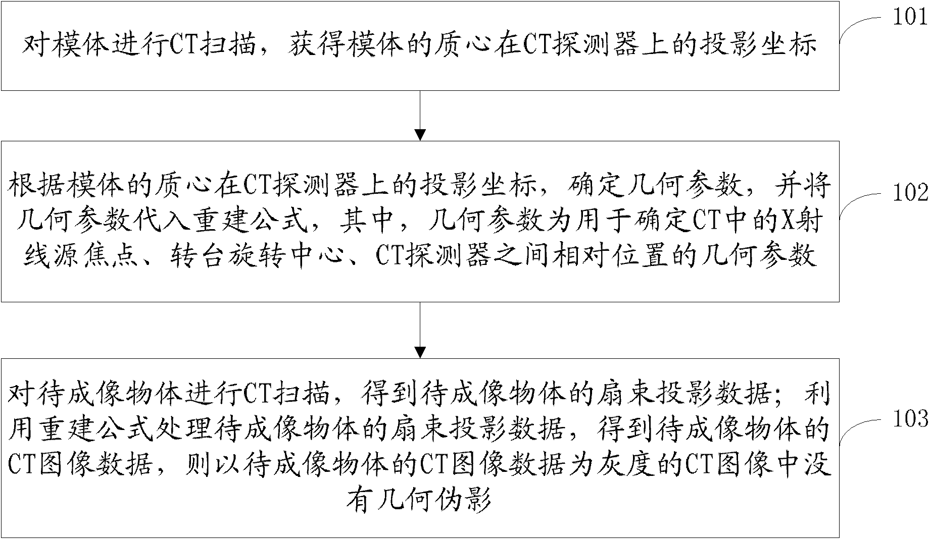
[0063] figure 1 It is a flowchart of a method for eliminating geometric artifacts in CT images provided by the present invention. Such as figure 1 As shown, the method includes:

[0064] Step 101: CT scan the phantom to obtain the projected coordinates of the phantom's centroid on the CT detector.

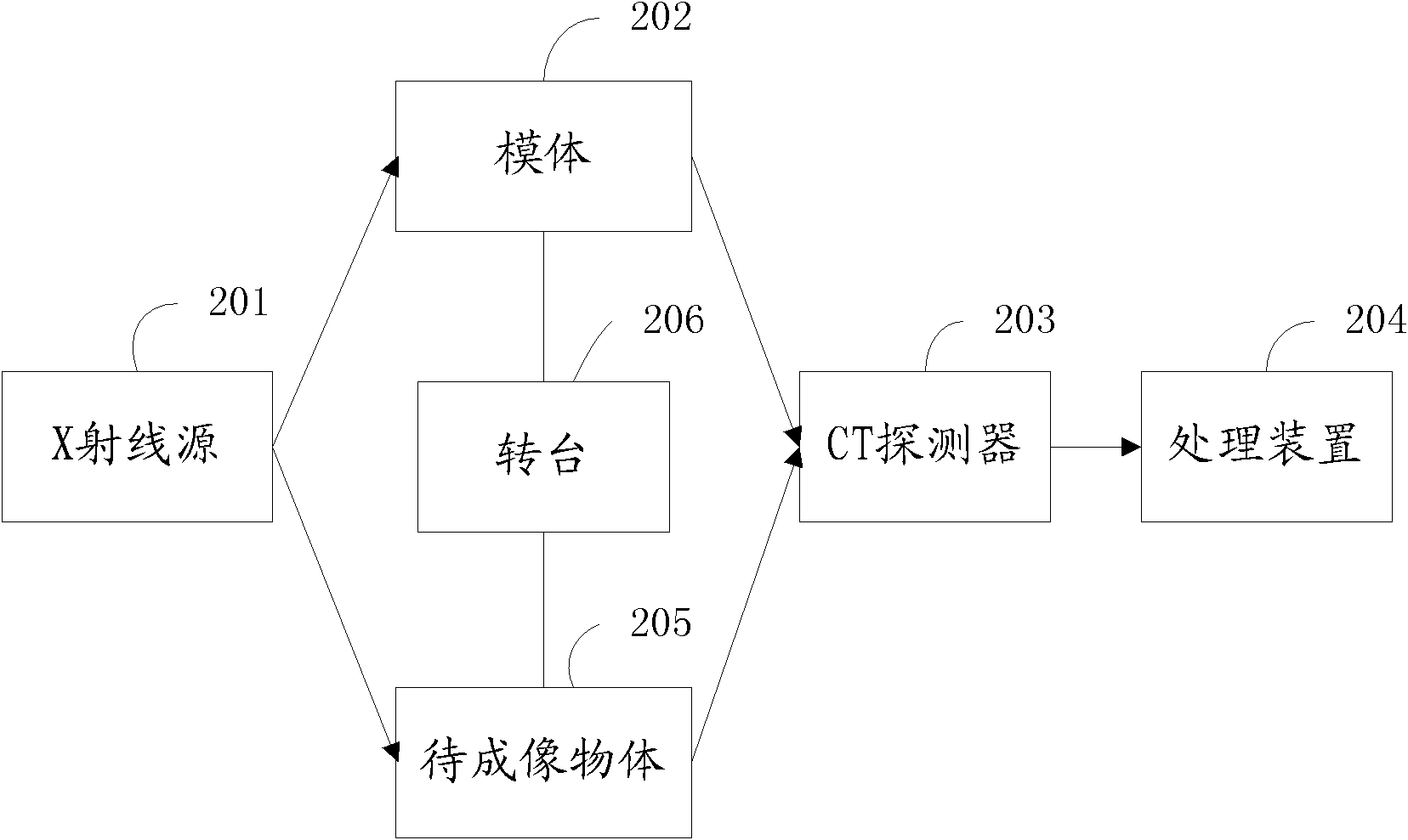
[0065] Here, CT includes an X-ray source, a turntable and a CT detector, wherein the X-ray source can emit X-rays passing through the phantom, and the CT detector can accurately detect the intensity of the X-rays passing through the phantom to itself. In this step, the CT scanning of the phantom and the CT scanning of the object to be imaged in step 103 are all through the relative movement between the CT detector and...