
Apparatus and method for characterization of acute otitis media
a technology of otitis media and appendix, which is applied in the field of devices for the detection of middle ear effusion, can solve the problems of delayed speech and language skills development, no one can achieve the diagnostic accuracy of invasive myringotomy and tympanocentisis, and the overall likelihood of obtaining an accurate diagnosis using any of the non-invasive methods is no better than 50%, so as to achieve the effect of determining the viscosity
Patent Information
- Authority / Receiving Office
- US · United States
- Patent Type
- Patents(United States)
- Current Assignee / Owner
- Publication Date
- 2020-05-26
Smart Images
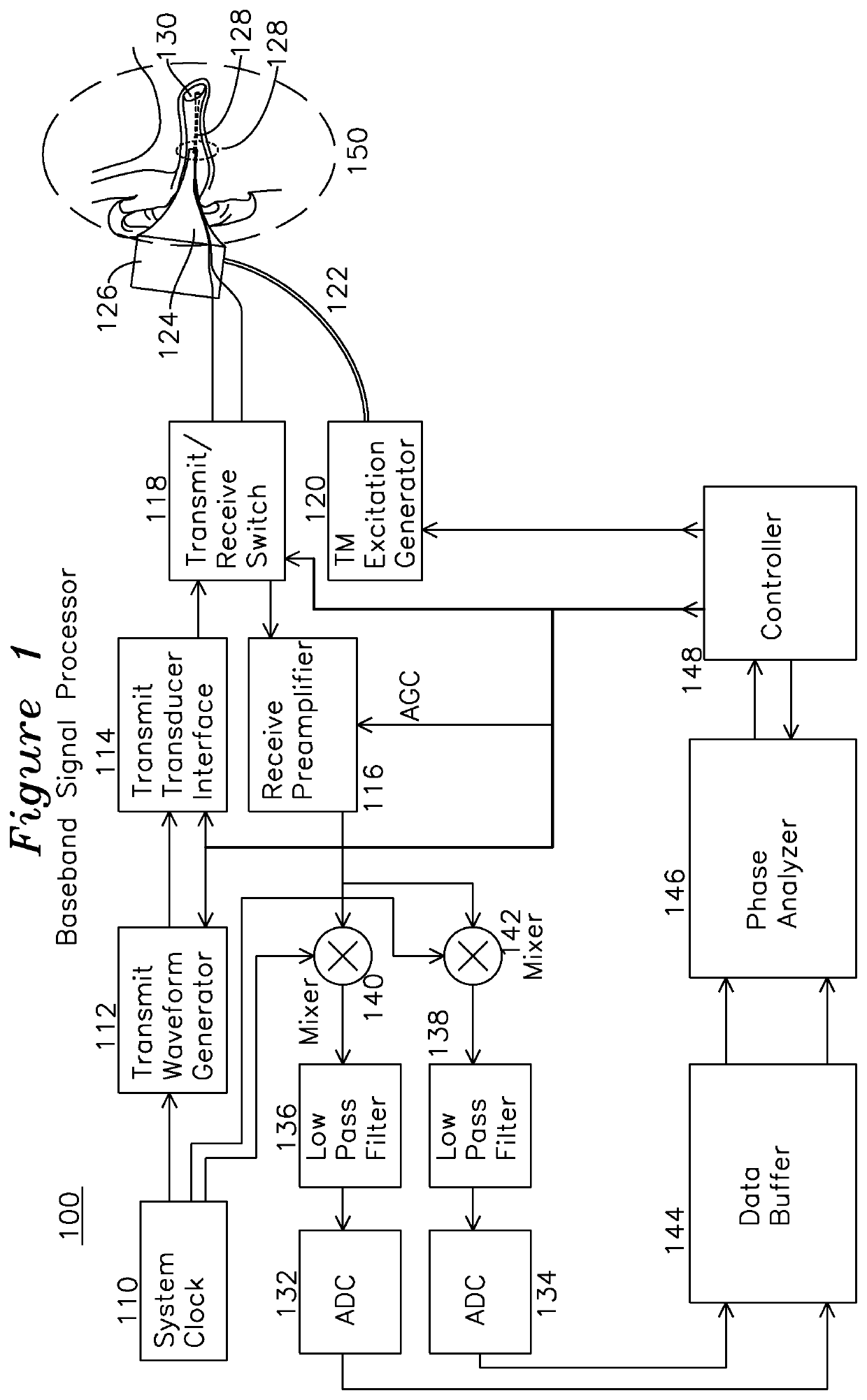
Figure 1 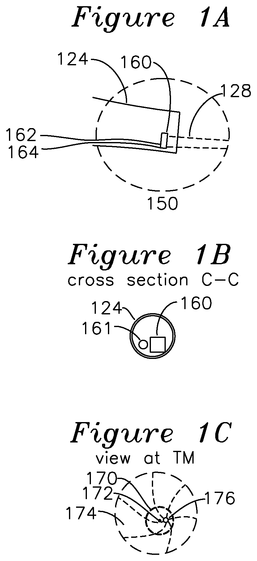
Figure 2 
Figure 3
Abstract
Description
FIELD OF THE INVENTION
[0001] The present invention relates to a device for the detection of middle ear effusion with discrimination of fluid type. In particular, the invention relates to the characterization of middle ear effusion behind the tympanic membrane by stimulating the tympanic membrane using a low frequency excitation such as acoustic and measuring the displacement behavior with a comparatively higher frequency excitation such as ultrasound.BACKGROUND OF THE INVENTION
[0002] Acute otitis media (AOM) is an inflammatory process in the middle ear and is the most common clinical condition seen by pediatricians in children fifteen years and younger. AOM is generally associated with the presence of a middle ear effusion and is considered a middle ear inflammation. Complications of undiagnosed AOM can include hearing loss. Left untreated in children, recurrent AOM can also lead to delays in the development of speech and language skills.
[0003] There are two key factors in the diagnosi...
Examples
Embodiment Construction
[0044]FIG. 1 shows a signal processor for an example embodiment of a tympanic membrane characterization system. Region 150 (shown in magnified view FIG. 1A) includes a cross section view of a middle ear and tympanic membrane 130 of a subject being examined. The tympanic membrane 130 is interrogated by an ultrasound beam 128 from an ultrasound transducer 160 (shown in FIG. 1A) which is optionally mounted on the inner surface of a speculum tip 124, and is detachable from an otoscope speculum mounting adapter 126. In one embodiment of the invention, an optical source 161 seen in the FIG. 1B cross section view of FIG. 1A, generates a visual indication the region of insonification by the ultrasound by illumination of a target or region of the tympanic membrane within the ear canal, as seen in FIG. 1C. FIG. 1C shows the view of the tympanic membrane as seen through the speculum, including the tympanic membrane 174, “cone of light”176, which is a reflective region of the TM which is normal...