
Transverse cavity device and method
a transverse cavity and device technology, applied in the field of bone compression fracture treatment, can solve the problems of reducing the structural integrity of the vertebrae, affecting the patient, and reducing the height of the vertebrae, so as to increase the area of the “footprint” and reduce the total distraction for
Patent Information
- Authority / Receiving Office
- US · United States
- Current Assignee / Owner
- Publication Date
- 2005-08-18
- Estimated Expiration
- Not applicable · inactive patent
Smart Images
Figure 1 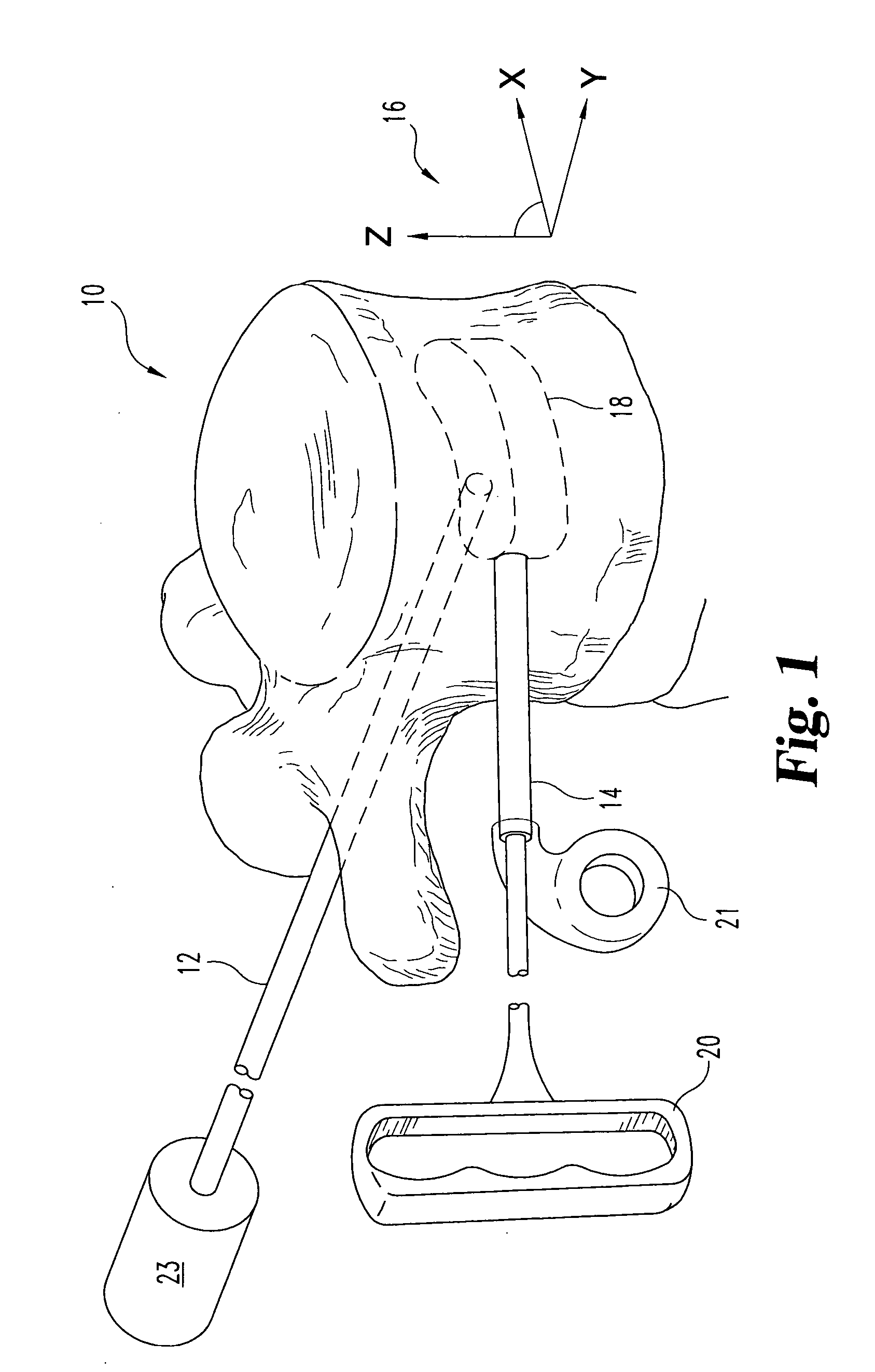
Figure 2 
Figure 3
Abstract
Description
CROSS-REFERENCE TO RELATED CASES
[0001] The present case claims the benefit of, and incorporates by reference the following U.S. provisional applications:
[0002] U.S. Provisional Patent Application Ser. No. 60 / 227,050 filed Aug. 21, 2000, entitled “Vertebroplasty Cavity Creation using an Expanding Tube” and,
[0003] U.S. Provisional Patent Application Ser. No. 60 / 225,191 filed Aug. 14, 2000, entitled “Vertebral Body Expander.”FIELD OF THE INVENTION
[0004] The present invention relates generally to the treatment of compression fractures in bones, and more specifically to a device and a method for cutting a “transverse” cavity in the bone as one part of a therapy. BACKGROUND OF THE INVENTION
[0005] The human spine consists of a complex set of interrelated anatomic elements including a set of bones called vertebral bodies. Intervertebral discs separate most vertebral bodies. These discs includes a “spongy” nucleus pulpous surrounded by an annulus fibrosis “membrane.” The annulus fibrosi...
Examples
Embodiment Construction
[0034]FIG. 1 is a phantom view of a vertebral body showing a transverse cavity 18 and a coordinate system 16. This figure shows a vertebral body 10 in isolation. Two possible surgical entry points into the vertebral body contemplated within the scope of the invention are illustrated. One entry point is “transpedicular.” This approach is indicated by the physical location of tube 12, traveling through the pedicle into the vertebral body 10. Another approach is “extra-pedicular.” This access approach is illustrated by tool 14 entering the vertebral body at a location lateral of the transpedicular approach on the posterolateral corner of the vertebral body.
[0035] The typical surgery will include a small incision in the back adjacent to the vertebral body. Next, a small gauge needle or guide-wire is introduced to confirm proper positioning under fluoroscopy. Physicians typically utilize an 11-gauge needle for the transpedicular approach and a larger needle or tube (up to 6 mm ID) for t...