
Positioning support and fetal heart rate registration support for ctg ultrasound transducers
a technology of ultrasound transducers and support posts, which is applied in the field of positioning support and fetal heart rate registration support of ctg ultrasound transducers, can solve the problems of difficult finding the optimal transducer position, long period of signal loss reported during fhr monitoring, and strong limitations in clinical practice to interpret fhr traces, etc., to achieve better assessment of fetal health status, facilitate the initial placement of the transducer, and facilitate the effect of fetal heart ra
Patent Information
- Authority / Receiving Office
- US · United States
- Patent Type
- Applications(United States)
- Current Assignee / Owner
- Publication Date
- 2019-05-09
Smart Images
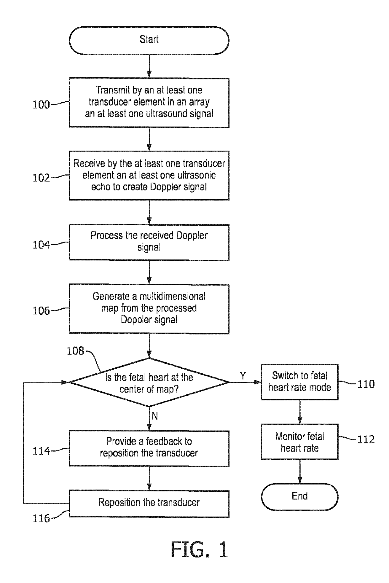
Figure 1 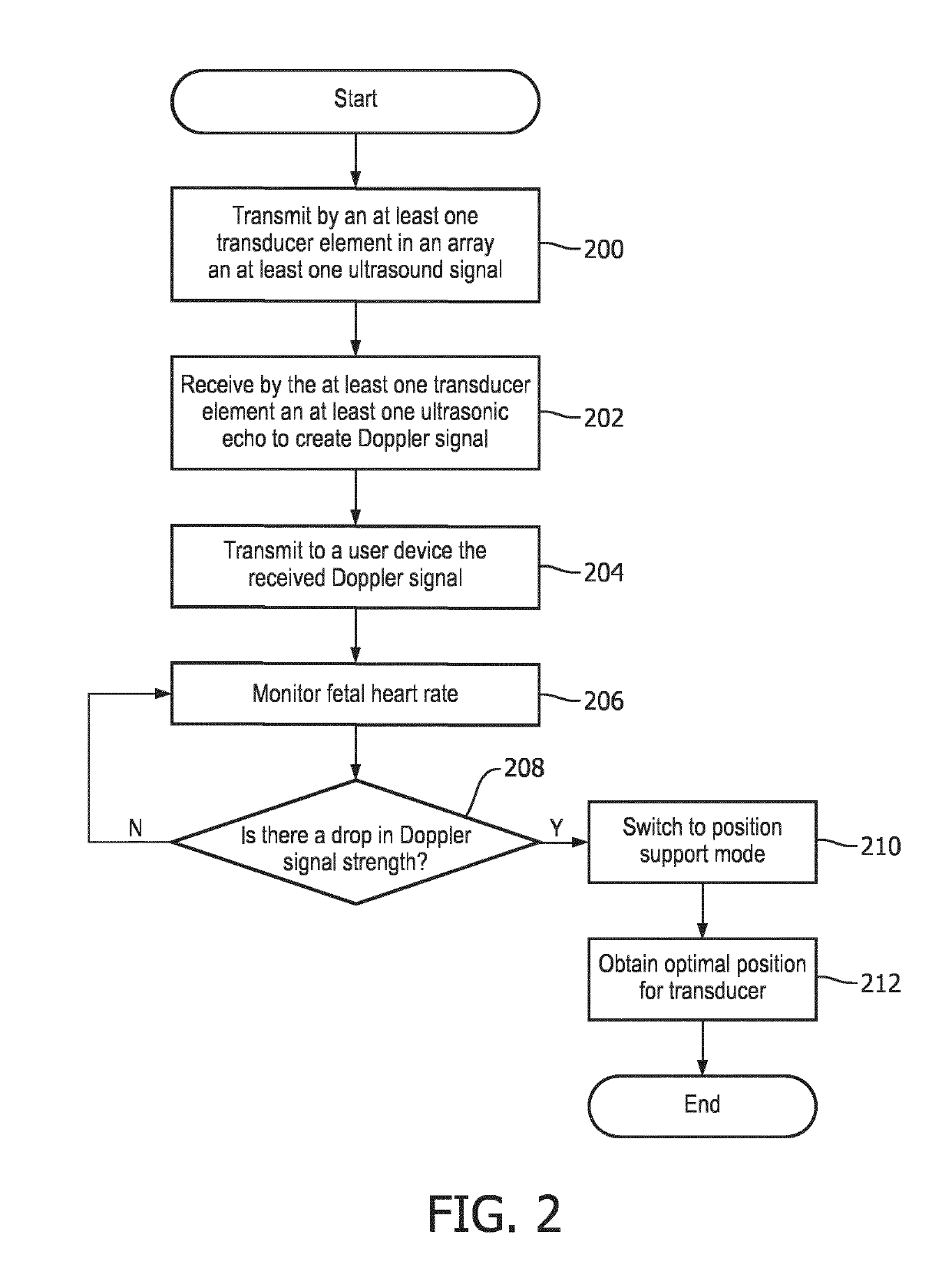
Figure 2 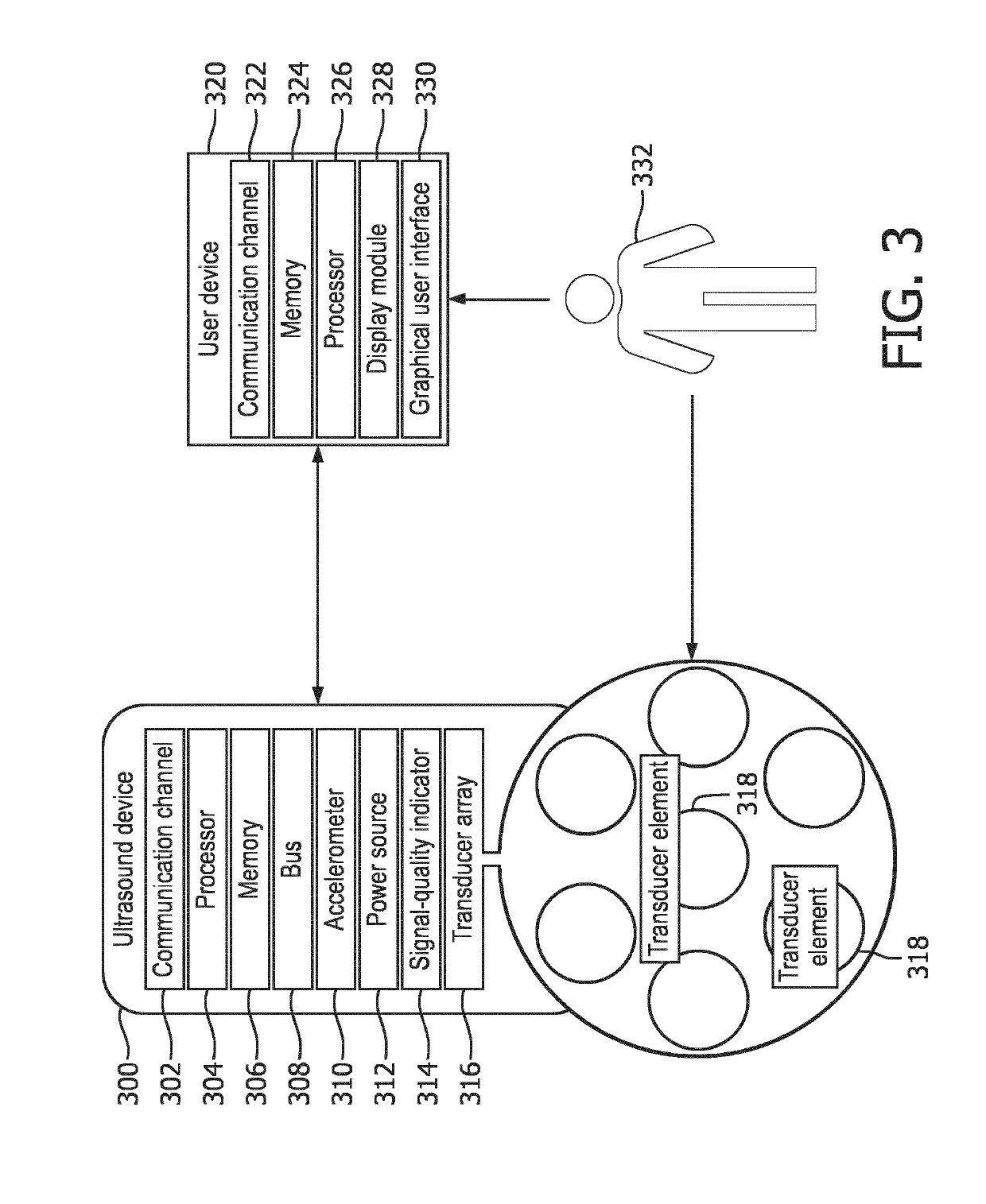
Figure 3
Abstract
Description
BACKGROUND OF THE INVENTION
[0001] Measuring the fetal heart rate (fHR) during labor and delivery is the most commonly applied obstetric procedure to assess the health state of the fetus and to determine if early intervention is required. However, for Doppler ultrasound (US) as well as for abdominal electrodes, long periods of signal loss have been reported during fHR monitoring. For the assessment of the health state of the fetus, these long signal loss periods strongly limit the interpretation of the fHR traces in clinical practice.
[0002] The ultrasound Doppler recording quality highly depends on the correct placement of the transducer on the maternal abdomen, since the fetal heart needs to be located within the ultrasound beam. However, finding the optimal transducer position can be a challenging task. In clinical practice, skilled nurses palpate the maternal abdomen and determine the fetal presentation from the outside. By relying on a signal-quality indicator and by listening to t...
Examples
Embodiment Construction
[0030]The following are definitions of terms as used in the various embodiments of the present disclosure.
[0031]The term “depth” as used herein refers to a distance measured from the transducer array plane to a point within the maternal abdomen volume that is being scanned. Depth is set to have a value of “0” at the inner wall of the maternal abdomen and a maximum value at the farthest point from which a Doppler signal can be obtained. The depth may be, in some embodiments, the middle of the sample volume from which the Doppler signal is calculated.
[0032]The term “channel” as used herein refers to a wired or wireless data pathway allocated to each transducer element in a transducer array disclosed in the present disclosure. One channel is set aside for processing the Doppler signal received from all transducer elements when the ultrasound device is used in the fetal heart rate mode (fUR-mode).
[0033]The term “feedback” as used herein refers to an audio, visual, or combined audio and ...